DOI:
10.1039/C4RA13553A
(Paper)
RSC Adv., 2014,
4, 64967-64976
Transcriptional regulation of the pregnane-X receptor by the Ayurvedic formulation Chandraprabha Vati
Received
31st October 2014
, Accepted 11th November 2014
First published on 24th November 2014
Abstract
Chandraprabha Vati (CPV), a multi-ingredient phyto formulation, is widely used in Ayurveda for the treatment of liver and kidney disorders. In this study, we attempt to elucidate the mode of action of CPV. We specifically focus on the effects of CPV on the transcriptional regulation of Pregnane-X-Receptor (PXR) and its subsequent effects on interleukins, Peroxisome Proliferator-Activated Receptor-γ (PPARγ) and type 4 Glucose Transporter (GLUT4). Our results show that CPV up-regulates PXR moderately in contrast to its individual ingredients such as chebulinic acid or linalool that down regulate PXR. Further, the expression of Cytochrome P450 3A4 (CYP3A4), the gene involved in drug elimination, is only moderately up-regulated by CPV, again in contrast to the effect of some of its ingredients. CPV down regulates the levels of pro-inflammatory cytokines and upregulates the levels of PPARγ, which in turn upregulates GLUT4 expression. These together suggest that the therapeutic properties of CPV can be attributed to its multi-pronged action, viz., prevention of inflammation, moderate expression of PXR that activates several downstream pathways and tight regulation of CYP3A4 thereby slowing down the elimination of the chemical constituents. In addition, these results emphasize on the need for multi-ingredient approach towards designing effective therapeutic formulations.
1. Introduction
Ayurvedic formulations have been traditionally used in India to treat various human disorders for many centuries. While Ayurvedic practitioners have emphasized the beneficial effects of these formulations, their molecular mechanisms of action remain unclear. Such an understanding is quintessential for its greater global acceptance, visibility and effective use.1 For example, the elucidation of piperine, an alkaloid found in the fruits of the Piperaceae family of plants, as a “bioenhancer” spurred the interest of integrating ancient knowledge with modern medicine.2 Subsequently, Risorine, an antitubercular formulation containing piperine, has been shown to be as effective as the commercial rifampin preparations.3 The elucidation of the mechanism of action of Withania somnifera in treating Alzheimer's disease has identified a unique strategy, viz., targeting the periphery, for the rapid elimination of amyloid beta (Aβ).4 Studies on the immunomodulatory properties of Tinospora cordifolia5,6 and Withania somnifera7 has now led to the development of an Ayurvedic product called Immuforte® that has been shown to be clinically safe.8 Thus, elucidation and documentation of the effects and mechanisms of action of Ayurvedic formulations is essential for their global recognition.
The efficacy of Ayurvedic formulations is usually attributed to the multiple phytochemical ingredients in it. It is believed that the numerous phytochemicals present in an Ayurvedic preparation bind to and modulate multiple targets thereby providing a clinical efficacy beyond the reach of ‘single-molecule drugs’.9,10 For example, the extracts of cannabis have been shown to have more effective antispastic effects than its main ingredient tetrahydrocannabinol.11–13 The beneficial antidepressant action of Hypericum perforatum extract is attributed to the cooperative action of several compounds in St. John's Wort.14 Further, it is also known that the number of active components in an Ayurvedic extract need not necessarily determine the number of targets.15,16 These studies, in addition to substantiating the role of synergism in the efficacy of Ayurvedic preparations, also demonstrate the complexities in elucidating the underlying mechanisms of action. While synergism in the beneficial roles of Ayurvedic formulations has been clearly substantiated, what is unclear is whether a single formulation can have multiple compounds that differentially regulate the same target. This is particularly important in the drug metabolic pathways since a critical balance is essential to regulate the elimination of a phytochemical and at the same time ensure sufficient residence time of the phytochemical in the system for its action.
The residence time of a molecule in the biological system is dictated by the extent of activation of a class of nuclear receptors. Pregnane-X-Receptor (PXR) is an important nuclear receptor whose primary function is to detect endogenous and exogenous ligands and stimulate their clearance mechanisms in the body.17,18 This ligand-elimination function of PXR also implicates it in various diseases and disorders. For example, the secretion of the bile acids, bilirubin and cholesterol by liver to the small intestine is mediated by PXR.19 The differential expression and activation of PXR regulates bile acids and phospholipid transporter genes required for the hepatobiliary transport systems.19,20 In case of hepatobiliary obstruction, it has been shown that the activation of PXR by rifampin, a known activator of PXR, improves biliary secretion which, otherwise, would lead to cholestatic liver disorder.21 PXR expression levels have also been implicated in kidney disorders such as glomerular sclerosis, tubule-interstitial disease, renal lipotoxicity, kidney fibrosis and hypertension.22 The activation of PXR alters the expression of several molecular markers such as TNF-alpha, interleukin-6, interleukin-1, NF-kappa-B, NADPH oxidase, TGF-beta1, LCAT (lecithin cholesterol acyl transferase). While activation of PXR reduces the expression of TNF-alpha, interleukin-6 and interleukin-1, it activates NF-kB, NADPH oxidase, TGF-beta1 and LCAT.22 This plays an important role in the prevention of kidney diseases such as renal fibrosis and glomerular sclerosis. PXR also has recently been identified to possess a role in regulating blood glucose levels via Peroxisome Proliferator-Activated Receptor-γ (PPARγ) mediated GLUT4 (type 4 Glucose Transporter) transport mechanism.23–25
Thus, it is clear that PXR is a good pharmacological target for several diseases in addition to being a xenobiotic receptor involved in “drug” elimination. Hence, modulation of PXR not only determines the residence times of the drugs but also has broader implications in the treatment of several diseases. In this study, we investigate the modulation of PXR by the ingredients of a well-known Ayurvedic formulation, Chandraprabha Vati (CPV). CPV is prepared from 29 plant ingredients namely, Psoralea corylifolia, Acorus calamus, Cyperus rotundus, Andrographis paniculata, Tinospora cordifolia, Cedrus deodara, Curcuma longa, Aconitum heterophyllum, Berberis aristata, Piper longum, Plumbago zeylanica, Coriandrum sativum, Terminalia chebula, Terminalia bellerica, Phyllanthus emblica, Piper nigrum, Embilia ribes, Scindapsus officinalis, Zingiber officinale, Hordeum vulgare, Operculina turpethum, Baliospermum montanum, Piper chaba, Cinnamomum tamala, Cinnamomum zeylanica, Elettaria cardamomum, Commiphora wightii, Bambusa bambos and Cinnamomum camphora. CPV has been used to treat a wide range of diseases that include urinary tract infections, urinary calculi, colic pain, rhinitis, bronchitis, asthma, eczema, dermatitis, liver disorders, spleen disorders, anemia, eye infections, diabetes, back ache etc.26 Water is the recommended vehicle for administering CPV. The use of a single formulation to treat different diseased conditions suggests that there may exist a common target that can be involved in activation of diverse pathways resulting in the curative properties of the formulation.
Recently, it has been reported that PXR exhibits anti-fibrotic nature by interfering with TGF-β and interleukin pathways.22 This may have an implication in the context of liver and kidney disorders, which has not been proved yet. The PXR receptor has now been recognized to play a major role for multi-drug formulations and may serve to improve their efficacy and mitigate toxicity.27 We believe that the multi-ingredient Chandraprabha Vati may have transcriptional regulation of this receptor that may serve to reveal its mechanism of therapeutic action. We used few representative ingredients of CPV for elucidating the mechanism of action. Our choice of ingredients is based on the predominant phytoconstituents reported in each of the plant ingredient.
2. Materials and methods
2.1. Materials
Rifampin, piperine and linalool were purchased from Alfa Aesar, USA and chebulinic acid was procured from Natural Remedies, India. Quercetin and ellagic acid were purchased from Sigma Aldrich, USA. Chandraprabha Vati (Impcops, Chennai) was procured from Ayurvedic medical store at Thanjavur.
2.2. In silico studies
The structure of PXR was obtained from the protein databank (pdb id: 1SKX). Hydrogen atoms were added to the protein consistent with pH 7.0 using the protein preparation wizard in the Schrödinger suite.28 Further, the protein's hydrogen bond network was also optimized using the wizard. The so-prepared structure was then subject to energy minimization and the termination condition for minimization was fixed as the step when the root mean square deviation of the heavy atoms in the structure relative to the starting structure exceeded 0.3 Å. This process also ensures that the hydrogen atoms are placed in optimized geometries. The protein thus prepared was used for docking of the ligands as described below.
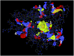
Potential binding sites in PXR were predicted using the SiteMap tool in the Schrödinger suite.29,30 Fig. 1 shows the probable binding sites in PXR labeled 1, 2, 3, 4 and 5. Five different binding sites were identified in PXR. Of these predicted sites, site 1 had the highest score.
 |
| Fig. 1 Predicted binding sites in PXR. | |
Table 1 gives the site scores and volumes of the predicted binding sites. Interestingly, site 1 also overlaps with the rifampin-binding site (Fig. 2).
Table 1 Site scores and volumes of predicted binding sites in PXR
Site |
Score |
Volume (Å3) |
1 |
1.20 |
452 |
2 |
0.89 |
192 |
3 |
0.75 |
170 |
4 |
0.70 |
129 |
5 |
0.68 |
85 |
 |
| Fig. 2 Overlap of rifampin-binding site and the site 1. | |
Rifampin is a molecule that is well known for its affinity to PXR. Hence binding of a ligand to site 1 is more likely to result in the activation of this receptor. Receptor grid was then generated for site 1 using Glide module (v 5.8) of the Schrödinger suite. The grid box and center were set to default.
The structures of the ligands were obtained from the pubchem database.31 LigPrep module (version 2.5) of the Schrödinger suite was used to generate conformers of the ligands. The ligands were then docked using the extra precision mode in the Glide module32–34 of the Schrödinger suite.
2.3. In vitro studies
As PXR is predominantly expressed in the liver, HepG2 (hepatocellular carcinoma) cells (NCCS, Pune) were used for all in vitro studies. The concentrations of the individual ligands used in the study were between 1 to 1000 nM. The choice of this range was based on the concentration of rifampin that was reported to activate PXR.35,36 As CPV is a multi-ingredient formulation, the amount of CPV was used as weight per volume ratios in the experiments. A concentration of 40 μg mL−1 of CPV corresponds to the therapeutic dose prescribed by traditional medical practitioners (250 mg per dose per person).
2.4. Cell viability assay
Cell viability was determined using MTS assay as reported in the literature.37 Briefly, 10
000 cells were seeded in a 96-well plate and cultured in Dulbecco's Modified Eagle Medium (DMEM, Invitrogen) supplemented with 4 mM L-glutamine (Invitrogen, USA) and 4.5 g L−1 glucose. The cells were incubated with the test samples 24 h after seeding. The incubation was carried out at 37 °C with 5% carbon dioxide for 24 h. At the end of this incubation the cells were washed with phosphate buffered saline (PBS) to remove non-adherent cells. To this, 25 μL of MTS reagent (CellTiter 96 AQueous one solution, Promega) and 1 mL of serum-free media were added and incubated at 37 °C for 2 h. The reaction was stopped by the addition of sodium dodecyl sulphate (SDS) solution. The absorbance was read at 490 nm using spectrophotometry (Lambda 25, Perkin-Elmer, USA).
2.5. Intracellular reactive oxygen species (ROS) assay
Ten thousand cells were incubated with different concentrations of the test samples. The intracellular ROS levels were determined after 8 hours of seeding using the following procedure. After the incubation period, cells were washed with PBS and followed by addition of 150 mL of 0.01 M dichlorofluorescein diacetate (DCF-DA) (Sigma Aldrich, USA) reagent and kept at 37 °C for 1 h in dark. The excess DCF-DA reagent was then removed and the cells were washed with PBS followed by addition of 100 mL of PBS. The absorbance was then measured at 485 nm using multimode reader (Infinite 200M, Tecan, USA).
2.6. Interleukin assay
The interleukin expression was measured using multi-analyte ELISA kit from Qiagen, USA using the manufacturer's protocol. Twenty five thousand cells were seeded in a 96-well plate and incubated with CPV (40 μg mL−1). After 24 h, the supernatant was collected and used for interleukin determination using sandwich ELISA procedure.
2.7. Gene expression studies
The expression of PXR and CYP3A4 genes was determined using quantitative RT-PCR. Hundred thousand cells were seeded and incubated with different concentrations of the test samples for 24 h. After incubation, the total RNA was isolated using trizol (Invitrogen, USA) following the procedure as described in the literature.38 In brief, 1 mL of trizol was added to the samples and kept for 0.5 h at room temperature. The solution was collected, and RNA was extracted with 0.2 mL of chloroform (Merck, India). The solution was centrifuged at 12
000 rpm for 15 minutes at 4 °C and the extracted RNA was stabilized using 70% ethanol prepared with nuclease-free water (Qiagen, USA). The RNA was centrifuged using a QIA shredder spin column (Qiagen) and dissolved in RNase-free water (Qiagen, USA). cDNA was obtained after a two-step reaction and subjected to a real-time RT-PCR (Eppendorf AG22331, Germany) using master mix from Kapa, USA. Table 2 shows the primers designed on the basis of published gene sequences. Quantitative values were determined by the δ–δ method and normalized with the house-keeping gene, β-actin, and the control.
Table 2 Forward and reverse primers used for the gene expression studies
Gene |
Forward primer |
Reverse primer |
PXR |
5′-CCCAGCCTGCTCATAGGTTC-3′ |
5′-CTGTGATGCCGAACAACTCC-3′ |
CYP3A4 |
5′-GGGCTTTTGTATGTTTGAC-3′ |
5′-GGTGTTGAGGATGGAATG-3′ |
β-Actin |
5′-GTCATCACCATTGGCAATGAG-3′ |
5′-CGTCATACTCCTGCTTGCTG-3′ |
2.8. Quantification of PPARγ and GLUT4
About 0.2 × 10⁶ cells were seeded and grown to 70% confluence in a 24-well tissue culture plate conditioned with 5% CO2 atmosphere at 37 °C. Cells were then treated with 10, 20, 30 and 40 μg mL−1 of Chandraprabha Vati for 48 h followed by cell lysis using Tween 80, the resultant mixture was then mixed and centrifuged for 10 min at 3000 rpm. Supernatant was collected and used as test sample for ELISA. Assay was performed using the ELISA kit (Qiagen QY 03235) by the addition of test samples at different concentration into respective wells which are already coated with anti-GLUT4 and anti-PPARγ antibodies, followed by the addition of secondary antibody conjugated to horseradish peroxidase except for the blank wells; this was then incubated for 60 minutes at 37 °C. Each well was then washed thrice with 1× PBS (washing buffer), followed by the addition of chromogen solution (o-phenylenediamine dihydrochloride) with gentle shaking and incubated in the dark for 10 min at room temperature. This reaction was terminated with the addition of 3 N HCl (stop solution). Absorbance were read at 450 nm using Microplate Reader (Epoch, BioTek, USA) within 15 min after reaction termination. Background absorbance obtained using blank well was subtracted from all values obtained from wells containing samples. Estimation of GLUT4 and PPARγ in the test samples was determined according to standard graph generated using various standard concentrations and their corresponding absorbance values.
3. Results and discussion
3.1. In vitro studies
3.1.1 Cell viability. In vitro studies were carried out to investigate the influence of CPV and the selected constituents on cell viability. Fig. 3 shows the results of the viability of HepG2 cells incubated with the test samples after 24 h. It is observed that cells exposed to CPV exhibit viabilities comparable to that of the untreated cells (negative control) at all doses studied (Fig. 3). Ellagic acid, chebulinic acid, linalool, piperine and quercetin did not alter the viability of cells at all doses. These results suggest that CPV and its constituents are not cytotoxic. Rifampin, however exhibited a small but significant reduction in the cell viability at a concentration of 1000 nM (1 μM). This is consistent with literature reports where rifampin has been demonstrated to be highly toxic at concentrations of 1 μM and above.39–41
 |
| Fig. 3 [A] Cell viability in presence of selected molecules. Rifampin alone exhibits toxicity at higher concentrations. [B] Cell viability as a function of CPV concentration shows no discernible toxicity. Values are expressed as mean ± S.D. n = 3; a indicates significant difference when compared to other molecules at the same concentration at 95% confidence (P < 0.05) determined using t-test. | |
3.1.2 Effect of CPV on intracellular ROS levels. Reactive oxygen species (ROS) levels in a cell are normally tightly regulated through the cellular anti-oxidant system comprising superoxide dismutase (SOD), catalase and glutathione. Interference of a molecule with the intracellular anti-oxidant system can lead to higher levels of ROS that may lead to deleterious effects on the cell functions and integrity. Fig. 4 shows the influence of the selected molecules and CPV on the intracellular ROS levels. It is observed that the intracellular ROS values in cells treated with ellagic acid, chebulinic acid and rifampin after 8 hours of incubation did not exhibit any significant changes when compared with the untreated cells that served as the negative control (Fig. 4). This indicates that these constituents do not elicit ROS production in cells at the concentrations used. Linalool, piperine and quercetin show a slight increase in the intracellular ROS levels when compared to the control cells. It has been reported using in vivo models that piperine can reduce levels of anti-oxidant enzymes in cells thereby contributing to enhanced intracellular ROS levels.42 Surprisingly, quercetin also exhibited an increase in the intracellular ROS levels despite its commonly reported anti-oxidant property.43 However, it has now been documented that quercetin can act both as an anti-oxidant and pro-oxidant depending on various factors.44 The ROS levels in cells incubated with CPV were comparable to the control. At the outset this suggests that CPV does not induce ROS formation. However, since we observe that linalool, piperine and quercetin (components of CPV) individually induce slight ROS formation while CPV does not (relative to control cells), we hypothesize that there may be several molecules that are potent anti-oxidants in CPV that exert their action through various mechanisms.45–47
 |
| Fig. 4 [A] ROS levels as a function of concentration shows a mixed response to various molecules; the straight line indicates the value for the control cells. [B] ROS levels do not show significant changes from the control values on treatment with different concentrations of CPV. Values are expressed as mean ± S.D. n = 3. | |
3.1.3 Influence of CPV on gene expression levels of PXR. Fig. 5 shows the influence of CPV and the selected constituents on the expression levels of PXR. Ellagic acid, linalool, chebulinic acid and quercetin were used in the experiment. From Fig. 5, it is seen that chebulinic acid and linalool down-regulates PXR expression when compared with the untreated cells (control). However, ellagic acid, rifampin, quercetin and CPV exhibit an up-regulation of PXR. Quercetin has been reported to activate PXR in several studies.48–51 However, the increase in PXR levels by quercetin is not as pronounced as observed with rifampin or ellagic acid. To the best of our knowledge, no previous reports have indicated the role of ellagic acid on PXR activation. Rifampin induces PXR expression levels both at low as well as high concentrations, which is on expected lines since rifampin is a well-known PXR agonist.21,52 Linalool does not activate PXR in the concentrations tested. Chebulinic acid, like linalool, does not enhance the PXR expression levels.
 |
| Fig. 5 [A] PXR gene expression levels treated with individual molecules. Rifampin shows high levels of PXR expression and [B] expression levels of PXR in cells exposed to CPV. Values are expressed as mean ± S.D. n = 3; a indicates significant difference when compared to other concentrations at 95% confidence (P < 0.05) determined using t-test. | |
CPV induces up-regulation of PXR expression, which is, however, considerably lower than rifampin suggesting that it is a moderate activator of PXR. The moderate levels of PXR activation by CPV is probably due to the net effect of the positive and negative activators present in this multi-ingredient preparation.
3.1.4 Influence on CYP3A4 expression. Activation of PXR can lead to initiation of many signaling cascades that can define the biological fate of a molecule. One of the pathways that is closely linked with the activation of PXR is the Phase I cytochrome p450 enzymes.19,52 CYP3A4 is a monooxygenase that belongs to the cytochrome p450 family whose activation results Phase 1 modification of the molecule and subsequent elimination of the molecule from the human body by Phase II conjugation and Phase III transport.53 In this context, we explored the influence of CPV and its selected constituents on the CYP3A4 gene expression levels (Fig. 6). It is observed that linalool and chebulinic acid down regulates CYP3A4 levels while quercetin, ellagic acid and rifampin up regulate CYP3A4 levels, which correlates with the PXR activation data. Surprisingly, CPV did not elevate CYP3A4 levels to the extent it increased the PXR expression levels. From the observations that some of the ingredients of CPV (linalool and chebulinic acid) individually down regulate CYP3A4 while some of the ingredients (quercetin, ellagic acid) individually upregulate CYP3A4, it can be concluded that CPV, as a formulation, contains activators and inhibitors of CYP3A4. The moderate increase in the CYP3A4 expression levels when compared to that of PXR expression level may also indicate a tight regulation of the xenobiotic pathway.
 |
| Fig. 6 [A] Cyp3A4 expression levels induced by ellagic acid, linalool, chebulinic acid, quercetin and rifampin; [B] Cyp3A4 levels induced by CPV. Values are expressed as mean ± S.D. n = 3. | |
3.2. In silico studies suggest potential strong and weak binders of PXR
The major phytoconstituents reported in the different plant ingredients present in CPV were chosen to explore their binding affinity to PXR. Table 3 shows the docking scores for each of the chosen ligands to PXR at site 1.
Table 3 XP Glide score (kcal mol−1) for the binding of the phytoconstituents to the PXR structures
Ligands |
Plant source |
Pubchem accession ID |
XP score (kcal mol−1) |
Piperine |
Piper nigrum |
CID 63804 |
−6.8 |
Piper longum |
Zingiberol |
Zingiber officinale |
CID 6455496 |
−5.3 |
Zingiberene |
Zingiber officinale |
CID 92776 |
−4.4 |
Phellanderene |
Berberis aristata |
CID 7460 |
−4.4 |
Linalool |
Cinnamomum tamala |
CID 6549 |
−4.5 |
Chebulinic acid |
Terminalia chebula |
CID 250396 |
NA |
Terminalia bellerica |
Ellagic acid |
Termanalia chebula |
CID 5281855 |
−7.0 |
Berberine |
Berberis aristata |
CID 2353 |
−2.0 |
Quercetin |
Coriandrum sativam |
CID 5280343 |
−5.4 |
β-Asarone |
Acorus calamus |
CID 5281758 |
−2.6 |
Curcumin |
Curcuma longa |
CID 969516 |
−6.7 |
Cyperone |
Cyperus rotundus |
CID 6452086 |
−4.5 |
Embelin |
Embilia ribes |
CID 3218 |
−5.2 |
Epicatechin |
Termanalia chebula |
CID 72276 |
−6.0 |
Pinene |
Cedrus deodara |
CID 6654 |
−3.8 |
Camphor |
Cinnamomum camphora |
CID 2537 |
−4.9 |
Gallic acid |
Termanalia chebula |
CID 370 |
−5.0 |
Gugglesterone E |
Commiphora wightii |
CID 6510278 |
−5.3 |
The molecules can be grouped under three major categories based on their docking scores to PXR viz. strong binders with XP Glide scores greater than −6.0 kcal mol−1, moderate binders with XP Glide scores between −5.0 kcal mol−1 and −6.0 kcal mol−1 and weak binders with scores below –5.0 kcal mol−1. The docking scores reveal the possibility for ellagic acid, epicatechin, piperine and curcumin to bind strongly to PXR than the rest of the molecules. Linalool, zingiberene, berberine, β-asarone, camphor, cyperone and pinene show poor binding affinity (based on XP Glide scores) to PXR and therefore represent the weak binders, Zingiberol, quercetin embelin, gallic acid and guggulsterone E show moderate binding affinity to PXR. Chebulinic acid, interestingly, does not bind to the predicted site 1 of PXR and binds to the predicted site 2 with high affinity (XP Glide score: −14.0 kcal mol−1). It is now recognized that mere binding to the receptor might not result in any biological activity. Rather, there are specific amino acid residues that need to be involved in the binding interactions with the ligand to activate the receptor (Fig. 7).
 |
| Fig. 7 Interactions of selected ligands with PXR and their interacting amino acid residues [A] ellagic acid; [B] linalool; [C] quercetin and [D] rifampin. | |
The amino acid residues that have been implicated in the activation of PXR receptor are Leu206, Leu209, Val211, Leu240, Met243, Met246, S247, Phe251, Phe281, Gln285, Phe288, Trp299, Tyr 306, Leu308, Leu324, His407, Phe429, Tyr306 and Ile414.54 Table 4 shows the list of PXR residues in site 1 interacting with each ligand. The residues that are known to activate PXR on binding with ligand are highlighted.
Table 4 List of amino acid residues in the predicted site 1 of PXR interacting with each ligand
Ligand |
Interacting residues |
Ellagic acid |
Val 211, Trp299, His407, Phe281, Ser247, Met246, Phe288, Tyr306 |
Piperine |
Met246, Gln285, Ser247, His407, Ile414, Phe420, Leu240, Ala244, Met425, Met243, Trp299, Tyr306 |
Linalool |
Trp299, Gln285, Met243, Tyr306, Val211, Phe288 |
Epicatechin |
Leu324, Phe288, Met243, Met246, Ser247, Tyr306, Trp299, His327, Met323 |
Curcumin |
Trp299, Phe288, Gln285, His327, Met323, Glu321, Leu324, Val211, Leu308, Cys284, Phe281, Phe251, Met243, His407, Leu411 |
Pinene |
Tyr306, Met243, His327, Gln285, Trp299, Phe288 |
β-Asarone |
Met246, Ser247, Phe281, His407, Gln285, Met323, His327, Tyr306, Trp299, Phe288, Met243 |
Berberine |
Met425, Phe420, Ile414, Leu240, Met323, His327, Gln285, Trp299, Phe288, Ser247, Met243, His407 Ala244 |
Camphor |
Gln285, Val211, Phe288, Trp299, Tyr306 |
Cyperone |
Gln285, Leu324, Met323, His327, Tyr306, Phe288, Trp299, Met246, Met243 |
Embelin |
His407, Met243, Met323, His327, Phe288, Trp299, Tyr306, Met246, Gln285, Cys284, Phe251, Phe281 |
Gallic acid |
Trp299, Val211, Phe288, His327, Met323, Gln285 |
Gugglesterone E |
Ser247, Gln285, Phe288, Met323, Leu324, Glu321, Val211, Leu308, Trp299, Met246, Met243 |
Quercetin |
Phe281, Leu411, His407, Phe429, Met243, Tyr306, Phe288, Met246, Trp299, Cys284, Phe251 |
Zingiberene |
Met323, Leu324, Phe288, Gln285, His327, Tyr306, Met243, Trp299, Val211, Leu308 |
Zingiberol |
Phe288, Trp299, His327, Met323, Ser247, His407, Phe281, Phe251, Gln285, Met243 |
Chebulinic acid |
Not applicable |
Rifampin |
Trp299, Gln285, Ser247, His407, Arg410, Leu308 |
Thus, the in silico studies suggest that the different molecules present in CPV have different binding affinities to PXR and based on their interactions with the amino acid residues in site 1, it is possible that they might activate PXR to different extents. To ascertain if PXR is indeed activated by the constituents in CPV, we monitored the levels of interleukins, PPARγ and GLUT4.
3.3. Effect on interleukin production
PXR activation has been suggested to influence NF-κB levels, which in turn can influence the inflammatory cytokine levels.55 Fig. 8 shows the levels of interleukins 1α, 1β, 6, TNF-α and GM-CSF in cells treated with CPV (40 μg mL−1). It is observed that the levels of interleukins 1α, 1β, 6 and TNF-α are significantly reduced on treatment with CPV. Interestingly, CPV up-regulates the GM-CSF (Granulocyte macrophage-colony stimulating factor) significantly when compared with the untreated cells. Increase in GM-CSF levels has been suggested to enhance regeneration of hepatocytes.22,56 According to classical literature, CPV can be used to treat liver disorders.26 This may be correlated with its ability to stimulate the GM-CSF levels. No prior knowledge on the influence of CPV or its ingredients on interleukins is available and hence the results presented here could open up entirely new vistas in deciphering the molecular targets of this traditional medicine (Fig. 8).
 |
| Fig. 8 Effect of CPV (40 μg mL−1) on the proinflammatory cytokine levels. Values are expressed as mean ± S.D. n = 3; a indicates significant difference when compared to the control at 95% confidence (P < 0.05) determined using t-test. | |
3.4. Effect on PPARγ and GLUT4
PPARγ is a nuclear receptor involved in glucose metabolism via GLUT4 transport system.48,49 It has been shown that PXR can activate PPARγ expression.23 The subsequent activation of PPARγ has been known to activate GLUT4 expression (Fig. 10). Our results show increased levels of PPARγ at higher doses of CPV (40 μg mL−1) (Fig. 9). The concentration of 40 μg mL−1 corresponds to the therapeutic dose prescribed by traditional medical practitioners (250 mg per dose per person). Correspondingly we also observe an increase in GLUT4 protein levels (Fig. 9). The results, taken together, seem to suggest that PXR is probably activated by CPV, which, in turn, increases PPARγ levels. It has been recognized that activation of PXR can inhibit FOXO-1 through expression of PPAR-γ.57 This activation of PXR can interfere with gluconeogenesis pathway thereby regulating glucose levels in the system (Fig. 10). One of the therapeutic uses of CPV is as an anti-diabetic agent. It is likely that this action could be a result of PXR activation by the constituents of CPV.
 |
| Fig. 9 Effect of different of concentrations of Chandraprabha Vati on the activity of PPARγ and GLUT4. Values are expressed as mean ± S.D. n = 3. P < 0.05. a indicates significant difference when compared to the values at other concentrations at 95% confidence (P < 0.05) determined using t-test. | |
 |
| Fig. 10 Schematic representation for probable mechanism of action of Chandraprabha Vati to treat liver disorders. Solid lines – our findings, dotted lines – from the literature. | |
Herbal drug interactions with liver targets have recently gained momentum with several reports highlighting the ability of plants used in traditional medicines such as St. John's Wort, Gingko biloba, Coleus forskohlii, Commiphora mukul, Humulus lupulus, Piper methysticum, Salvia miltiorrhiza, etc., to activate PXR.58 The transcriptional activation of PXR by CPV emphasizes the importance of optimal activation as over-expression of PXR might trigger the elimination of the drug. However, identification of this critical threshold for PXR has not been accomplished yet. PXR activation may trigger signaling cascades that could be responsible for the pharmacological activities of CPV. Fig. 10 depicts the possible implications of PXR activation by CPV on its reported therapeutic activity.23,59–61
4. Conclusions
In this work, we attempted to elucidate for the first time, the molecular mechanism underlying the beneficial effect of an Ayurvedic formulation, Chandraprabha Vati. We focused on CPV's action on the transcriptional regulation of PXR and its effects on CYP3A4, interleukins, PPARγ and GLUT4. The study revealed that CPV upregulates PXR, an important step in the activation of several subsequent therapeutic pathways. CPV showed down regulation of CYP3A4 indicating that the xenobiotic elimination pathway is suppressed, which would facilitate longer residence times of the constituents. Further, the suppression of pro-inflammatory cytokines, the stimulation of GM-CSF and PPARγ indicates that CPV might act on multiple targets to confer its beneficial effect. Also, interestingly, the effect of CPV is not the same as that of some of its ingredients tested individually. This clearly underscores the importance of multi-ingredients for the efficacy of Ayurvedic formulations. The study, in essence, has shed light into one of the possible modes by which CPV elicits beneficial effects.
Acknowledgements
This work was supported by the Centre of Excellence in National Facility for the Scientific Manufacturing of Ayurvedic and Siddha – Rasa Aushadhaies and Bhasmas (Z.15015/1/2010-COE), Indian Council for Medical Research (ICMR), New Delhi and also thank SASTRA University for the infrastructural support. We also thank the anonymous reviewers whose comments helped improve the manuscript.
References
- Y. Liu and M.-W. Wang, Life Sci., 2008, 82, 445–449 CrossRef CAS PubMed.
- C. K. Atal, R. K. Dubey and J. Singh, J. Pharmacol. Exp. Ther., 1985, 232, 258–262 CAS.
- G. Randhawa, J. Kullar and Rajkumar, Int. J. Appl. Basic Med. Res., 2011, 1, 5–10 CrossRef CAS PubMed.
- N. Sehgal, A. Gupta, R. K. Valli, S. D. Joshi, J. T. Mills, E. Hamel, P. Khanna, S. C. Jain, S. S. Thakur and V. Ravindranath, Proc. Natl. Acad. Sci. U. S. A., 2012, 109, 3510–3515 CrossRef CAS PubMed.
- U. M. Thattet and S. A. Dahanukar, Phytother. Res., 1989, 3, 43–49 CrossRef.
- S. A. Dahanukar, U. M. Thatte, N. Pai, P. B. More and S. M. Karandikar, Indian J. Gastroenterol., 1988, 7, 21–23 CAS.
- L. Davis and G. Kuttan, J. Ethnopharmacol., 2000, 71, 193–200 CrossRef CAS PubMed.
- G. Vanage, M. Vahlia, M. Chawda, N. Selkar, P. Patil and R. Dhumal, Toxicol. Int., 2013, 20, 87–94 CrossRef PubMed.
- B. M. Schmidt, D. M. Ribnicky, P. E. Lipsky and I. Raskin, Nat. Chem. Biol., 2007, 3, 360–366 CrossRef CAS PubMed.
- S. Bent, J. Gen. Intern. Med., 2008, 23, 854–859 CrossRef PubMed.
- J. D. Wilkinson, B. J. Whalley, D. Baker, G. Pryce, A. Constanti, S. Gibbons and E. M. Williamson, J. Pharm. Pharmacol., 2003, 55, 1687–1694 CrossRef CAS PubMed.
- E. M. Williamson and F. J. Evans, Drugs, 2000, 60, 1303–1314 CrossRef CAS PubMed.
- A. W. Zuardi, I. Shirakawa, E. Finkelfarb and I. G. Karniol, Psychopharmacology, 1982, 76, 245–250 CrossRef CAS PubMed.
- U. Simmen, J. Higelin, K. Berger-Büter, W. Schaffner and K. Lundstrom, Pharmacopsychiatry, 2001, 34, 137–142 CrossRef.
- G. Ulrich-Merzenich, D. Jobst, H. Zeitler, J. Müller and H. Vetter, Planta Med., 2007, 73, SL_026 Search PubMed.
- G. Ulrich-Merzenich, D. Panek, H. Zeitler, H. Wagner and H. Vetter, Phytomedicine, 2009, 16, 495–508 CrossRef CAS PubMed.
- M. W. Sinz, Drug Metab. Rev., 2013, 45, 3–14 CrossRef CAS PubMed.
- S. A. Kliewer, B. Goodwin and T. M. Willson, Endocr. Rev., 2002, 23, 687–702 CrossRef CAS PubMed.
- J. W. Jonker, C. Liddle and M. Downes, J. Steroid Biochem. Mol. Biol., 2012, 130, 147–158 CrossRef CAS PubMed.
- J. He, S. Nishida, M. Xu, M. Makishima and W. Xie, Gastroenterology, 2011, 140, 2095–2106 CrossRef CAS PubMed.
- H. U. Marschall, M. Wagner, G. Zollner, P. Fickert, U. Diczfalusy, J. Gumhold, D. Silbert, A. Fuchsbichler, L. Benthin, R. Grundström, U. Gustafsson, S. Sahlin, C. Einarsson and M. Trauner, Gastroenterology, 2005, 129, 476–485 CrossRef PubMed.
- C. Tovar-Palacio, N. Torres, A. Diaz-Villaseñor and A. Tovar, Genes Nutr., 2012, 7, 483–498 CrossRef CAS PubMed.
- J. Zhou, M. Febbraio, T. Wada, Y. Zhai, R. Kuruba, J. He, J. H. Lee, S. Khadem, S. Ren, S. Li, R. L. Silverstein and W. Xie, Gastroenterology, 2008, 134, 556–567 CrossRef CAS PubMed.
- M. Armoni, C. Harel and E. Karnieli, Trends Endocrinol. Metab., 2007, 18, 100–107 CrossRef CAS PubMed.
- G. R. Gandhi, A. Stalin, K. Balakrishna, S. Ignacimuthu, M. G. Paulraj and R. Vishal, Biochim. Biophys. Acta, Gen. Subj., 2013, 1830, 2243–2255 CrossRef CAS PubMed.
- B. Sushama and K. Nishteswar, International Journal of Pharmaceutical and Biological Archive, 2012, 3, 1368–1375 Search PubMed.
- S. S. Ong, Y.-M. Wang, S. C. Chai and T. Chen, in Drug Development – A Case Study Based Insight into Modern Strategies, ed. D. C. Rundfeldt, InTech Open, 2011, pp. 337–363 Search PubMed.
- G. Madhavi Sastry, M. Adzhigirey, T. Day, R. Annabhimoju and W. Sherman, J. Comput.-Aided Mol. Des., 2013, 27, 221–234 CrossRef CAS PubMed.
- T. A. Halgren, J. Chem. Inf. Model., 2009, 49, 377–389 CrossRef CAS PubMed.
- T. Halgren, Chem. Biol. Drug Des., 2007, 69, 146–148 CAS.
- E. E. Bolton, Y. Wang, P. A. Thiessen and S. H. Bryant, in Annual Reports in Computational Chemistry, ed. A. W. Ralph and C. S. David, Elsevier, 2008, vol. 4, pp. 217–241 Search PubMed.
- R. A. Friesner, R. B. Murphy, M. P. Repasky, L. L. Frye, J. R. Greenwood, T. A. Halgren, P. C. Sanschagrin and D. T. Mainz, J. Med. Chem., 2006, 49, 6177–6196 CrossRef CAS PubMed.
- T. A. Halgren, R. B. Murphy, R. A. Friesner, H. S. Beard, L. L. Frye, W. T. Pollard and J. L. Banks, J. Med. Chem., 2004, 47, 1750–1759 CrossRef CAS PubMed.
- R. A. Friesner, J. L. Banks, R. B. Murphy, T. A. Halgren, J. J. Klicic, D. T. Mainz, M. P. Repasky, E. H. Knoll, M. Shelley, J. K. Perry, D. E. Shaw, P. Francis and P. S. Shenkin, J. Med. Chem., 2004, 47, 1739–1749 CrossRef CAS PubMed.
- Y. H. Liu, S. L. Mo, H. C. Bi, B. F. Hu, C. G. Li, Y. T. Wang, L. Huang, M. Huang, W. Duan, J. P. Liu, M. Q. Wei and S. F. Zhou, Xenobiotica, 2011, 41, 259–280 CrossRef CAS PubMed.
- X. Song, M. Xie, H. Zhang, Y. Li, K. Sachdeva and B. Yan, Drug Metab. Dispos., 2004, 32, 35–42 CrossRef CAS PubMed.
- D. Sundaramurthi, K. S. Vasanthan, P. Kuppan, U. M. Krishnan and S. Sethuraman, Biomed. Mater., 2012, 7, 045005 CrossRef PubMed.
- P. Kuppan, K. S. Vasanthan, D. Sundaramurthi, U. M. Krishnan and S. Sethuraman, Biomacromolecules, 2011, 12, 3156–3165 CrossRef CAS PubMed.
- S. A. Emami, N. Vahdati-Mashhadian, R. Vosough and M. B. Oghazian, Pharmacologyonline, 2009, 3, 327–339 Search PubMed.
- N. Vahdati-Mashhadian, M. Jaafari and A. Nosrati, Pharmacologyonline, 2007, 3, 405–413 Search PubMed.
- N. Vahdati-Mashhadian, M. R. Jafari, N. Sharghi and T. Sanati, Iran. J. Pharm. Res., 2013, 12, 141 CAS.
- S. C. D'Cruz and P. P. Mathur, Asian J. Androl., 2005, 7, 363–368 CrossRef PubMed.
- A. W. Boots, G. R. M. M. Haenen and A. Bast, Eur. J. Pharmacol., 2008, 585, 325–337 CrossRef CAS PubMed.
- T. Geetha, V. Malhotra, K. Chopra and I. P. Kaur, Indian J. Exp. Biol., 2005, 43, 61–67 CAS.
- R. Gopalakrishna and U. Gundimeda, J. Nutr., 2002, 132, 3819S–3823S Search PubMed.
- T.-y. Kao, Y.-c. Chung, Y.-c. Hou, Y.-w. Tsai, C.-h. Chen, H.-p. Chang, J.-l. Chou and C.-p. Hsu, Anticancer Res., 2012, 32, 4413–4418 CAS.
- T. Böttcher, J. Gerber, A. Wellmer, A. V. Smirnov, F. Fakhrjanali, E. Mix, J. Pilz, U. Klaus Zettl and R. Nau, J. Infect. Dis., 2000, 181, 2095–2098 CrossRef PubMed.
- S. Jian-Han, Z. Yi-Fan, Z. Hui-Juan and Z. Xin-Qian, Int. J. Autom. Comput., 2004, 18, 219–223 Search PubMed.
- S. J. Han, Z. Zheng, Y. Fan, H. Juan and Z. X. Qiang, Chin. J. Pharmacol. Toxicol., 2004, 3, 011 Search PubMed.
- M. N. Jacobs, G. T. Nolan and S. R. Hood, Toxicol. Appl. Pharmacol., 2005, 209, 123–133 CrossRef CAS PubMed.
- T. K. Chang, AAPS J., 2009, 11, 590–601 CrossRef CAS PubMed.
- T. Li and J. Y. L. Chiang, Drug Metab. Dispos., 2006, 34, 756–764 CrossRef CAS PubMed.
- C. Xu, C.-T. Li and A.-N. Kong, Arch. Pharmacal Res., 2005, 28, 249–268 CrossRef CAS.
- Y.-D. Gao, S. H. Olson, J. M. Balkovec, Y. Zhu, I. Royo, J. Yabut, R. Evers, E. Y. Tan, W. Tang, D. P. Hartley and R. T. Mosley, Xenobiotica, 2007, 37, 124–138 CrossRef CAS PubMed.
- X. Gu, S. Ke, D. Liu, T. Sheng, P. E. Thomas, A. B. Rabson, M. A. Gallo, W. Xie and Y. Tian, J. Biol. Chem., 2006, 281, 17882–17889 CrossRef CAS PubMed.
- A. Eroğlu, S. Demirci, H. Akbulut, N. Sever, S. Demirer and A. Ünal, HPB, 2002, 4, 67–73 CrossRef PubMed.
- S. Kodama, C. Koike, M. Negishi and Y. Yamamoto, Mol. Cell. Biol., 2004, 24, 7931–7940 CrossRef CAS PubMed.
- L. B. Moore, B. Goodwin, S. A. Jones, G. B. Wisely, C. J. Serabjit-Singh, T. M. Willson, J. L. Collins and S. A. Kliewer, Proc. Natl. Acad. Sci. U. S. A., 2000, 97, 7500–7502 CrossRef CAS PubMed.
- J. Hukkanen, J. Hakkola and J. Rysä, Drug Metab. Drug Interact., 2014, 29, 3–15 CAS.
- S. Kakizaki, Y. Yamazaki, D. Takizawa and M. Negishi, Curr. Drug Metab., 2008, 9, 614–621 CrossRef CAS PubMed.
- J. Sonoda, L. W. Chong, M. Downes, G. D. Barish, S. Coulter, C. Liddle, C.-H. Lee and R. M. Evans, Proc. Natl. Acad. Sci. U. S. A., 2005, 102, 2198–2203 CrossRef CAS PubMed.
|
This journal is © The Royal Society of Chemistry 2014 |
Click here to see how this site uses Cookies. View our privacy policy here.