Lipid coated mesoporous silica nanoparticles as an oral delivery system for targeting and treatment of intravacuolar Salmonella infections

Rajeev J. Mudakaviabc, Ashok M. Raichur*bc and Dipshikha Chakravortty*ac
aDepartment of Microbiology and Cell Biology, Indian Institute of Science, Bangalore, 560012, India. E-mail: dipa@mcbl.iisc.ernet.in; Fax: +91 80 23602697; Tel: +91 80 22922842
bDepartment of Materials Engineering, Indian Institute of Science, Bangalore, 560012, India. E-mail: amr@materials.iisc.ernet.in; Fax: +91 80 23600472; Tel: +91 80 22933238
cThe Bioengineering Program, Indian Institute of Science, Bangalore, 560012, India. E-mail: mudakavi@platinum.materials.iisc.ernet.in; Tel: +91 80 22932624

Received 23rd October 2014 , Accepted 6th November 2014

First published on 7th November 2014


Abstract

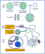
Lipid coated mesoporous silica nanoparticle (L-MSN) were synthesized for oral delivery of ciprofloxacin for intracellular elimination of Salmonella pathogen. The particle size was found to be between 50–100 nm with a lipid coat of approximately 5 nm thickness. The lipid coating was achieved by sonication of liposomes with the MSN particles and evaluated by CLSM and FTIR studies. The L-MSN particles exhibited lower cytotoxicity compared to bare MSN particles. Ciprofloxacin, a fluoroquinolone antibiotic, loaded into the L-MSN particles showed enhanced antibacterial activity against free drug in in vitro assays. The lipid coat was found to aid in intravacuolar targeting of the drug cargo as observed by confocal microscopy studies. We also observed that a lower dose of antibiotic was sufficient to clear the pathogen from mice and increase their survivability using the L-MSN oral delivery system.


1. Introduction

Bacterial pathogens are known to develop ingenious methods for better survival in intracellular environments. Some of the intracellular pathogens exhibiting such characteristics are Mycobacterium tuberculosis, Chlamydia trachomatis, Salmonella, Listeria etc. Salmonella is the causative organism of gastroenteritis, typhoid and paratyphoid diseases. The usual route of Salmonella infection occurs through consumption of contaminated food or drink. The ingested bacteria is sampled by the M-cells and dendritic cells present in the Peyer's patches in the small intestine.1,2 The bacterium employs clever molecular machinery to invade the gut epithelial barrier. Once inside, the bacterium replicates inside the host cell in specialized vacuoles called Salmonella containing vacuoles (SCV) and evades the immune system.1–4 Treatment of Salmonella infection by conventional therapy is difficult due to barriers created by the bacteria in order to evade the antibacterial regime.5 The treatment involves seven days of antibiotic therapy administered orally or by intravenous route. Existing carrier systems include liposomes,6–12 microparticles13,14 and nanoparticles.15 These systems suffer from the limitation that they are not robust enough to be delivered orally. Their antibacterial activity has been studied by delivering the system via intravenous (IV) or intraperitoneal (IP) injection routes.15 The injection route reduces compliance and entails administration by a trained person. We have developed a silica particulate system to deliver higher payload of the drug within the infected cell for oral administration. The carrier system follows the bacterial route of internalization which contributes to its targeting capability (Scheme 1).
image file: c4ra12973c-s1.tif
Scheme 1 (A) Design: the drug is entrapped within the mesoporous silica particle, protected by a lipid coating. (B) Delivery: particle system is robust for oral delivery for treatment of Salmonella (STM) infection. (C) Mechanism: the particle enters into the infected gastrointestinal cell and releases the antibiotic cargo to eliminate the pathogen.

We used mesoporous silica nanoparticles (MSN) which were synthesized by the modified Stober's process. The MSN system is an extremely robust nanoparticle system used for drug delivery. The main advantage is its ease of synthesis, reproducibility and stability. However, toxicity and accumulation in tissues remain a limitation for its use. In order to overcome the existing toxicity of the MSN, lipid was coated around the particle. The biomimetic lipid coat around the nanoparticle reduces the toxicity of the inorganic particles and improves biocompatibility,16–18 since the cellular membrane is also composed of lipids such as phosphatidylcholine. Some pathogens also induce the accumulation of lipids of the host cell to mediate the process of intracellular survival and replication.4,19–21 Hence, we engineered lipid based particle system to target intracellular pathogens and prolong the antibacterial activity of existing antibiotics. Some of the other lipid coated systems have been developed for treatment of cancer,22,23 gout,24 neuropathic pain,25 diabetes26 etc.

The existing treatment for Salmonella infection includes administration of fluoroquinolones or a third generation cephalosporin antibiotics. We selected ciprofloxacin as the model drug to be encapsulated in the particles in our studies. It is known that particle size plays a very important role in the internalization into cells. Submicron sized particles have been reported to be taken up by M-cells and macrophages present in Peyer's patches.27–30 Since the particles get internalized by the same route as the Salmonella, we expected that the particles will be recruited near the intracellular bacteria.

2. Experimental

2.1. Materials

1,2-Dipalmitoyl-sn-glycero-3-phosphocholine (DPPC) was obtained from Lipoid Gmbh (Ludwigshafen, Germany). Cholesterol, DMSO and hydrochloric acid (37%) were obtained from S. D. Fine. Chem. (Mumbai, India). Tetraethylorthosilicate (TEOS, 99%), Dulbecco's modified Eagles' medium (DMEM), fetal bovine serum (FBS), ciprofloxacin, curcumin, gentamicin and trypsin–EDTA solution were purchased from Sigma-Aldrich (Bangalore, India). Hexadecyltrimethylammonium bromide (CTAB ≥99%) was purchased from SRL chemicals (Mumbai, India). DiI stain (DiI) was obtained from Invitrogen BioServices (Bangalore, India). Carbon coated TEM grids (CF300-Cu) were purchased from EMS (Hatfield, PA, USA). Autoclaved MiliQ water (Milipore, Billirica, MA, USA) was used for all the experiments.

2.2. Synthesis of mesoporous silica nanoparticles (MSN) and lipid coating

The synthesis technique used was a modification of the Stober process.31,32 1 g of CTAB was added to 480 ml of deionised water along with 2 ml of 2.5 M sodium hydroxide solution and stirred at 80 °C for 30 minutes until completely dissolved, and cooled to 25 °C. Then 5 ml of TEOS was added slowly (1 ml min−1) to the CTAB solution and stirred rapidly at 700 rpm for 3 hours. The resulting particles were then centrifuged at 10[thin space (1/6-em)]000 rpm in a REMI centrifuge for 15 minutes and the particles collected and dried for further processing. The particles were refluxed for 6 hours in acidified ethanolic solution followed by calcination at 550 °C for 6 hours in a tube furnace. Calcination and acid refluxing was carried out to remove the surfactant and other organic additives. The lipid coating was achieved by ultrasonication of MSN particles with liposomes. The liposomes were prepared by film hydration method described earlier.33 For lipid coating, equal aliquots of 2 mM liposome dispersion (70[thin space (1/6-em)]:[thin space (1/6-em)]30, DPPC[thin space (1/6-em)]:[thin space (1/6-em)]Chol) and 10 mg ml−1 MSN particle in Mili-Q was sonicated for 10 seconds using a probe sonicator. The resultant lipid coated MSN (L-MSN) particles were used for further studies.

2.3. Drug loading and release from L-MSN particles

Drug loading was carried out at pH 4 by incubating 5 mg of MSN particles with ciprofloxacin solution having a concentration of 5 mg ml−1 for 12 hours. The drug loaded particles were centrifuged and the supernatant was removed. The percentage of ciprofloxacin loaded was determined by measuring the concentration of the supernatant solution containing the unentrapped drug. The particles were dried and stored for further studies. The ciprofloxacin loaded MSN particles were then subjected to lipid coating, to form Ciprofloxacin loaded lipid MSN particles (Cip L-MSN). The Cip L-MSN particles were centrifuged and washed twice with water to remove the excess drug and liposomes.

The release studies were carried out using 10 mg of the Cip L-MSN particles incubated in 1 ml of release media. Release media consisted of acidic media pH 2.5 and neutral media pH 7.4 simulating the gastrointestinal conditions of the stomach and of the intestine respectively. Aliquots from the release media were obtained at various time intervals to determine the ciprofloxacin release. The ciprofloxacin release was monitored by measuring absorbance at 275 nm using a spectrophotometer (Nanodrop, ND1000, Thermo Scientific, Wilmington, DE, USA).

2.4. Characterization of particles

The particle size and morphology of the prepared particles were characterized by using Technai G2 T20 Transmission Electron Microscope (FEI, The Netherlands) and Sirion Scanning Electron Microscope (FEI, The Netherlands). For TEM imaging, the particles at a concentration of 1 mg ml−1 were dispersed in water and sonicated for 30 minutes, and 5 μl of this dispersion was transferred to the TEM grids. The TEM grids were dried overnight at 40 °C, and imaged at 200 kV. For SEM imaging, the dispersion was deposited on a silica wafer, air dried and sputter coated with gold and imaged at 8–10 kV. Size distribution of the nanoparticles and liposomes were determined using a Zetasizer Nano (Malvern, Worcesterchire, UK) equipped with a diode laser operated at 659 nm. Surface area measurement was carried out by Nitrogen adsorption method on ASAP-200 surface area and porosity analyzer (Micromeritics Instrument Corporation; USA) and surface area was determined using the Brunauer–Emmet–Teller model. FTIR spectra of the samples were recorded in the range 400 cm−1 to 4000 cm−1 with a resolution of 4 cm−1 using FTIR spectrometer (Bruker, Germany).

2.5. Cell culture conditions

RAW macrophage cell line and HeLa cells were cultured in DMEM with 10% FBS maintained at 37 °C in CO2 (5%) atmosphere. In all studies, the cells were allowed to adhere to the culture plates for at least 12 hours after trypsinization. Salmonella typhimurium 14028 (STM) was used throughout the work. In confocal studies, green fluorescent protein expressing STM (STM-GFP) was used.

2.6. Cellular viability assay

Cytotoxicity of bare MSN particles, lipid coated particles and liposomes were evaluated by MTT assay. RAW 264.7 cells were seeded at a density of 5 × 10⁴ cells per ml in 96 well microtiter plate. The cells were treated with MSN particles in the concentration range of 3 mg ml−1 to 0.01 mg ml−1. After 24 hours, 20 μl of MTT dye (5 mg ml−1) was added to each well and kept for 4 hours at 37 °C. The resulting formazan crystals were dissolved by DMSO and absorbance of treated and untreated samples were recorded at 570 nm. Viable cells reduce the tetrazolium compound to insoluble formazan. DMSO dissolves the formazan forming a purple colored solution. The relative absorbance of the colored solution is used as a relative measure of percentage viability.

2.7. In vitro studies

The antibacterial activity of Cip L-MSN and free ciprofloxacin was compared in the concentration range of 1–0.1 μg ml−1 using overnight culture of STM. To 96-well plate containing different concentrations of ciprofloxacin and Cip L-MSN particles in Luria–Bertani broth media (LB medium), 1 × 10⁶ bacteria was added and incubated for 12 hours. After incubation, 10 μl of the broth was plated onto Salmonella-Shigella agar (SS-agar) plate and CFU determined. The intracellular survival assay was carried out according to procedure by Gnanadhas et al.34 RAW 264.7 or HeLa cells at a cellular density of 1 × 10⁵ cells per well were seeded in a 24-well plate and infected with Salmonella (STM) in the ratio of (1[thin space (1/6-em)]:[thin space (1/6-em)]10, MOI) and incubated for 30 minutes. DMEM containing Gentamicin (100 mg ml−1) was added to the infected cells and incubated further for 1 h to remove extracellular bacteria. Cip L-MSN and ciprofloxacin (50 μg ml−1) was then added to the media containing gentamicin (10 mg ml−1) and incubated for 2 h and 16 h post infection. Phosphate buffered saline (PBS, pH 7.4) was added to cells along with gentamicin but without ciprofloxacin. This set was used as the untreated control (UT). After the incubation period, the cells were lysed with 0.1% Triton-X 100 to release the intracellular bacteria. The lysate was suitably diluted and plated onto SS-agar for determining colony forming units (CFU). The fold change in intracellular replication of STM was determined by calculating the ratio of CFU counts at 16 h to 2 h i.e., CFU (16 h)/CFU (2 h). Concentration lower than the clinical dose of (50 μg ml−1) were investigated similarly using the assay on RAW 264.7 cells.

2.8. Cellular uptake and infection studies by CLSM

RAW 264.7 cells at a cellular density of 1 × 10⁵ cells per well were seeded on coverslips placed inside a 24-well plate for 12 hours prior to the experiment. The cells were infected with Salmonella (STM-GFP) in the ratio of (1[thin space (1/6-em)]:[thin space (1/6-em)]10, MOI) and incubated for 30 minutes. The infected cells were washed repeatedly to remove the extracellular bacteria. The cells were incubated with the L-MSN particles for 2 hours and 4 hours to observe the cellular uptake and localization. The confocal microscopy images were acquired using a Zeiss LSM 710 (Carl Zeiss Microimaging Inc., Thornwood, USA) using a 63×, 1.4-NA oil immersion objective. Zen 2009 Light Edition software was used for processing and overlaying channels of the image.

2.9. In vivo survival assay

The in vivo survival assay was carried out to determine the survival of BALB/c mice after challenge with 1 × 10⁶ inoculum of Salmonella typhimurium administered by oral gavage. 12 hours post infection, two treatment regimens were started with ciprofloxacin at 10 mg kg−1 body weight and Cip L-MSN with 5 mg kg−1 body weight given orally twice a day for 3 days. The morbidity and mortality of the mice was observed for 15 days post infection.

3. Results and discussion

3.1. Synthesis and characterization of L-MSN particles

L-MSN particles were designed to carry and deliver the antibiotic payload to the bacteria infected cells to combat the infection. The particle sizes of the MSN were in the range of 80–100 nm measured by DLS and confirmed by TEM (Fig. 1). The surface area of the MSN particles was found to be greater than 1000 m2 g−1 measured by N2 adsorption method. The high surface area of MSN particles enabled increased loading capacity of antibiotic molecules. Liposomes were formulated using phospholipids using the film hydration technique.35 The phospholipid composition consisted of DPPC and cholesterol in the molar ratio of 70[thin space (1/6-em)]:[thin space (1/6-em)]30. This ratio was selected as it had the ideal size around 51 ± 14 nm with the polydispersity index (PDI) of 0.29. The lipid coating procedure involved transient rupture of liposome vesicle and its deposition on MSN surface. Since the liposomes are smaller than the MSN particles, sonication procedure was expected to deposit liposome on MSN particles. TEM images show that the lipid coating was approximately 5 nm in thickness.
image file: c4ra12973c-f1.tif
Fig. 1 Transmission electron microscopy image of mesoporous silica nanoparticles (a), and after liposome fusion (b). Scale bar 50 nm (a) and 20 nm (b).

The lipid coating by liposome fusion was confirmed by sonicating silica microparticles (5 μm) with liposomes containing a fluorescent membrane dye (curcumin and DiI) and observed using confocal microscopy (Fig. 2). The lipid coating on the nanoparticles was further ascertained by FTIR studies. The IR spectrum shows characteristic peaks at 1078 cm−1 due to asymmetric vibration of Si–O groups present in the MSN. The characteristic peaks observed liposomes are present at 2900–2850 cm−1 and 1730 cm−1 which correspond to the stretching vibrations of alkyl groups and ester groups respectively. Similar peaks were identified in the L-MSN particles which confirmed lipid forming a coat over the MSN particles (Fig. 3).


image file: c4ra12973c-f2.tif
Fig. 2 Scanning electron microscopy (a) and CLSM image (b) of silica microparticle. CLSM image of curcumin (lipid marker) adsorbed on bare silica microparticle (c) and after liposome fusion (d). CLSM image of fluorescent lipid dye (DiI) adsorbed onto bare silica microparticle (e) and after liposome fusion (f). Scale bar is 10 μm.

image file: c4ra12973c-f3.tif
Fig. 3 FTIR spectra of MSN, liposome and lipid coated MSN (L-MSN) nanoparticles.

3.2. Drug loading and release from L-MSN particles

Ciprofloxacin was loaded into the particles by incubating the drug solution with MSN at pH 4 for 12 hours. The loading pH was selected based on electrostatic attraction between ciprofloxacin and MSN. Ciprofloxacin is an amphoteric molecule having acidic groups as well as basic groups. It would be positively charged (pKa1, 6.4 and pKa2, 8.2) while MSN would be negatively charged at pH 4 leading to electrostatic attraction.36 The amount of ciprofloxacin loaded was 0.99 ± 0.09 mg for every 5 mg of MSN. However, the drug loading decreased during the lipid coating procedure to nearly 9% i.e. 0.45 mg/5 mg of L-MSN. The decrease in the drug loading was attributed to the vigorous sonication procedure which may have led to leaking out of drug prior to lipid encapsulation.

The antibiotic delivery system was designed to deliver the drug thorough the oral route. The delivery system encounters extremely low pH in the stomach before encountering the intestinal environment. Hence, we studied the release behavior of ciprofloxacin from Cip L-MSN particles in both acidic and neutral pH. Nearly 90% of the drug released in 30 minutes after incubation in case of bare MSN loaded ciprofloxacin, compared to 30% release in Cip L-MSN particles. The increased drug release in acidic media of L-MSN particles was higher compared to neutral pH probably due to increased solubility of ciprofloxacin at lower pH. In both cases however the drug release continued upto 12 hours (Fig. 4).

3.3. Cellular viability studies of MSN and L-MSN

The MSN particles exhibited significant cytotoxicity above 1 mg ml−1 as evaluated by MTT assay carried out on RAW 264.7 macrophage cell line (Fig. 5). Hence we wanted to compare the cellular viability of L-MSN particles with bare MSN particles above the cytotoxic concentration. We found that MSN exhibited higher toxicity compared to L-MSN at concentrations above 1 mg ml−1. At 3 mg ml−1, bare MSN particles showed almost 50% viability as compared to 75% viability seen in L-MSN system. Similarly, liposomes used for the lipid coating were also subjected to MTT assay and found to be nontoxic (data not shown).
image file: c4ra12973c-f4.tif
Fig. 4 Ciprofloxacin release from MSN particles and L-MSN particles in release medium (pH 2.5 and pH 7.4).

image file: c4ra12973c-f5.tif
Fig. 5 Cellular viability studies by MTT assay of L-MSN particles at various concentrations compared to untreated cells (UT) (a). Cytotoxicity comparison between L-MSN particles and bare MSN particles (b).

3.4. In vitro antibacterial activity

The antibacterial effect of the Cip L-MSN particle was compared to free ciprofloxacin (Cip) by in vitro studies. Cip L-MSN particles showed increased antibacterial activity compared to free drug against STM bacteria (Fig. 6a) incubated overnight in LB medium.
image file: c4ra12973c-f6.tif
Fig. 6 In vitro antibacterial study: CFU analysis of Cip L-MSN and free drug in LB medium (a). Dose based comparison of ciprofloxacin and Cip L-MSN in RAW macrophage cells (b). Intracellular proliferation assay of STM infection after treatment with free ciprofloxacin and Cip L-MSN particles at a concentration of 50 μg ml−1 in RAW 264.7 cells (c) and HeLa cells (d).

This could be attributed to the prolonged release of the antibiotic from L-MSN system. The intracellular proliferation assay which is a measure of intracellular replication potential of pathogens was carried out. In untreated samples (UT) Salmonella undergoes replication inside the host cell resulting in an increased fold change. After antibiotic treatment the fold change is expected to decrease due to bacterial cell death. The fold change was compared between the treatments. We observed lower fold change in Cip L-MSN as compared to free drug in both RAW 264.7 macrophage cells (Fig. 6c) and HeLa cells (Fig. 6d). To further investigate whether lower doses of Cip L-MSN was sufficient to clear the infection, we compared lower concentrations of ciprofloxacin between treatments. Cip L-MSN particle system was found to be more effective compared to free drug (Fig. 6b) even at lower doses.

3.5. Cellular uptake studies and SCV targeting of L-MSN

The cellular uptake of the L-MSN particles was studied by incubating fluorescent L-MSN particles with the Salmonella infected cell using confocal microscopy (Fig. 7). The DiI loaded L-MSN particles (red) were observed for their intracellular localization with Salmonella. We observed increased uptake of the L-MSN at 4 h as compared to 2 h. Even more surprising observation was that the GFP expressing Salmonella was ensheathed in red fluorescence. The red fluorescence was due to the lipid dye which was initially present in L-MSN particle system which led us to suspect that the increased antibacterial activity could be due to the localization of our L-MSN system around the intracellular bacteria.
image file: c4ra12973c-f7.tif
Fig. 7 CLSM studies: L-MSN uptake and its localization studies in STM infected RAW 264.7 cells at 2 h (A) and 4 h (B) post infection. DiI loaded L-MSN particles (i), intracellular STM-GFP (ii), merged image (iii) with brightfield (iv) is shown.

3.6. In vivo antibacterial activity

The antibacterial activity of lipid coated MSN particles were tested in an in vivo survival assay. Salmonella typhimurium (STM) was administered orally to mice to model the infection occurring in humans. In mouse model, infection with STM is fatal at an oral dose of 1 × 10⁶ CFU.

Two treatment regimens were administered to Salmonella infected mice: First treatment regime included ciprofloxacin administered at 20 mg kg−1 body weight while the second treatment consisted of Cip L-MSN system at 10 mg kg−1 given orally twice a day for 3 days to maintain therapeutic concentration of the drug.37 The survival of the mice was observed till 15 days post infection (Fig. 8). Our data indicated that even a lower dose of ciprofloxacin administrated by the lipid coated particulate system was as effective as free drug which was similar to the in vitro findings. This indicates that the developed lipid coated MSN particulate system can be administered orally instead of intravenous routes requiring lower dosage of the antibiotic to clear the intracellular Salmonella infection.


image file: c4ra12973c-f8.tif
Fig. 8 In vivo antibacterial assay: mice were treated with ciprofloxacin (20 mg kg−1, p.o, BID) and with Cip L-MSN (10 mg kg−1, p.o, BID) for 3 days after Salmonella infection and assessed for mortality (n = 4).

4. Conclusions

Lipid coated MSN particles (L-MSN) encapsulated with ciprofloxacin showed improved antibacterial activity in clearing intravacuolar Salmonella infection. The L-MSN particle system exhibited controlled release of the antibiotic, which could aid in prolonged antibacterial effect. The lipid coat around the particle improves its biocompatibility and aided in intravacuolar targeting of the drug cargo leading to lower requirement of antibiotic dose as observed in the in vivo model. The oral route of administration helped in ease of administration and delivery. The lipid based system for targeting Salmonella has the potential for treatment of other pathogens which also show an intracellular lifestyle.

Acknowledgements

Financial assistance from DBT and LSRB is gratefully acknowledged. The Central animal facility, Confocal facility and The “Bioengineering Program” are acknowledged.

Notes and references

  1. Y. P. M. van der Meer-Janssen, J. van Galen, J. J. Batenburg and J. B. Helms, Prog. Lipid Res., 2010, 49, 1–26 CrossRef CAS PubMed.
  2. F. Lafont and F. G. van der Goot, Cell. Microbiol., 2005, 7, 613–620 CrossRef CAS PubMed.
  3. J. Deiwick, S. P. Salcedo, E. Boucrot, S. M. Gilliland, T. Henry, N. Petermann, S. R. Waterman, J.-P. Gorvel, D. W. Holden and S. Méresse, Infect. Immun., 2006, 74, 6965–6972 CrossRef CAS PubMed.
  4. T. J. Pucadyil, P. Tewary, R. Madhubala and A. Chattopadhyay, Mol. Biochem. Parasitol., 2004, 133, 145–152 CrossRef CAS PubMed.
  5. K. Melzak, S. Melzak, E. Gizeli and J. Toca-Herrera, Materials, 2012, 5, 2306–2325 CrossRef CAS PubMed.
  6. C. Cordeiro, D. J. Wiseman, P. Lutwyche, J. C. Evans, B. B. Finlay, M. S. Webb and M. Uh, Antimicrob. Agents Chemother., 2000, 44, 533–539 CrossRef CAS.
  7. T. Tadakuma, N. Ikewaki, T. Yasuda, M. Tsutsumi, S. Saito and K. Saito, Antimicrob. Agents Chemother., 1985, 28, 28–32 CrossRef CAS.
  8. J. V. Desiderio and S. G. Campbell, Infect. Immun., 1985, 48, 658–663 CAS.
  9. C. E. Swenson, K. A. Stewart, J. L. Hammett, W. E. Fitzsimmons and R. S. Ginsberg, Antimicrob. Agents Chemother., 1990, 34, 235–240 CrossRef CAS.
  10. J. Gubernator, D.-K. Zuzanna, A. Dorotkiewicz-Jach, W. Doroszkiewicz and A. Kozubek, Lett. Drug Des. Discovery, 2007, 4, 297–304 CrossRef CAS.
  11. O. Balland, H. Pinto-Alphandary, A. Viron, E. Puvion, A. Andremont and P. Couvreur, J. Antimicrob. Chemother., 1996, 37, 105–115 CrossRef CAS PubMed.
  12. M. S. Webb, N. L. Boman, D. J. Wiseman, D. Saxon, K. Sutton, K. F. Wong, P. Logan and M. J. Hope, Antimicrob. Agents Chemother., 1998, 42, 45–52 CAS.
  13. E. Fattal, S. Pecquet, P. Couvreur and A. Andremont, Int. J. Pharm., 2002, 242, 15–24 CrossRef CAS.
  14. W. Liu, Y. Yang, N. Chung and J. Kwang, Avian Dis., 2001, 45, 797–806 CrossRef CAS.
  15. M. N. Seleem, P. Munusamy, A. Ranjan, H. Alqublan, G. Pickrell, N. Sriranganathan, A. Ranjan and N. Sriranganathan, Antimicrob. Agents Chemother., 2009, 53, 4270–4274 CrossRef CAS PubMed.
  16. M. M. van Schooneveld, E. Vucic, R. Koole, Y. Zhou, J. Stocks, D. P. Cormode, C. Y. Tang, R. E. Gordon, K. Nicolay, A. Meijerink, Z. A. Fayad and W. J. M. Mulder, Nano Lett., 2008, 8, 2517–2525 CrossRef CAS PubMed.
  17. S. Ramishetti and L. Huang, Ther. Delivery, 2012, 3, 1429–1445 CrossRef CAS PubMed.
  18. X. Zhang, F. Li, S. Guo, X. Chen, X. Wang, J. Li and Y. Gan, Biomaterials, 2014, 35, 3650–3665 CrossRef CAS PubMed.
  19. M. J. Garner, R. D. Hayward and V. Koronakis, Cell. Microbiol., 2002, 4, 153–165 CrossRef CAS.
  20. K. M. Cadigan, D. M. Spillane and T. Y. Chang, J. Cell Biol., 1990, 110, 295–308 CrossRef CAS.
  21. D. M. Catron, M. D. Sylvester, Y. Lange, M. Kadekoppala, B. D. Jones, D. M. Monack, S. Falkow and K. Haldar, Cell. Microbiol., 2002, 4, 315–328 CrossRef CAS.
  22. C. E. Ashley, E. C. Carnes, K. E. Epler, D. P. Padilla, G. K. Phillips, R. E. Castillo, D. C. Wilkinson, B. S. Wilkinson, C. A. Burgard, R. M. Kalinich, J. L. Townson, B. Chackerian, C. L. Willman, D. S. Peabody, W. Wharton and C. J. Brinker, ACS Nano, 2012, 6, 2174–2188 CrossRef CAS PubMed.
  23. J. Liu, X. Jiang, C. Ashley and C. J. Brinker, J. Am. Chem. Soc., 2009, 131, 7567–7569 CrossRef CAS PubMed.
  24. V. Cauda, H. Engelke, A. Sauer, D. Arcizet, C. Bräuchle, J. Rädler and T. Bein, Nano Lett., 2010, 10, 2484–2492 CrossRef CAS PubMed.
  25. E. C. Dengler, J. Liu, A. Kerwin, S. Torres, C. M. Olcott, B. N. Bowman, L. Armijo, K. Gentry, J. Wilkerson, J. Wallace, X. Jiang, E. C. Carnes, C. J. Brinker and E. D. Milligan, J. Controlled Release, 2013, 168, 209–224 CrossRef CAS PubMed.
  26. V. J. Mohanraj, T. J. Barnes and C. A. Prestidge, Int. J. Pharm., 2010, 392, 285–293 CrossRef CAS PubMed.
  27. A. T. Florence, A. M. Hillery, N. Hussain and P. U. Jani, J. Controlled Release, 1995, 36, 39–46 CrossRef CAS.
  28. O. Borges, A. Cordeiro-da-Silva, S. G. Romeijn, M. Amidi, A. de Sousa, G. Borchard and H. E. Junginger, J. Controlled Release, 2006, 114, 348–358 CrossRef CAS PubMed.
  29. D. J. Brayden, M. A. Jepson and A. W. Baird, Drug Discovery Today, 2005, 10, 1145–1157 CrossRef CAS.
  30. E. Gullberg, M. Leonard, J. Karlsson, A. M. Hopkins, B. David, A. W. Baird and P. Artursson, Biochem. Biophys. Res. Commun., 2000, 279, 808–813 CrossRef CAS PubMed.
  31. W. Stober and A. Fink, J. Colloid Interface Sci., 1968, 26, 62–69 CrossRef.
  32. Y. Yang, W. Song, A. Wang, P. Zhu, J. Fei and J. Li, Phys. Chem. Chem. Phys., 2010, 12, 4418–4422 RSC.
  33. A. Güven, M. Ortiz, M. Constanti and C. K. O'Sullivan, J. Liposome Res., 2009, 19, 148–154 CrossRef PubMed.
  34. D. P. Gnanadhas, M. Ben Thomas, M. Elango, A. M. Raichur and D. Chakravortty, J. Antimicrob. Chemother., 2013, 68, 2576–2586 CrossRef CAS PubMed.
  35. B. Ruozi, D. Belletti, A. Tombesi, G. Tosi, L. Bondioli, F. Forni and M. A. Vandelli, Int. J. Nanomed., 2011, 6, 557–563 CrossRef CAS PubMed.
  36. F. Varanda, M. J. Pratas de Melo, A. I. Caço, R. Dohrn, F. A. Makrydaki, E. Voutsas, D. Tassios and I. M. Marrucho, Ind. Eng. Chem. Res., 2006, 45, 6368–6374 CrossRef CAS.
  37. M. LeBel, Pharmacother. J. Hum. Pharmacol. Drug Ther, 1988, vol. 8, pp. 3–30 Search PubMed.

This journal is © The Royal Society of Chemistry 2014
Click here to see how this site uses Cookies. View our privacy policy here.