DOI:
10.1039/C4RA12207K
(Paper)
RSC Adv., 2014,
4, 61525-61536
Retracted Article: Investigation of anticorrosive, antibacterial and in vitro biological properties of a sulphonated poly(etheretherketone)/strontium, cerium co-substituted hydroxyapatite composite coating developed on surface treated surgical grade stainless steel for orthopedic applications
Received
11th October 2014
, Accepted 21st October 2014
First published on 21st October 2014
Abstract
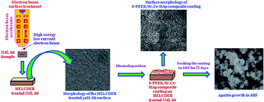
In the present investigation, a sulphonated poly(etheretherketone)/strontium, cerium co-substituted hydroxyapatite (S-PEEK/Sr,Ce-HAp) composite coating is obtained on high energy low current DC electron beam (HELCDEB) treated 316L stainless steel (316L SS) by electrodeposition. The surface of the 316L SS was treated using HELCDEB with an energy of 500 keV and a beam current of 1.5 mA. The as-formed coatings on HELCDEB treated 316L SS were characterised by X-ray diffraction (XRD), Fourier transform infrared spectroscopy (FT-IR) and high resolution scanning electron microscopy (HRSEM). Electrochemical results show that the S-PEEK/Sr,Ce-HAp coating with an optimum 2 wt% S-PEEK concentration on HELCDEB treated 316L SS possesses maximum corrosion resistance in Ringer’s solution. The antibacterial activity and in vitro bioactivity of the composite coatings were investigated. The results revealed that the HELCDEB treatment of the 316L SS improved anticorrosion performance and also that the combination of S-PEEK and Sr,Ce-HAp in the coating greatly improved the bioactivity and biocompatibility of the as-developed composite coating on HELCDEB treated 316L SS.
1. Introduction
Hydroxyapatite (HAp, Ca10(PO4)6(OH)2) is a bioceramic material, which promotes osteointegration and thus accelerates tissue fixation at the implant surface during the early stages of implantation.1–4 In recent years, several studies have been reported on the synthesis and coating of various bivalent and trivalent mineral (Sr2+, Mg2+, Zn2+, Ce3+, La3+, Y3+, Gd3+, Tb3+, etc.) substituted HAp which revealed that the substitution of minerals improved its bioactivity, biocompatibility, osteoconductivity and structural stability.5–19 Among all the minerals, strontium is considered as one of the essential and necessary minerals. It has been revealed that strontium (Sr2+) is present at higher concentrations in the regions of high metabolic turnover in bone.20 Sr2+ has some attractive properties like modifying the bone balance towards osteogenesis and is extensively used in the treatment of osteoporosis. Strontium ranelate shows an excellent prospective approach for the treatment of osteoporosis and it has provided huge amounts of data on the mechanism of action of Sr2+ in bone cells.21 Sr2+ plays a dual role of inhibiting osteoclast activity and stimulating osteoblast differentiation and reduces the incidences of fractures in osteoporotic patients and bone resorption.22–24 In orthopedics and the dental field, the majority of infections after implantation occur due to the microorganisms present in the implanted materials, and the problem generally requires the removal of the implant.22
In order to overcome this obstacle, researchers attempted to develop the antibacterial property of HAp in terms of divalent and trivalent cation substitution (Ce3+, Ag+, Zn2+ and Cu2+) at the calcium site. Among these ions, Ce3+ has been used as antibacterial agent in different biomedical fields for a long time owing to its high safety and broad range of antibacterial activity. Ce3+ ions in small quantities are imperative for a variety of metabolic processes in the majority of the living microorganisms. Several in vitro studies have reported that the Ce3+ ions in implant coatings play a significant role in preventing or minimizing initial bacterial adhesion.25 There are three important mechanisms for the antibacterial properties of Ce3+ ions. First, Ce3+ ions bind to proteins and deactivate them. Second, Ce3+ ions interact with the microbial membrane causing structural and permeability changes. Finally, Ce3+ ions can interact with microbial nucleic acids, preventing microbial replication.26–28
Thus Sr2+ and Ce3+, as important metal ions were found to be very effective in enhancing the structural stability, and biological and antibacterial properties of Ca-HAp. Recently Gopi et al., have achieved the synthesis of Sr2+ and Ce3+ co-substituted HAp (Sr,Ce-HAp) nanoparticles with increased bioactivity and antibacterial activity.22
Considerable attention has recently been directed towards the mixing of polymers with inorganic materials which show excellent features with homogenous mechanical properties.29 The addition of a polymer to the HAp at an ambient temperature reduces its brittleness. Among all the polymers, poly(etheretherketone) (PEEK) is a biocompatible, stable and safe thermoplastic polymer suitable for orthopedic applications.30,31 It is perfectly matched for in vivo medical device applications as it combines excellent chemical and hydrolysis resistance and outstanding tribological properties with extensive biocompatibility and high strength.30 Sulfur containing polymers have excellent biocompatibility and optical properties.32 The introduction of sulfur into PEEK (S-PEEK) increases its thermal stability, biocompatibility and affinity towards metals in comparison with the analogous non-sulfur containing PEEK.33,34 Despite the attractive advantages and progress in the preparation of S-PEEK, the employment of Sr,Ce-HAp for the formation of a S-PEEK/Sr,Ce-HAp composite is preferred which combines both the antibacterial and bioactivity properties for orthopaedic applications. It has advantages over other polymer composites with HAp due to its biocompatibility, high fracture toughness, excellent mechanical properties, chemical inertness, ease of processing, lack of toxicity and excellent resistance to moisture.5,35,36
Among different techniques exploited for composite orthopaedic coatings, electrodeposition (ED) is particularly attractive because it can be utilized to produce uniform coatings with controllable properties on complexly-shaped and porous structures, at an ambient temperature and without the need for expensive processing equipment.30,32,37–39 ED is based on the controlled electric-field-induced deposition of charged particles in an organized way onto an implant surface. Co-deposition of bioceramics and polymers is also another fascinating feature of ED.39,40 Surgical grade 316L stainless steel (316L SS) is the most commonly used orthopaedic implant substrate that has gained significant advantages due to its immense mechanical and corrosion properties.41,42 Though 316L SS shows an excellent biocompatibility, it cannot form direct chemical bonds to bone cells.4 A bioactive HAp coating on 316L SS implants is able to stimulate bone growth and affixation of the bone to the implant surface during the early stage of implantation.7 However, due to the continuous interaction with the harsh environment, the HAp coating degrades during long-term implantation and results in the corrosion of the underlying metallic alloy. Hence, surface treatment of a metal alloy prior to the development of coating is essential in the prevention of metallic corrosion and to withstand long-term implantation conditions. Several surface modification techniques have been applied to improve the corrosion resistance and adhesion strength of the implant material in body fluids.43–46 The electron beam treatment has emerged as a powerful surface modification technique as it exhibits essential advantages over conventional methods by its high efficiency, simplicity and reliability.47–49 Low energy high current pulsed electron beam (LEHCPEB) treatment induces crater eruptions in the 316L SS surface that preferentially occur at MnS inclusions and produce a surface purification effect. The mixing in the melting process and the subsequent solute trapping effect during the fast solidification process results in the homogenisation of elements in the melted layer.47,50 The LEHCPEB irradiation has several advantages such as (1) strong interfacial bonding between the melted region and the substrate, (2) the prevention of surface oxidation and intrusion of the inclusions because of a short irradiation time, and (3) the prevention of pores or cracks because of homogeneous heating and cooling. The irradiations of the metallic substrate by the electron beam modifies the surface of the metallic substrate by controlling its microstructure. High energy low current DC electron beams (HELCDEBs) possess all the advantages of LEHCPEBs, in addition to the in-depth energy deposition, very high power levels, and shock generation capabilities.51,52
In the present work we have achieved the S-PEEK/Sr,Ce-HAp composite coating on HELCDEB treated 316L SS by the ED method. The effect of S-PEEK concentrations on the crystallinity and morphology of the S-PEEK/Sr,Ce-HAp composite coating on HELCDEB treated 316L SS for orthopedic applications was analyzed. The anticorrosion performance of the S-PEEK/Sr,Ce-HAp composite coating on HELCDEB treated 316L SS in Ringer’s solution was studied by different electrochemical techniques. The in vitro bioactivity of the S-PEEK/Sr,Ce-HAp composite coating at an optimum concentration of 2 wt% S-PEEK was examined by soaking it in Simulated Body Fluid (SBF) for avarious days. Finally, the results revealed that the as-developed composite coating can serve as a potential candidate for major orthopedic applications with good anticorrosion performance, antibacterial activity and bone bonding ability.
2. Materials and methods
2.1 Chemicals
All chemicals were of analytical grade and used without further purification throughout the experiments. The chemicals used for the S-PEEK synthesis were p-dihydroxybenzene, sulfobenzide, di-terbutyl peroxide, potassium carbonate (K2CO3), sodium carbonate (Na2CO3) and sulphuric acid (H2SO4). For the Sr,Ce-HAp synthesis, calcium nitrate tetrahydrate (Ca(NO3)2·4H2O), strontium nitrate hexahydrate (Sr(NO3)2·6H2O), cerium nitrate hexahydrate (Ce(NO3)3·6H2O), diammonium hydrogen phosphate ((NH4)2HPO4) and ammonia solution (NH4OH) were used. Deionized water and ethanol were employed as the solvents throughout the experiment.
2.2 Synthesis of S-PEEK
In this preparation of S-PEEK, 60 g of di-terbutyl peroxide, sulfobenzide and p-dihydroxybenzene with a mass ratio of 1
:
5
:
2 were charged in a 250 mL four-necked flask equipped with a reflux condenser. The above mixture was first stirred for 30 min at an ambient temperature, and then heated to 180 °C at 5 °C min−1 under constant stirring in a pure nitrogen atmosphere. Then 2.5 g of K2CO3 and 1.25 g of Na2CO3 were added into the above mixture at 180 °C, and the reaction mixture was further heated to 320 °C at 5 °C min−1 under stirring. The resulting mixture was sulphonated in 98 wt% H2SO4 for about 5 h under vigorous mechanical stirring at 65 °C. Finally, the obtained S-PEEK solution was decanted into excess ice-cold water. The precipitated S-PEEK was filtered and washed several times with deionized water and acetone until the pH was neutral and was dried under vacuum at 140 °C for 24 h. The above synthesis was carried out according to the procedure reported earlier.29,53,54 The schematic representation of the sulphonation process of PEEK is described in Fig. 1.
 |
| Fig. 1 Schematic representation of the sulphonation process of PEEK. | |
2.3 316L SS specimen preparation
Type 316L SS (procured from the Steel Authority of India Ltd., (SAIL), India) with elemental composition (wt%) C – 0.0222, Si – 0.551, Mn – 1.67, P – 0.023, S – 0.0045, Cr – 17.05, Ni – 11.65, Mo – 2.53, Co – 0.136, Cu – 0.231, Ti – 0.0052, V – 0.0783, N – 0.0659 with a remaining composition of Fe, was used as the metal substrate for the ED.2,4 The 316L SS specimens with a size of 10 × 10 × 3 mm were embedded in the epoxy resin, leaving an area of 1 cm2 for exposure to the electrolyte solution.42,43 Before ED, these specimens were abraded with different grades of silicon carbide (SiC) emery papers from 400 to 1200 grits. After polishing, these samples were ultrasonically cleaned and thoroughly washed with acetone and deionized water for 20 min, dried in air and then used for further studies.
2.4 Surface treatment of the 316L SS specimens
The 316L SS specimen was surface treated with a 500 keV electron beam of energy and a 1.5 mA beam current using a 700 keV DC Accelerator55 developed at the Raja Ramanna Centre for Advanced Technology (RRCAT), Indore (MP). The sample was placed on the conveyer system in air. A DC electron beam, scanning at a frequency of 100 Hz was allowed to fall on the sample. The sample was allowed to pass through the beam at the speed of 20 m min−1 and a total of two passes were done with a 30 s separation.
2.5 ED of the S-PEEK/Sr,Ce-HAp composite on surface treated 316L SS
The experiments were carried out at room temperature under a DC voltage in a three electrode cell. A platinum electrode was used as the anode and the HELCDEB treated 316L SS was used as the cathode with the saturated calomel electrode (SCE) as the reference electrode. The electrolyte used for the deposition of (0, 1, 1.5 and 2 wt%) S-PEEK/Sr,Ce-HAp composites onto the HELCDEB treated 316L SS electrode by ED, consisted of aqueous solutions of 0.2 M Ca(NO3)2·4H2O, 0.2 M Sr(NO3)2·6H2O, 0.1 M Ce(NO3)2·6H2O, 0.3 M (NH4)2HPO4 and the S-PEEK (0–2 wt%) at room temperature (28 ± 1 °C) under magnetic stirring. The deposition was carried out galvanostatically using an electrochemical workstation (CHI 760C, CH Instruments, USA) at a constant current density of 7 mA cm−2 for a 60 min duration. All the samples were carefully and slowly pulled out of the electrolyte solution after ED. After deposition, the composite coating was gently rinsed with deionized water, dried and subsequently stored in desiccators for further characterization.
2.6 Surface characterisation
The characteristic functional groups of S-PEEK/Sr,Ce-HAp composite specimens were identified using FT-IR (Nicolet 380 FT-IR Spectrometer (Perkin Elmer, USA)) over the frequency range from 4000 cm−1 to 400 cm−1 with a number of 32 scans and a spectral resolution of 4 cm−1. The phase composition of the S-PEEK/Sr,Ce-HAp composite coated samples was analyzed by XRD (Bruker D8 Advance diffractometer) operated at a voltage of 40 kV and a current of 30 mA was used with the scanning angle ranging from 20 to 60°, with a scan rate (2θ) of 0.02°. The surface morphology and elemental composition of the composite coatings on surface treated 316L SS were examined by a HRSEM (JSM 840A scanning microscope, JEOL-Japan) equipped with EDAX. Before taking the HRSEM images, the specimens were sputter-coated with gold.
2.7 Electrochemical evaluation of the composite coatings
The electrochemical experiments were carried out through potentiodynamic polarisation and electrochemical impedance spectroscopy (EIS) in Ringer’s physiological solution (NaCl – 8.6 g L−1, CaCl2·2H2O – 0.66 g L−1 and KCl 0.6 g L−1) at a pH of 7.4 and a temperature of 37 ± 1 °C. The electrochemical measurements were performed with a three-necked cell using the CHI 760 electrochemical workstation (USA), consisting of a saturated calomel electrode (SCE), 316L SS and platinum electrodes as the reference, working and counter electrodes, respectively. During the polarisation studies, the potential was measured at a scan rate of 1 mV s−1 in the potential range between −1000 to 1000 mV (vs. SCE). The EIS experiments were performed in the frequency range of 10 mHz to 100 kHz with a perturbation amplitude of 5 mV. All the measured potential values are related to the SCE and all the electrochemical parameters are normalized with respect to the area of the 316L SS electrode (1 cm2). The obtained electrochemical data were recorded using internally available software and the corrosion tests were repeated at least three times to ensure reliability and reproducibility and the results were found to be reproducible.
2.8 Antibacterial activity
The in vitro antibacterial activity of the S-PEEK/Sr,Ce-HAp composite at an optimum concentration of S-PEEK (2 wt%) has been investigated against two prokaryotic strains, E. coli and S. aureus, by the agar disc diffusion method. The inoculums of the E. coli and S. aureus were prepared from fresh overnight broth cultures (Tripton soy broth with 0.6% yeast extract – Torlak, Serbia) that were incubated at 37 °C with constant stirring and the resulting broth cultures were used for the diffusion test. For comparison purposes, the antibacterial activities of S-PEEK/Sr-HAp and S-PEEK/Ce-HAp composite coatings were also evaluated.
The agar disc diffusion test was performed on Muller-Hinton agar. The test was carried out by pouring agar into Petri dishes to form 4 mm thick layers and adding 2 mL dense inoculums of the test organisms of two prokaryotic strains in order to obtain semi confluent growth. Petri plates were dried for 10 min in air and after that, discs (6 mm) were prepared from Whatman filter paper, immersed into different volumes of (25, 50, 75, 100, 125 μL) composite samples. Then the test discs were incubated at 37 °C for 24 h and were placed at equal distances. After the test, the antibacterial activity was measured as the zone of inhibition (mm) around the disc which was produced by the composite samples against the two prokaryotic strains.
2.9 In vitro analysis
2.9.1 Cell viability test by MTT assay. The cytotoxicity of the S-PEEK/Sr-HAp, S-PEEK/Ce-HAp and S-PEEK/Sr,Ce-HAp composite (at an optimum concentration of S-PEEK (2 wt%)) coatings to human osteosarcoma MG63 cells (purchased from National Centre for Cell Science (NCCS), Pune, India (HOS MG63, ATCC CRL-1427TM)) was determined by using a MTT assay. The cell viability tests were examined by using an indirect method, where the immersion extracts collected from the immersion test were used for culturing HOS MG63 cells. The test cells (HOS MG63) were cultured in Dulbecco’s Modified Eagle Medium (DMEM, GIBCO), which consisted of a minimal essential medium (Hi Media Laboratories), supplemented with 10% fetal bovine serum (FBS, Biowest, France) and 12-well tissue culture plates at 104 cells per mL. The cultured cell was then incubated for 1, 4 and 7 days at 37 °C under the humidified atmosphere of 5% CO2 and 95% air. The samples under examination were sterilised in an autoclave at 140 °C for 2 h and then placed in 12-well tissue culture plates. The HOS MG63 cell culture medium in each 12-well tissue culture plate was renewed every 2 days with immersion extracts supplemented with 10% FBS, and incubated at 37 °C in an atmosphere of 95% air and 5% CO2. To determine the cell proliferation of the composite samples, the MTT assay tests were carried out as a function of incubation time for 1, 4 and 7 days. 10 mL of MTT solution (5 mg thiazolyl blue tetrazolium bromide powder containing 1 mL phosphate buffered saline (PBS, OXOID Limited, England)) was then added into each 12-well tissue culture plates on the 1st, 4th and 7th day. After incubation for 1, 4 and 7 days, 100 mL of 10% sodium dodecyl sulphate (SDS, Sigma-Aldrich, USA) in 0.01 M hydrochloric acid (HCl acid, Sigma-Aldrich, UK) was added into each 12-well tissue culture plate and then incubated at 37 °C in an atmosphere of 95% air and 5% CO2 for 20 h. The absorbance of viability was recorded by using the ELISA microplate reader at a 570 nm wavelength, with a reference wavelength of 640 nm to determine the cell viability of the composite sample and its cell viability (%) was calculated in comparison with the control wells using the following equation:
% Cell viability = [A]test/[A]control × 100. |
2.9.2 Immersion of S-PEEK/Sr,Ce-HAp composites in simulated body fluid. The bone-bonding ability of the S-PEEK/Sr-HAp, S-PEEK/Ce-HAp and S-PEEK/Sr,Ce-HAp composite (at an optimum concentration of S-PEEK (2 wt%)) coatings on HELCDEB treated 316L SS samples can be evaluated by examining the apatite formation on their surfaces in simulated body fluid (SBF) at 37 °C for 14 and 21 days. The composition and preparation of SBF solution, which was buffered with hydrochloric acid to maintain a pH of 7.4, has been described in the literature.22,56 Then, the composite coated samples were soaked in 50 mL of SBF solution in a beaker with an airtight lid for 14 and 21 days, where the temperature was maintained at 37 ± 1 °C in an incubator. The test solutions were renewed every three days for a period of 14 and 21 days to avoid any change in the cationic concentration that may occur due to degradation of the composite coated samples. The immersed samples were then gently washed with de-ionized water and the formation of a bone-like apatite on the as-coated HELCDEB treated 316L SS samples was evaluated by HRSEM. Also, the antibacterial activity of the apatite grown composite samples were evaluated using the procedure adopted in Section 2.8.
3. Results and discussion
3.1 Surface characterisation
3.1.1 FT-IR spectra. FT-IR spectra of Sr,Ce-HAp, S-PEEK and the S-PEEK/Sr,Ce-HAp (with 2 wt% S-PEEK) composite coatings are shown in Fig. 2(a)–(c). The composite coated (Fig. 2(c)) material shows that the spectrum is the overlapped ones of the S-PEEK and Sr,Ce-HAp components. The characteristic band at 3462 cm−1 is assigned to the vibration of the O–H groups; thus, the sulphonic acid group in S-PEEK interacted with a H2O molecule. The aromatic C–C band was split into two peaks and appeared at 1470 cm−1 and 1489 cm−1 owing to the substitution achieved by sulphonation. The presence of a sulphonic group in the backbone is confirmed by the peaks at 1079 cm−1 and 1229 cm−1 which are due to the symmetric and asymmetric O–S–O stretching vibrations, respectively. Whereas, the peak found at 1012 cm−1 is due to the stretching vibration of the S–O group in the S-PEEK. The strong peak at 1635 cm−1 is ascribed to the carbonyl group located along the backbone of the polymer (S-PEEK) chain. All the above said peaks confirm the presence of S-PEEK in the composite (S-PEEK/Sr,Ce-HAp) material. The peaks for the phosphate group appeared at 949 cm−1 (ν1), 471 cm−1 (ν2), 1012 (ν3), 1079 cm−1 (ν3), 596 and 567 cm−1 (ν4), respectively. The broad bands at 3462 and 1635 cm−1 depict the stretching and bending modes of water molecules, while the bands at 3582 and 637 cm−1 are attributed to the stretching and bending vibration modes of the OH− group of Sr,Ce-HAp. Compared with those of S-PEEK (Fig. 2(b)) and Sr,Ce-HAp (Fig. 2(a)), the spectrum of the S-PEEK/Sr,Ce-HAp composite (Fig. 2(c)) demonstrates that the peaks neither shift nor new absorption peaks are identified with the addition of S-PEEK, suggesting that the composites are the mixture of these two compounds without forming identifiable new interfacial chemical bonds.
 |
| Fig. 2 FTIR spectra of (a) Sr,Ce-HAp (b) S-PEEK and (c) S-PEEK/Sr,Ce-HAp (with an optimum 2 wt% S-PEEK) composite coatings. | |
3.1.2 XRD analysis. X-ray diffraction measurements were carried out to investigate the effect of S-PEEK and Sr,Ce-HAp on the phase structure of the S-PEEK/Sr,Ce-HAp composite at an optimum concentration of 2 wt% S-PEEK (Fig. 3). The composite coated (Fig. 3(c)) material illustrates characteristic peaks of S-PEEK and Sr,Ce-HAp. The peak (2θ) values of S-PEEK at 18.9°, 20.5°, and 21.9° (Fig. 3(b)) match well with the previous report.29 The main diffraction peaks for S-PEEK are mainly in the range of 15–30°, whereas those in the range of 30–45° are ascribed to the Sr,Ce-HAp phase.31 The diffraction peaks (Fig. 3(c)) of Sr,Ce-HAp were observed at 2θ values of 25.8°, 31.9°, 32.5° and 32.8° (ref. 22) and no other secondary peaks were found. The peaks identified for Sr,Ce-HAp are in good agreement with the standard data for HAp and also consistent with the International Centre for Diffraction Data (ICDD card no. 09-0432). Whereas for the Sr,Ce-HAp coating, the peak positions deviated slightly which may be owing to crystal lattice distortion which occurred as the product of substitution of mineral (Sr and Ce) ions in HAp. As a result, the patterns of the S-PEEK/Sr,Ce-HAp (2 wt% S-PEEK) composite illustrates that no appreciable new interfacial crystalline phases were lost or formed with the addition of S-PEEK.
 |
| Fig. 3 XRD patterns of (a) Sr,Ce-HAp (b) S-PEEK and (c) S-PEEK/Sr,Ce-HAp (with an optimum 2 wt% S-PEEK) composite coatings. | |
3.1.3 Scanning electron microscopic and elemental investigations. The HRSEM morphology of the HELCDEB treated, Sr,Ce-HAp coated and S-PEEK/Sr,Ce-HAp composite (with 1, 1.5 and 2 wt% of S-PEEK) coated on HELCDEB treated 316L SS by electrodeposition at 7 mA cm−2 for 60 min are presented in Fig. 4(a)–(e) and the elemental composition of the composite with an optimum 2 wt% of S-PEEK is depicted in Fig. 4(f). Fig. 4(a) represents the morphology of HELCDEB treated 316L SS which revealed a rough surface with a uniformly distributed microstructure. An implant with a rough surface is known to have a favorable effect for a strong and adherent coating on the 316L SS. The surface morphology of the Sr,Ce-HAp coating on HELCDEB treated 316L SS (Fig. 4(b)) exhibited the formation of leaf-like microstructure. The HRSEM images of the S-PEEK/Sr,Ce-HAp composite coatings deposited on HELCDEB treated 316L SS at three different concentrations of S-PEEK (1, 1.5 and 2 wt%) are shown in Fig. 4(b)–(d). The morphology of 1 wt% of S-PEEK in the S-PEEK/Sr,Ce-HAp composite surface is entirely covered with the uniform arrangement of leaf-like structures (Fig. 4(c)). On the addition of 1.5 wt% S-PEEK, a needle-type interconnected (Fig. 4(d)) morphology of the S-PEEK/Sr,Ce-HAp coating on HELCDEB treated 316L SS is obtained. On further increasing the S-PEEK concentration up to 2 wt% in the composite, the image (Fig. 4(e)) revealed the presence of a needle-like structure that is formed compactly and uniformly on the HELCDEB treated 316L SS substrate, and micropores formed between those structure. It is worth noting that most of the needle-type structures are interconnected which should be encouraging for bone ingrowths on the surface of the S-PEEK/Sr,Ce-HAp composite coated 316L SS. These types of interconnected pores permit the proliferation and attachment of diverse cell types accountable for the development of new tissues.
 |
| Fig. 4 HRSEM micrographs of (a) HELCDEB treated 316L SS (b) Sr,Ce-HAp coated on HELCDEB treated 316L SS (c) S-PEEK/Sr,Ce-HAp (with 1 wt% S-PEEK) (d) S-PEEK/Sr,Ce-HAp (with 1.5 wt% S-PEEK), (e) S-PEEK/Sr,Ce-HAp (with 2 wt% S-PEEK) and the (f) EDAX spectrum of S-PEEK/Sr,Ce-HAp (with optimum 2 wt% S-PEEK) composite coatings on HELCDEB treated 316L SS. | |
The EDAX spectrum showing the constituent elements of the S-PEEK/Sr,Ce-HAp coated 316L SS is presented in Fig. 4(f), which indicates the presence of Ca, Sr, Ce, C, O, N, S and P in the relative coating.
3.2 Electrochemical characterization
3.2.1 Potentiodynamic polarisation studies. In order to determine the corrosion resistance ability of the coatings in physiological solution, the typical potentiodynamic polarization curves of the uncoated 316L SS, HELCDEB treated, Sr,Ce-HAp and S-PEEK/Sr,Ce-HAp (2 wt% of S-PEEK) composite coated on HELCDEB treated 316L SS in Ringer’s solution for long term applications were studied (Fig. 5(a)). The electrochemical values of the polarisation parameters such as corrosion potential (Ecorr), breakdown potential (Eb) and repassivation potential (Epp) were calculated from the polarization curves. From the obtained potentiodynamic polarization curves, the Ecorr, Eb and Epp values for the uncoated 316L SS specimen were found to be −870, +449 and −90 mV vs. SCE, respectively. While the polarisation curve recorded for the HELCDEB treated 316L SS specimen showed Ecorr, Eb and Epp values of −730, +463 and 148 mV vs. SCE, respectively. For the Sr,Ce-HAp coated on HELCDEB treated 316L SS specimen, polarization testing revealed Ecorr, Eb and Epp values of −575, +535 and 183 mV (vs. SCE), respectively. Thus, the polarization values obtained for Sr,Ce-HAp coated on HELCDEB treated 316L SS were found to be nobler than that of the HELCDEB treated 316L SS specimen. These higher Ecorr, Eb and Epp values obtained for Sr,Ce-HAp coated on HELCDEB treated 316L SS are due to the formation of a leaf-shaped interconnected flake-like coating. The polarisation curve of the S-PEEK/Sr,Ce-HAp (with 2 wt% S-PEEK) composite coated on HELCDEB treated 316L SS specimen showed Ecorr and Eb values of −473 and +696 mV (vs. SCE) respectively, while the Epp value was 252 mV (vs. SCE). These Ecorr, Eb and Epp values of the S-PEEK/Sr,Ce-HAp (with 2 wt% S-PEEK) composite coated on HELCDEB treated 316L SS specimen showed a maximum shift in the noble direction when compared to that of the S-PEEK/Sr,Ce-HAp (with 1 and 1.5 wt% S-PEEK) composite coating and also that of the uncoated and the Sr,Ce-HAp coated on HELCDEB treated 316L SS specimens (Table 1). The shift of the Ecorr, Eb and Epp values towards the noble direction is an indication that the S-PEEK/Sr,Ce-HAp (with 2 wt% S-PEEK) composite coating on HELCDEB treated 316L SS specimen possessed maximum anticorrosion performance in Ringer’s solution. The improvement in corrosion resistance observed for the S-PEEK/Sr,Ce-HAp (with an optimum 2 wt% S-PEEK) composite coating on HELCDEB treated 316L SS specimen is believed to be due to the thick, compact and uniform surface coverage of the needle-like composite structure on the HELCDEB treated 316L SS specimen.
 |
| Fig. 5 (a) Potentiodynamic polarisation curves and (b) Nyquist plots obtained for untreated, HELCDEB treated, Sr,Ce-HAp coated and S-PEEK/Sr,Ce-HAp (with optimum 2 wt% S-PEEK) composite coated on HELCDEB treated 316L SS in Ringer’s solution. | |
Table 1 Electrochemical parameters for untreated, HELCDEB treated, Sr,Ce-HAp and S-PEEK/Sr,Ce-HAp (with 1, 1.5 and 2 wt% S-PEEK) composite coated HELCDEB treated 316L SS
Samples |
Ecorr (mV vs. SCE) |
Eb (mV vs. SCE) |
Epp (mV vs. SCE) |
Rp (Ω cm2) |
Untreated 316L SS |
−870 |
449 |
−90 |
41.1 |
HELCDEB treated 316L SS |
−730 |
463 |
148 |
1156 |
Sr,Ce-HAp coated on HELCDEB treated 316L SS |
−575 |
535 |
183 |
1846 |
S-PEEK/Sr,Ce-HAp (with 1 wt% S-PEEK) composite coated on HELCDEB treated 316L SS |
−492 |
615 |
212 |
2490 |
S-PEEK/Sr,Ce-HAp (with 1.5 wt% S-PEEK) composite coated on HELCDEB treated 316L SS |
−486 |
632 |
239 |
2548 |
S-PEEK/Sr,Ce-HAp (with 2 wt% S-PEEK) composite coated on HELCDEB treated 316L SS |
−473 |
696 |
252 |
2689 |
3.2.2 Electrochemical impedance spectroscopy studies. EIS is the most powerful technique which can provide the most valuable information about the resistance of all the as-coated specimens in the Ringer’s solution. The Nyquist plots obtained for the uncoated, HECDEB treated, Sr,Ce-HAp and S-PEEK/Sr,Ce-HAp composite coated on HELCDEB treated 316L SS specimens in Ringer’s solution are shown in Fig. 5(b). For the uncoated 316L SS specimen the polarisation resistance (Rp) value is 41.1 Ω cm2. For the HELCDEB treated 316L SS specimen the polarisation resistance (Rp) is found to be 1156 Ω cm2. The Rp value obtained for the Sr,Ce-HAp coating on HELCDEB treated 316L SS specimen is found to be 1846 Ω cm2 which is greater than that for the HELCDEB treated 316L SS. This is ascribed to the effective anticorrosion performance of the Sr,Ce-HAp coating formed on the HELCDEB treated 316L SS. From Fig. 5(b), the Rp values for S-PEEK/Sr,Ce-HAp (with 1, 1.5 and 2 wt% S-PEEK) composite coatings on HELCDEB treated 316L SS are found to be 2490 Ω cm2, 2548 Ω cm2 and 2692 Ω cm,2 respectively which show the highest Rp value for the S-PEEK/Sr,Ce-HAp (2 wt% S-PEEK) composite coating (Table 1). The maximum Rp value of this S-PEEK/Sr,Ce-HAp (2 wt% S-PEEK) composite coating on HELCDEB treated 316L SS is due to the more effective barrier of the as-formed composite layer. From the EIS results, it could be well ascertained that the S-PEEK/Sr,Ce-HAp (2 wt% S-PEEK) composite coated on HELCDEB treated 316L SS is more corrosion protective than the other as-formed coated and uncoated 316L SS specimens.
3.3 Antibacterial assessment
The antibacterial activity of the S-PEEK/Sr-HAp, S-PEEK/Ce-HAp and S-PEEK/Sr,Ce-HAp composite (at an optimum concentration of S-PEEK (2 wt%)) coatings was evaluated against the two prokaryotic strains by the disc diffusion method. These two prokaryotic strains, E. coli and S. aureus, are the model Gram-negative bacteria and Gram-positive bacteria and are the general bacteria that are found in contaminated wounds. The zone of inhibition around the composite samples at five different concentrations (25, 50, 75, 100 and 125 μL) against E. coli and S. aureus is shown in Fig. 6(a)–(f). When compared to S-PEEK/Sr-HAp and S-PEEK/Ce-HAp coated samples, the S-PEEK/Sr,Ce-HAp composite coated sample showed enhanced anti-bacterial activity which is clearly evident from the zone of inhibition (Fig. 6(a)–(f)). For the S-PEEK/Sr,Ce-HAp composite sample, the inhibition zones for E. coli and S. aureus were found to be 11 & 10 mm for 25 μL, 12 & 11 mm for 50 μL, 13 & 12 mm for 75 μL, 14 & 13 mm for 100 μL and 15 & 14 mm for 125 μL volumes, respectively. The antibacterial results revealed that the highest concentration of the S-PEEK/Sr,Ce-HAp composite sample showed excellent antibacterial activity against the two prokaryotic strains. The reason may be that the incorporation of minerals (Sr2+ and Ce3+) plays a vital role in enhancing the antibacterial activity. In particular, the activity of the S-PEEK/Sr,Ce-HAp composite against the E. coli strain was slightly higher when compared to that of S. aureus, which means that the composite sample is more reactive against E. coli. This result is well supported by the photographs of the plates showing the zone of inhibition (Fig. 6(e) and (f)). From the results it is evident that there is an effective influence on Gram-negative bacteria rather than Gram-positive bacteria which is due to the differences in the structures of cell walls.57
 |
| Fig. 6 Antibacterial activity of the S-PEEK/Sr-HAp composite coating against (a) E. coli and (b) S. aureus, S-PEEK/Ce-HAp composite coating against (c) E. coli and (d) S. aureus and S-PEEK/Sr,Ce-HAp composite coating against (e) E. coli and (f) S. aureus bacteria. | |
3.4 In vitro biological studies of the composite coatings
3.4.1 Cell viability test with HOS MG63 cells. The cell proliferation of HOS MG63 cells on the S-PEEK/Sr-HAp, S-PEEK/Ce-HAp and S-PEEK/Sr,Ce-HAp composite (at optimum concentration of S-PEEK (2 wt%)) coated samples was determined using a MTT assay and the results are presented in Fig. 7. The absorbance of cell viability at a wavelength of 570 nm is directly proportional to the number of living cells in the HOS MG63 cell culture medium. The % cell viability of 100 μg mL−1 composite coated samples was calculated with respect to control wells at 1, 4 and 7 days. It is evident from Fig. 7 that the S-PEEK/Sr,Ce-HAp composite coating on HELCDEB treated 316L SS samples exhibited an enhanced cell viability over that of the control samples (S-PEEK/Sr-HAp and S-PEEK/Ce-HAp composite coatings, respectively on HELCDEB treated 316L SS specimens). The superior cell viability of the S-PEEK/Sr,Ce-HAp composite coating is attributed to the presence of both Sr and Ce substituted HAp and S-PEEK in the composite. The viability of the S-PEEK/Sr,Ce-HAp coating is also found to improve on increasing the days in the cell culture medium from 1 to 7. The cell viability results of the composite coatings are in good agreement with the observed optical microscopic images.
 |
| Fig. 7 Bar diagram showing the % viability of HOS MG63 cells on S-PEEK/Sr-HAp, S-PEEK/Ce-HAp and S-PEEK/Sr,Ce-HAp composite coatings for 1, 4 and 7 days of incubation. | |
The optical microscopic cell viability results of the S-PEEK/Sr-HAp, S-PEEK/Ce-HAp and S-PEEK/Sr,Ce-HAp composite (at an optimum concentration of S-PEEK (2 wt%)) coated samples in 12-well tissue cultured plates at 1, 4 and 7 days are shown in Fig. 8(a)–(i). It can be clearly seen from the images (Fig. 8(g)–(i)) that a number of cells were found to be viable in the S-PEEK/Sr,Ce-HAp composite coated sample. The composite (S-PEEK/Sr,Ce-HAp) coating at 7 days in the culture medium (Fig. 8(i)) shows the presence of more viable cells which is evidence that the biocompatibility of the composite (S-PEEK/Sr,Ce-HAp) coating has been improved by the presence of both Sr and Ce ions and also S-PEEK in the composite. The cell viability results revealed that the composite (S-PEEK/Sr,Ce-HAp) coating showed excellent promotion of the cell viability (99.61%) which is encouraging for orthopaedic applications.
 |
| Fig. 8 Optical images showing the viability of HOS MG63 cells on S-PEEK/Sr-HAp composite coating for (a) 1, (b) 4 and (c) 7 days, S-PEEK/Ce-HAp composite coating for (d) 1, (e) 4 and (f) 7 days and S-PEEK/Sr,Ce-HAp composite coating for (g) 1, (h) 4 and (i) 7 days of incubation. | |
3.4.2 In vitro apatite forming ability of the composite coating in SBF. In the present work, the evaluation of the apatite forming ability of S-PEEK/Sr-HAp, S-PEEK/Ce-HAp and S-PEEK/Sr,Ce-HA composite (at an optimum concentration of S-PEEK (2 wt%)) coatings on HELCDEB treated 316L SS for various days at 37 ± 0.5 °C was carried out. Fig. 9(a)–(f) show the surface morphology of the S-PEEK/Sr-HAp, S-PEEK/Ce-HAp and S-PEEK/Sr,Ce-HAp composite coatings on HELCDEB treated 316L SS after being soaked in SBF for 14 and 21 days. The surface of the composite coated specimens showed apatite formation at 14 and 21 days of immersion in SBF but the surface coverage and the amount of apatite formation varied. The bone-like apatite, exhibiting a flower like structure with pores in-between, formed on the S-PEEK/Sr,Ce-HAp composite coating on HELCDEB treated 316L SS after 14 days of immersion in SBF is shown in Fig. 9(e). When the number of days of immersion in SBF was increased to 21 days, the apatite formation turned into a dense flower like morphology with pores in-between them (Fig. 9(f)). Moreover, Fig. 9(e) and (f) show the enhanced apatite growth on the S-PEEK/Sr,Ce-HAp composite coating on HELCDEB treated 316L SS sample for 14 and 21 days of immersion in SBF, respectively when compared to that on the control samples of S-PEEK/Sr-HAp and S-PEEK/Ce-HAp composite coatings on HELCDEB treated 316L SS samples soaked in SBF for 14 and 21 days (Fig. 9(a)–(d)). The reason for the enhanced growth of apatite on the S-PEEK/Sr,Ce-HAp composite coating on HELCDEB treated 316L SS may be the combined effect of mineral ions (Sr2+ and Ce3+) and S-PEEK present in the composite and also the improved anticorrosion performance of HELCDEB treated 316L SS. The apatite layer formed on the S-PEEK/Sr,Ce-HAp composite coating can improve the osteoconduction and osteointegration properties.
 |
| Fig. 9 HRSEM images of apatite growth on S-PEEK/Sr-HAp composite coating on HELCDEB treated 316L SS at (a) day 14 and (b) day 21, S-PEEK/Ce-HAp composite coating on HELCDEB treated 316L SS at (c) day 14 and (d) day 21, and S-PEEK/Sr,Ce-HAp composite coating on HELCDEB treated 316L SS at (e) day 14 and (f) day 21, in SBF solution. | |
3.4.2.1 Antibacterial assessment of apatite grown composite coatings. The antibacterial activity of the apatite grown S-PEEK/Sr-HAp, S-PEEK/Ce-HAp and S-PEEK/Sr,Ce-HAp composite coatings on HELCDEB treated 316L SS against the two prokaryotic strains by the disc diffusion method was evaluated. The zone of inhibition around the apatite grown S-PEEK/Sr,Ce-HAp composite samples (for 14 and 21 days of immersion in SBF) at five different concentrations (25, 50, 75, 100 and 125 μL) against E. coli and S. aureus, supports its antibacterial activity as shown in Fig. 10(a) and (b). Moreover, it is evident from Fig. 10(a) and (b) that the apatite grown S-PEEK/Sr,Ce-HAp composite coating showed better antibacterial activity than the apatite grown S-PEEK/Sr-HAp and S-PEEK/Ce-HAp composite coatings for 14 and 21 days immersion in SBF. This may be due to the release of Sr2+ and Ce3+ ions from the S-PEEK/Sr,Ce-HAp composite coating through the pores that exist in-between the flower like apatite morphology (Fig. 9(e) and (f)) which plays a vital role in enhancing the antibacterial activity by forming bonds with the micro organisms leading to cell death. Hence the as-developed S-PEEK/Sr,Ce-HAp composite coating can protect the implant from bacterial infections.
 |
| Fig. 10 Antibacterial activity of the S-PEEK/Sr-HAp, S-PEEK/Ce-HAp and S-PEEK/Sr,Ce-HAp composite coatings on HELCDEB treated 316L SS after being soaked in SBF for (a) 14 and (b) 21 days. | |
4. Conclusions
This investigation reported the successful development of a S-PEEK/Sr,Ce-HAp composite coating on the HELCDEB treated 316L SS by electrodeposition to improve the corrosion resistance and surface bioactivity. The compositional and phase analysis using FT-IR and XRD spectra confirmed the formation of the S-PEEK/Sr,Ce-HAp composite coating on the HELCDEB treated 316L SS. Electrochemical corrosion studies demonstrated that the S-PEEK/Sr,Ce-HAp (with 2 wt% S-PEEK) composite coating on HELCDEB treated 316L SS showed enhanced corrosion resistance which is attributed to the HELCDEB treated 316L SS. The cell viability results revealed that the composite (S-PEEK/Sr,Ce-HAp) coating showed excellent promotion of the cell viability. The in vitro studies of the composite coated 316L SS implant in SBF confirmed that the composite coating of S-PEEK/Sr,Ce-HAp with 2 wt% S-PEEK concentration exhibited excellent bone-like apatite formation after 21 days of immersion. The as-formed S-PEEK/Sr,Ce-HAp with 2 wt% S-PEEK coating possessed a very good antibacterial property before and after apatite growth on its surface. Thus the results of anticorrosion, antibacterial and bioactivity analysis suggest that the S-PEEK/Sr,Ce-HAp composite-coating on HELCDEB treated 316L SS containing 2 wt% of S-PEEK possessed improved corrosion resistance, antibacterial activity and bioactivity. Further, the mechanical properties of the as-developed coatings on HELCDEB treated 316L SS will be carried out in the future.
Acknowledgements
One of the authors D. Gopi acknowledges the major financial support from the Defence Research and Development Organisation, New Delhi, India, (DRDO, no. ERIP/ER/1103949/M/01/1513), Department of Science and Technology, New Delhi, India (DST-TSD, Ref. no. DST/TSG/NTS/2011/73), DST-EMEQ, Ref. no. SB/EMEQ-185/2013) and Council of Scientific and Industrial Research (CSIR, Ref. no. 01(2547)/11/EMR-II, Dated: 12.12.2011). Also, D. Gopi and L. Kavitha acknowledge the UGC (Ref. no. F. 30-1/2013 (SA-II)/RA-2012-14-NEW-SC-TAM-3240 and Ref. no. F. 30-1/2013(SA-II)/RA-2012-14-NEW-GE-TAM-3228) for the Research Awards. D. Rajeswari acknowledges the major financial support from the DST ((DST-Ref. no. SR/WOS-A/PS-26/2012 (G)). The authors acknowledge Industrial and Medical Accelerator Section, Raja Ramanna Centre for Advanced Technology (RRCAT), Indore (MP), India for the support to carry out high energy electron beam surface treatment.
Notes and references
- D. Wang, C. Chen, T. He and T. Lei, J. Mater. Sci.: Mater. Med., 2008, 19, 2281–2286 CrossRef CAS PubMed.
- Q. Yuan and T. D. Golden, Thin Solid Films, 2009, 518, 55–60 CrossRef CAS PubMed.
- D. Gopi, V. Collins Arun Prakash, L. Kavitha, S. Kannan, P. R. Bhalaji, E. Shinyjoy and J. M. F. Ferreira, Corros. Sci., 2011, 53, 2328–2334 CrossRef CAS PubMed.
- D. Gopi, J. Indira, L. Kavitha and J. M. F. Ferreira, J. Appl. Electrochem., 2013, 43, 331–345 CrossRef CAS.
- D. Gopi, S. Ramya, D. Rajeswari and L. Kavitha, Colloids Surf., B, 2013, 107, 130–136 CrossRef CAS PubMed.
- K. L. Wong, C. T. Wong, W. C. Liu, H. B. Pan, M. K. Fong, W. M. Lam, W. L. Cheung, W. M. Tang, K. Y. Chiu, K. D. K. Luk and W. W. Lu, Biomaterials, 2009, 30, 3810–3817 CrossRef CAS PubMed.
- L. Yang, S. P. Amodio, F. Y. F. Barrere-de Groot, V. Everts, C. A. van Blitterswijk and P. Habibovic, Biomaterials, 2010, 3, 2976–2989 CrossRef PubMed.
- F. Bir, H. Khireddine, A. Touati, D. Sidane, S. Yala and H. Oudadesse, Appl. Surf. Sci., 2012, 38, 4885–4893 Search PubMed.
- Y. Huang, Q. Ding, X. Pang, S. Han and Y. Yan, Appl. Surf. Sci., 2013, 282, 456–462 CrossRef CAS PubMed.
- T. J. Webster, E. A. Massa Schlueter, J. L. Smith and E. B. Slamovich, Biomaterials, 2004, 25, 2111–2121 CrossRef CAS PubMed.
- W. L. Suchanek, K. Byrappa, P. Shuk, R. E. Riman, V. F. Janas and K. S. Ten Huisen, J. Solid State Chem., 2004, 177, 793–799 CrossRef CAS PubMed.
- R. V. Suganthi, K. Elayaraja, M. I. Ahymah Joshy, V. Sarath Chandra, E. K. Girija and S. Narayana Kalkura, Mater. Sci. Eng., C, 2011, 31, 593–599 CrossRef CAS PubMed.
- S. Kalita, S. Bose, H. Hosick and A. Bandyopadhyay, Biomaterials, 2004, 25, 2331–2339 CrossRef CAS PubMed.
- J. T. Webster, A. Elizabeth, M. Schuleter, L. J. Smith and B. E. Slamovich, Biomaterials, 2004, 25, 2111–2121 CrossRef PubMed.
- M. I. Ahymah Joshy, K. Elayaraja, R. V. Suganthi, V. Sarath Chandra and S. Narayana Kalkura, Curr. Appl. Phys., 2011, 11, 1100–1106 CrossRef PubMed.
- A. Yasukawa, K. Kandori, H. Tanaka and K. Gotoh, Mater. Res. Bull., 2012, 47, 1257–1263 CrossRef CAS PubMed.
- A. Yasukawa, K. Gotoh, H. Tanaka and K. Kandori, Colloids Surf., A, 2012, 393, 53–59 CrossRef CAS PubMed.
- C. Yang, P. Yang, W. Wang, J. Wang, M. Zhang and J. Lin, J. Colloid Interface Sci., 2008, 328, 203–210 CrossRef CAS PubMed.
- V. Aina, L. Bergandi, G. Lusvardi, G. Malavasi, F. E. Imrie, I. R. Gibson, G. Cerrato and D. Ghigo, Mater. Sci. Eng., C, 2013, 33, 1132–1142 CrossRef CAS PubMed.
- G. M. Blake, M. A. Zivanovic, A. J. McEwan and D. M. Ackery, Eur. J. Nucl. Med., 1986, 12, 447–454 CAS.
- P. J. Marie, Bone, 2007, 40, S5–S8 CrossRef CAS PubMed.
- D. Gopi, S. Ramya, D. Rajeswari, P. Karthikeyan and L. Kavitha, Colloids Surf., A, 2014, 451C, 177–186 Search PubMed.
- C. Capuccini, P. Torricelli, F. Sima, E. Boanini, C. Ristoscu, B. Bracci, G. Socol, M. Fini, I. N. Mihailescu and A. Bigi, Acta Biomater., 2008, 4, 1885–1893 CrossRef CAS PubMed.
- C. J. Chung and H. Y. Long, Acta Biomater., 2011, 7, 4081–4087 CrossRef CAS PubMed.
- V. Stanic, D. Janackovic, S. Dimitrijevic, S. B. Tanaskovic, M. M. Mirjana, S. Pavlovic, A. Krstic, D. Jovanovic and S. Raicevic, Appl. Surf. Sci., 2011, 257, 4510–4518 CrossRef CAS PubMed.
- Q. L. Feng, T. N. Kim, J. Wu, E. S. Park, J. O. Kim, D. Y. Lim and F. Z. Cui, Thin Solid Films, 1998, 335, 214–219 CrossRef.
- T. N. Kim, Q. L. Feng, J. O. Kim, J. Wu, H. Wang, G. C. Chen and F. Z. Cui, J. Mater. Sci.: Mater. Med., 1998, 9, 129–134 CrossRef CAS.
- H. Yang, L. Zhang and K. W. Xu, Ceram. Int., 2009, 35, 1595–1601 CrossRef CAS PubMed.
- R. Ma, L. Weng, X. Bao, Z. Ni, S. Song and W. Cai, Mater. Lett., 2012, 71, 117–119 CrossRef CAS PubMed.
- C. Wang, J. Ma and W. Cheng, Surf. Coat. Technol., 2003, 173, 271–275 CrossRef CAS.
- L. Wang, L. Weng, S. Song, Z. Zhang, S. Tian and R. Ma, Mater. Sci. Eng., A, 2011, 528, 3689–3696 CrossRef PubMed.
- Y. Nagao, A. Takasu and A. R. Boccaccini, Macromolecules, 2012, 45, 3326–3334 CrossRef CAS.
- S. S. Sundar and D. Sangeetha, J. Mater. Sci., 2012, 47, 2736–2742 CrossRef PubMed.
- A. Kalambettu and D. Sangeetha, Mater. Chem. Phys., 2014, 147, 168–177 CrossRef CAS PubMed.
- M. S. Abu Bakar, P. Cheang and K. A. Khor, Compos. Sci. Technol., 2003, 63, 421–425 CrossRef CAS.
- M. S. Abu Bakar, M. H. W. Cheng, S. M. Tang, S. C. Yu, K. Liao, C. T. Tan, K. A. Khor and P. Cheang, Biomaterials, 2003, 24, 2245–2250 CrossRef CAS.
- F. Pishbin, V. Mourin, S. Flor, S. Kreppel, V. Salih, M. P. Ryan and A. R. Boccaccini, ACS Appl. Mater. Interfaces, 2014, 6, 8796–8806 CAS.
- D. Gopi, N. Murugan, S. Ramya and L. Kavitha, J. Mater. Chem. B, 2014, 2, 5531–5540 RSC.
- D. Gopi, S. Nithiya, E. Shinyjoy, D. Rajeswari and L. Kavitha, Ind. Eng. Chem. Res., 2014, 53, 7660–7669 CrossRef CAS.
- F. Pishbin, V. Mouriño, J. B. Gilchrist, D. W. McComb, S. Kreppel, V. Salih, M. P. Ryan and A. R. Boccaccini, Acta Biomater., 2013, 9, 7469–7479 CrossRef CAS PubMed.
- D. Gopi, S. Ramya, D. Rajeswari, M. Surendiran and L. Kavitha, Colloids Surf., B, 2014, 114, 234–240 CrossRef CAS PubMed.
- D. Gopi, D. Rajeswari, S. Ramya, M. Sekar, R. Pramod, J. Dwivedi, L. Kavitha and R. Ramaseshan, Appl. Surf. Sci., 2013, 286, 83–90 CrossRef CAS PubMed.
- T. M. Yue, J. K. Yu and H. C. Man, Surf. Coat. Technol., 2001, 137, 65–71 CrossRef CAS.
- M. B. González and S. B. Saidman, Corros. Sci., 2011, 53, 276–282 CrossRef PubMed.
- K. Das, S. Bose and A. Bandyopadhyay, Acta Biomater., 2007, 3, 573–585 CrossRef CAS PubMed.
- D. Krupa, J. Baszkiewicz, J. Kozubowski, A. Barcz, J. Sobczak and A. Biliniski, Biomaterials, 2001, 22, 2139–2151 CrossRef CAS.
- J. X. Zou, K. M. Zhang, S. Z. Hao, C. Dong and T. Grosdidier, Thin Solid Films, 2010, 519, 1404–1415 CrossRef CAS PubMed.
- V. P. Rotshtein, Y. F. Ivanov, D. I. Proskurovsky, K. V. Karlik, I. A. Shulepov and A. B. Markov, Surf. Coat. Technol., 2004, 180–181, 382–386 CrossRef CAS PubMed.
- S. Z. Hao, P. S. Wu, J. X. Zou, T. Grosdidier and C. Dong, Appl. Surf. Sci., 2007, 253, 5349–5354 CrossRef CAS PubMed.
- K. M. Zhang, J. X. Zou, T. Grosdidier, C. Dong and D. Z. Yang, Surf. Coat. Technol., 2006, 201, 1393–1400 CrossRef CAS PubMed.
- V. R. Dave, D. L. Goodman, T. W. Eagar and K. C. Russell, Proc. of AWS High Energy Electron Beam Welding and Materials Processing, American Welding Society, Miami, Fl, 1993, pp. 156–186 Search PubMed.
- K. Lee, D.-H. Nam, S. Lee and N. J. Kim, Surf. Coat. Technol., 2006, 201, 1620–1628 CrossRef CAS PubMed.
- J. C. Tsai and C.-K. Lin, J. Power Sources, 2011, 196, 9308–9316 CrossRef CAS PubMed.
- S. M. J. Zaidi, S. D. Mikhailenko, G. P. Robertson, M. D. Guiver and S. Kaliaguine, J. Membr. Sci., 2000, 173, 17–34 CrossRef CAS.
- S. C. Bapna, R. Banwari, S. V. Venkateswaran, A. Kasliwal, R. Promod, K. Pankaj, A. Upadhyay, N. Goyal, C. K. Ramachandran and K. Ajay, Indian Particle Accelerator Conference, VECC, Kolkata, March 2005 Search PubMed.
- T. Kokubo and H. Takafama, Biomaterials, 2006, 27, 2907–2915 CrossRef CAS PubMed.
- W. Chen, S. Oh, A. P. Ong, N. Oh, M. Appleford and J. L. Ong, J. Biomed. Mater. Res., 2007, 82, 899–906 CrossRef CAS PubMed.
|
This journal is © The Royal Society of Chemistry 2014 |
Click here to see how this site uses Cookies. View our privacy policy here.