Crystal structure and substrate-binding mode of the mycoestrogen-detoxifying lactonase ZHD from Clonostachys rosea

Wei Peng ab, Tzu-Ping Koc, Yunyun Yangab, Yingying Zhengb, Chun-Chi Chenb, Zhen Zhub, Chun-Hsiang Huangb, Yi-Fang Zenge, Jian-Wen Huanggh, Andrew H.-J. Wangc, Je-Ruei Liu*def and Rey-Ting Guo*b
aCollege of Biotechnology, Tianjin University of Science and Technology, Tianjin 300457, China
bTianjin Institute of Industrial Biotechnology, Chinese Academy of Sciences, Tianjin 300308, China. E-mail: guo_rt@tib.cas.cn
cInstitute of Biological Chemistry, Taipei 115, Taiwan
dAgricultural Biotechnology Research Center, Academia Sinica, Taipei 115, Taiwan. E-mail: jrliu@ntu.edu.tw
eInstitute of Biotechnology, National Taiwan University, Taipei 106, Taiwan
fDepartment of Animal Science and Technology, National Taiwan University, Taipei 106, Taiwan
gGenozyme Biotechnology Inc., Taipei 106, Taiwan
hAsiaPac Biotechnology Co., Ltd, Dongguan 523808, China

Received 10th October 2014 , Accepted 7th November 2014

First published on 11th November 2014


Abstract

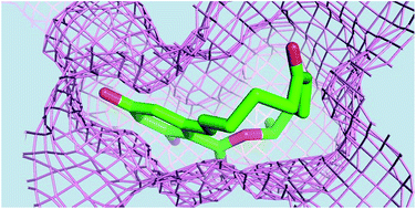
The mycotoxin zearalenone has been contaminating maize and other grains. It can be hydrolyzed and inactivated by the lactonase ZHD, which belongs to the α/β-hydrolase family. Besides the catalytic core domain, the enzyme comprises an α-helical cap domain. Zearalenone differs from other quorum-sensing lactones in its chemical structure. As revealed by the complex structure, the substrate binds into a deep pocket between the core and cap domains, adjacent to the catalytic triad Ser102–His242–Glu126. The enzyme–substrate interactions include three direct hydrogen bonds and several nonpolar contacts. In particular, the Trp183 side chain is engaged in both hydrogen bonding and T-stacking interactions with the benzoate ring. The central role of Trp183 in substrate binding was verified by the mutants W183A, W183H and W183F. Several mutants were also produced to investigate the roles of nearby amino-acid residues. Interestingly, mutants that destabilize the dimer had adverse functional effects on ZHD.


Many fungi produce secondary metabolites, encompassing not only antibiotics that are useful in fighting diseases but also mycotoxins that are harmful to humans and animals. Mycotoxins such as aflatoxin and ochratoxin A, found in contaminated foodstuff, can cause diseases upon ingestion and are not welcomed by feed and breeding industry.1 The Fusarium mycotoxin zearalenone (ZEN), a mycoestrogen found in contaminated maize and other grains, is implicated in reproductive disorders of swine and other domestic animals.2 The chemical structure of ZEN contains a lactone derived from 2,4-dihydroxybenzoic acid (a resorcylic acid; Fig. 1A),3 and it is not affected by cooking.1 When metabolized into zearalenols (ZOLs) the isomer α-ZOL shows increased estrogenic activity in human.4 This mycotoxin inhibits HSP90 and protein kinase activity.5,6 It also leads to DNA fragmentation and apoptosis.7 ZEN can be removed by chemical adsorption or inactivated by biotransformation. Recently, a ZEN-detoxifying gene zhd101 from Clonostachys rosea encoding a protein of 264 amino acids, named ZHD here, was isolated and successfully used in genetic modification of other organisms.8,9 ZHD is a novel lactonohydrolase that cleaves the ester bond of ZEN (and ZOLs), producing a dihydroxyphenyl derivative with an open side chain upon subsequent loss of CO2 (Fig. 1A).
image file: c4ra12111b-f1.tif
Fig. 1 Structures of ZEN and ZHD. In (A) the substrate ZEN is shown on the top, along with its corresponding hydrolysis intermediate (middle) and product (bottom). The carbon atoms are numbered 1–6 in the phenyl ring and 1′–12′ in the lactone loop. In (B) the ZHD monomer is shown as a cartoon drawing. The central β-sheet and the flanking α-helices in the core domain are coloured green and orange. The cap-domain α-helices are in magenta. The side chains of Ser102, His242, Glu126, Asp31 and Asp223, as well as the bound substrate from the S102A/ZEN complex, are shown as stick models. PDB codes: 3WZL (ZHD); 3WZM (S102A/ZEN).

Lactonases are receiving much attention because they can interfere with quorum sensing systems, which regulate microbial growth and pathogenesis.10 A major mechanism of “quenching” quorum signals is by cleaving and thus inactivating bacterial N-acyl homoserine lactones (AHL) by lactonase and acylase.11 The chemical structures of ZEN and AHL are very different, and so are the corresponding ring-opening reactions and the catalyzing enzymes (Fig. S1). AHL lactonases include enzymes with metallo-β-lactamase fold, (β/α)8-amidohydrolase fold, and α/β-hydrolase fold. ZHD belongs to the latter.8 Unlike some other AHL lactonases such as AiiA,12 ZHD does not require metal ion for activity.9 In addition to the catalytic core domain that contains the Ser/Cys-His-Asp/Glu triad, lactonases with α/β-hydrolase fold such as the enol-lactone hydrolase PcaD (Fig. S1) contain an α-helical cap domain,13 which covers the active site and determines the substrate specificity. Between the core and cap domains lies a deep tunnel-like cavity to accommodate the substrate. Detailed enzyme–substrate interactions in this class of lactonases have been illustrated by the complex structures of AidH.14 The substrate AHL is recognized via both hydrogen bonds (to the homoserine lactone ring) and hydrophobic interactions (with the acyl side chain). The cap domain is also involved in dimer formation of PcaD.13

A few mycotoxin-degrading enzymes have been discovered,1 but to our knowledge no mycotoxin-bound protein structure has been solved so far. To investigate the interactions by which ZHD recognizes its substrate, we expressed the gene in Escherichia coli, purified the recombinant protein (named rZHD here) and had it crystallized for structural analyses by X-ray diffraction. BLAST searches (http://blast.ncbi.nlm.nih.gov/) using the ZHD sequence only showed α/β-hydrolases with 30% identity in the N-terminal part (1–120), including those of PDB 2XUA (PcaD)13 and 3OM8. The latter protein shows 43% sequence identity to PcaD but only 24% to ZHD. Attempts to solve the structure of rZHD by molecular replacement (MR) were unsuccessful, and it prompted us to obtain rZHD crystals that contained selenium-methionine (SeMet) for structure determination by single or multi-wavelength anomalous diffraction (SAD or MAD). We also crystallized an inactive mutant S102A in complex with ZEN and analysed the substrate binding mode in conjunction with a number of other mutants.

The crystal structure of rZHD was solved by SAD using the peak data set of SeMet. The electron density map showed two well-defined protein molecules (designated monomers A and B) and a weak region that could only be interpreted as coils by automatic model-building programs. After preliminary refinement of monomers A and B, a third monomer C was correctly located by MR, using a native data set. The average temperature factor of monomer C is significantly higher than those of A and B (Fig. S2). The root-mean-square deviation (RMSD) between the backbone atoms of monomer A and B is 0.19 Å, whereas monomer C shows a larger RMSD of 0.50 Å. The mutant complex crystal of S102A/ZEN is isomorphous to the native (RMSD = 0.25 Å), and the initial difference Fourier map indicates the presence of a bound ZEN molecule in the active site of each monomer (Fig. S3A, S3B, S3C). After refinement, the conformation of the bound ligand can be determined unambiguously in the map (Fig. S3D).

The rZHD protein of 264 original amino acids plus a 14-residue N-terminal His-tag folds into a core α/β-hydrolase domain with a central β-sheet of eight parallel strands (β1–β8), which is flanked by seven α-helices (α1–α4 and α9–α11). Over the C-terminal edge of the β-sheet, a cap domain is formed by four α-helices (α5–α8; Fig. 1B). A comparison of rZHD with PcaD and AidH showed large difference in the cap domain (Fig. S4A). Superposition with PcaD (PDB 2XUA) yielded an RMSD of 2.1 Å for 135 Cα atoms in the core domain (Fig. S4B). For AidH (PDB 4G9E) only 93 Cα atoms in the N-terminal 120 residues were matched with an RMSD of 1.5 Å (Fig. S4C). The rZHD cap domain features a long but bent helix α8, which spans across the core domain on top of the β-sheet. The substrate ZEN is bound to a deep pocket between the core and cap domains (Fig. 1B and S5A). Monomer A in the native crystal and the S102A/ZEN complex differed by an RMSD of 0.14 Å for 244 Cα atoms, indicating no significant change in the overall protein conformation upon substrate binding. The ligand is bound in a bent mode, with the 14-membered lactone ring folded up by about 60° relative to the dihydroxybenzoate ring (Fig. S5B).

The oligomerization status of rZHD in solution was investigated by using size exclusion chromatography, which showed a peak with apparent molecular weight of 55.3 kDa, strongly suggestive of a dimer (Fig. S6A). There are three Cys residues in rZHD, but none of them are located on the protein surface. Cys101 and Cys124 are found in two neighbouring β-strands but facing different sides of the β-sheet. Consequently, no disulphide bond is likely to form within an rZHD monomer or between different monomers. In the rZHD crystal, monomers A and B are related by a non-crystallographic dyad axis, forming a dimer (Fig. S6B). The third monomer C also forms a similar dimer with its crystallographic symmetry related neighbour C′. The dimer interface is formed by 14 residues in loop β7-α10, helix α10 and strand β8, and covers 630 Å2 surface areas on each monomer. It is different from that of PcaD (Fig. S6C)13 and it does not involve the cap domain. Although not very large in area, the interface encompasses the nonpolar side chains of Phe222, Ile225, Val226, Ile235 and Leu237, which come together and form a stable hydrophobic patch (Fig. S6D). In addition, there are four hydrogen bonds between the backbone atoms of Ala214, Thr216, Thr218 and Glu219 (Fig. S6E).

As a member of α/β-hydrolase family, ZHD has its active site located on the C-terminal edge of the central β-sheet. The catalytic triad consists of Ser102, His242 and Glu126 (Fig. 1B). The acidic residue of catalytic triad is Glu126 rather than Asp223 as proposed previously.8 Nor is it Asp31 as predicted along with Glu126 in a recent phylogenetic study.15 The mutant E126A is inactive but D223A still retains 37% activity (Fig. 2). Glu126 is located at the C-terminal end of strand β6, close to Ser102 in the adjacent strand β5. From the nearby β8-α11 loop the His242 side chain intercalates between Ser102 and Glu126, making hydrogen bonds to both (Fig. 3A). In the S102A/ZEN complex, the NE2 atom of His242 is 3.4 Å from the ester oxygen (O10′) of ZEN, in a good position for proton transfer to the substrate. When the native structure is superposed on the mutant, the OG atom of Ser102 is 2.2 Å from the carbonyl carbon (C12) of ZEN, with a high potential for nucleophilic attack. The carbonyl oxygen (O12) makes a hydrogen bond to the peptide NH group of Gly32 (Fig. 3B), which is supposed to stabilize the developing negative charge in transition state.


image file: c4ra12111b-f2.tif
Fig. 2 Mutant activity. The specific activity of each ZHD mutant for ZEN hydrolysis is plotted as a percentage of the wild-type. Each solid bar corresponds to the average of three measurements and the standard deviation is shown as an error bar. LH/AA denotes the double mutant L132A/H134A. ND means that the activity could not be determined.

image file: c4ra12111b-f3.tif
Fig. 3 Interactions between ZEN and ZHD. The bound substrate of S102A/ZEN complex is shown as thick sticks and coloured pink. The protein is coloured green. In (A) the view is largely parallel to the phenyl ring of ZEN. The protein backbone is depicted as worm tracings and the selected side chains as thin sticks. The side chain of Ser102 from the wild-type structure is also shown, coloured cyan. Potential nucleophilic interaction and hydrogen bonds are shown as dashed lines. In (B) a different view shows the other surrounding amino acids in the active-site pocket. Most are nonpolar.

On the other side of the bound substrate, the indole plane of Trp183 (from helix α8) is perpendicular to the dihydroxybenzoate ring of ZEN, making a T-stacking interaction between the two aromatic groups (Fig. 3A). The indole NH group (NE1) is hydrogen bonded to the ortho hydroxyl group (O2) of ZEN, which also makes a hydrogen bond to the side chain (OG) of Ser103 (Fig. 3B). The other enzyme-substrate interactions are mostly non-polar. In addition to the indole of Trp183, the aliphatic side chains of Leu33, Val153, Met154, Val158, Leu135, Phe221, Pro128, Ile191 and Pro192 are in direct contact with the substrate. Pro192 has its CG atom at 3.1 Å from the para hydroxyl group (O4) of ZEN. Together these amino acids make a topologically complementary binding pocket, in which the substrate molecule is snugly accommodated (Fig. S5B). The ketone group (O6′) of ZEN is facing the entrance to the pocket, where the Leu132 and His134 side chains are 4.5 Å away from the substrate, not making any direct bond. Because the S102A/ZEN complex crystal was obtained by soaking, the substrate could diffuse freely into the pocket without significant conformational change of the enzyme. In the presence of the nucleophile (Ser102), the lactone can be cleaved and converted to the dihydroxyphenyl product with concomitant release of CO2, and readily removed from the active site.

The substrate binding pocket of ZHD is larger than those of PcaD and AidH (Fig. S5C). The chemical structure of ZEN has two closed rings, whereas those of enol-lactone and AHL have only one (Fig. S1). The larger 14-membered ring (or loop) of ZEN contains the lactone to be cleaved (opened) in the ZHD-catalysed reaction, whereas the smaller aromatic ring of dihydroxyphenyl group is retained. On the five-membered enol-lactone ring a short carboxymethyl “side group” is attached. In PcaD the side chains of Trp135 and Trp158 are important in defining the shape of active site.13 The AHL ring is also five-membered, but it has a longer acyl extension. In AidH, the substrate-binding tunnel is guarded by Phe189 and Phe192 at the entrance.14 Likewise, Phe64 and Phe68 constitute a “phenylalanine clamp” in AiiA, a metal-containing AHL lactonase, and the double mutant F64C/F68C showed different kinetic characteristics depending on disulphide bond formation.12 In comparison, the larger pocket of ZHD is more open, but the precise three-dimensional arrangement of the surrounding amino acids defines its specificity for ZEN. As shown above, the protruding side chain of Trp183 is especially important in shaping the pocket and interacting with the substrate (Fig. 3A). Interestingly, the AiiA mutant F107W in complex with the C10 product of AHL also had the indole NH group of Trp107 hydrogen bonded to the hydroxyl group of homoserine.12

As shown above, ZHD forms a complementary binding pocket for ZEN, suggesting that it is a highly specialized lactonase. We also hope to reveal the underlying basis of substrate binding and catalysis for future industrial applications in detoxifying the mycoestrogen. Therefore, instead of substrate analogues such as para-nitrophenyl esters that turn out readily detectable products, the ZHD activity was measured by using the authentic substrate, ZEN. In addition to S102A and E126A, several other mutants in and about the active-site pocket were constructed. As expected, the third catalytic-triad mutant of H242A also lost most activity. The residual 14% activity suggests that the general acid-base role of His242 in catalysis can be replaced, possibly by a solvent molecule. Likewise, although S102A of ZHD is inactive, the equivalent nucleophile mutant can be partially active in other α/β-hydrolases. For example, S172A of a β-diketone hydrolase still retained 10–20% of the wild-type activity.16 In contrast to H242A being partially active, E126A turned out to be inactive. Glu126 forms hydrogen bonds not only with His242 but also with Gly213 (N) and Thr216 (OG1) in loop β7-α10. Because the loop constitutes a major part of the ZHD dimer interface, Glu126 may play a structural role in addition to serving as the acidic residue in the catalytic triad.

Interestingly, the “wrong” catalytic-acid mutant D223A also lost more than 60% activity. Asp223 is found in helix α10. It makes two hydrogen bonds with Thr129 (OG1) and Asn224 (ND2), and is 4.5 Å away from Lys130 (NZ), implicating an electrostatic coupling. Removing the carboxylate group disrupts these interactions and presumably destabilizes the dimeric structure of ZHD. The dimer interface is close to the active-site pocket. Phe221 at the N-terminus of helix α10 directly participates in substrate binding. To investigate the correlation between dimer formation and catalytic activity of ZHD, the double mutant of I225E/V226E and the triple mutant of F222R/I225E/V226E were constructed, with the hope to obtain a monomer. Perhaps it would be more efficient in substrate binding and product release without the other monomer. However, these mutants could not be properly expressed, again suggesting that the functional unit of ZHD is a homodimer.

In the very middle of the substrate-binding pocket, the aromatic side chain of Trp183 is engaged in both T-stacking and hydrogen bonding interactions with ZEN. To investigate the role of Trp183 by breaking down its interactions with ZEN, three mutants W183F, W183H and W183A were made and tested. They turned out to be all inactivated (Fig. 2), clearly indicating that the combination of T-stacking and hydrogen bonding is essential in substrate binding. The former could be present in W183F and the latter in W183H, but neither mutant retained the enzymatic activity, not to mention the case of W183A in which both were eliminated. T-stacking is likely more important than hydrogen bonding, because the mutant W183F still showed some residual activity. In other α/β-hydrolases, such as lipases and the above-mentioned β-diketone hydrolase, the bulky indole side chain of a tryptophan is frequently observed to cover the active site.17 Tryptophan is usually found in a lid-like structure that separates the catalytic residues from the bulk solvent. However, by comparison, the role of the tryptophan is not so critical for activity in those enzymes as it is in ZHD.

Two other direct hydrogen bonds to ZEN are contributed by Gly32 (N) and Ser103 (OG). While Gly32 functions as the oxyanion hole, the role of Ser103 is probably not as important, because the mutant S103A turned out to be fully active. Ser103 and Trp183 bind to the same hydroxyl group of ZEN (O2). With an attempt to introduce an additional hydrogen bond between ZHD and ZEN, another mutant P192S was made. However, it showed only about 10% activity. Presumably Pro192, together with the adjacent Pro193, plays a structural role. These two consecutive proline residues are located at the C-terminus of the longest helix α8 in the cap domain.

Next, our attention was turned to the open side of the substrate-binding pocket. First, the two nonpolar residues Val153 and Val158 were made more polar, with a hope to facilitate substrate entry and product release, as well as possible hydrogen bond formation with the ketone group (O6′) of ZEN. As shown in Fig. 2, ZHD activity was retained in the mutants V153A and V153H, but it was reduced in V153Q (80%) and V153D (45%). Comparatively lower activity was observed in V158H (64%) and V158D (12%). Because Val153 is close to the ketone group, the contacts with substrate can be replaced by polar interactions. Val158 is packed against the adjacent hydrocarbon moiety of the lactone loop, and the interactions should remain nonpolar for good activity. Second, Leu132 and His134 were replaced by alanine. While L132A retained the original activity, H134A lost about one third, and the double mutant L132A/H134A lost more than 80% of the activity. Although both amino acids are not in direct contact with ZEN, the imidazole group of His134 appears to be more important than the aliphatic group of Leu132 in substrate binding, and when both are removed, exposure of the Leu135 side chain to the bulk solvent would further weaken the ZHD–ZEN interactions.

The lactonase ZHD contains a specialized active-site pocket, to which the substrate ZEN is bound in a bent conformation. On the other hand, ZHD functions as a homodimer in solution. The high-resolution crystal structure of ZHD–ZEN complex provides the groundwork for further protein engineering to enhance the enzymatic activity and dimer stability of ZHD under harsh conditions as frequently employed in industrial processes.

Acknowledgements

This work was supported by grants from the National Basic Research Program of China (2011CB710800 and 2011CBA00805), the National High Technology Research and Development Program of China (2012AA022200), the National Natural Science Foundation of China (31200053, 31300615 and 31400678), the Ministry of Science and Technology of Taiwan (MOST 102-2628-B002-007-MY2 and MOST 103-2314-B002-004-MY2) and Chinese Academy of Sciences (KSZD-EW-Z-015-2). We thank the National Synchrotron Radiation Research Centre of Taiwan for beam-time allocation and data collection assistance.

References

  1. S. Marin, A. J. Ramos, G. Cano-Sancho and V. Sanchis, Food Chem. Toxicol., 2013, 60, 218–237 CrossRef CAS PubMed.
  2. C. Cortinovis, F. Pizzo, L. J. Spicer and F. Caloni, Theriogenology, 2013, 80, 557–564 CrossRef CAS PubMed.
  3. N. Winssinger and S. Barluenga, Chem. Commun., 2007, 22–36 RSC.
  4. W. T. Shier, A. C. Shier, W. Xie and C. J. Mirocha, Toxicon, 2001, 39, 1435–1438 CrossRef CAS.
  5. S. Wang, Y. Xu, E. A. Maine, E. M. Wijeratne, P. Espinosa-Artiles, A. A. Gunatilaka and I. Molnár, Chem. Biol., 2008, 15, 1328–1338 CrossRef CAS PubMed.
  6. J. Lu, J. Y. Yu, S. S. Lim, Y. O. Son, D. H. Kim, S. A. Lee, X. Shi and J. C. Lee, Toxicol. In Vitro, 2013, 27, 1007–1017 CrossRef CAS PubMed.
  7. I. Ayed-Boussema, Z. Ouanes, H. Bacha and S. Abid, J. Biochem. Mol. Toxicol., 2007, 21, 136–144 CrossRef CAS PubMed.
  8. N. Takahashi-Ando, M. Kimura, H. Kakeya, H. Osada and I. Yamaguchi, Biochem. J., 2002, 365, 1–6 CrossRef CAS PubMed.
  9. N. Takahashi-Ando, S. Ohsato, T. Shibata, H. Hamamoto, I. Yamaguchi and M. Kimura, Appl. Environ. Microbiol., 2004, 70, 3239–3245 CrossRef PubMed.
  10. W. Fast and P. A. Tipton, Trends Biochem. Sci., 2012, 37, 7–14 CrossRef CAS PubMed.
  11. B. LaSarre and M. J. Federle, Microbiol. Mol. Biol. Rev., 2013, 77, 73–111 CrossRef CAS PubMed.
  12. C. F. Liu, D. Liu, J. Momb, P. W. Thomas, A. Lajoie, G. A. Petsko, W. Fast and D. Ringe, Biochemistry, 2013, 52, 1603–1610 CrossRef CAS PubMed.
  13. J. Bains, L. Kaufman, B. Farnell and M. J. Boulanger, J. Mol. Biol., 2011, 406, 649–658 CrossRef CAS PubMed.
  14. A. Gao, G. Y. Mei, S. Liu, P. Wang, Q. Tang, Y. P. Liu, H. Wen, X. M. An, L. Q. Zhang, X. X. Yan and D. C. Liang, Acta Crystallogr., Sect. D: Biol. Crystallogr., 2013, 69, 82–91 CAS.
  15. D. Popiel, G. Koczyk, A. Dawidziuk, K. Gromadzka, L. Blaszczyk and J. Chelkowski, BMC Microbiol., 2014, 14, 82 CrossRef PubMed.
  16. Y. Yang, T. P. Ko, L. Liu, J. Li, C. H. Huang, H. C. Chan, F. Ren, D. Jia, A. H. Wang, R. T. Guo, J. Chen and G. Du, ChemBioChem, 2014, 15, 1882–1886 CrossRef CAS PubMed.
  17. Y. Yang, T. P. Ko, L. Liu, J. Li, C. H. Huang, J. Chen, R. T. Guo and G. Du, Biochem. Biophys. Res. Commun., 2014, 452, 509–514 CrossRef CAS PubMed.

Footnotes

Electronic supplementary information (ESI) available: Experimental procedures for structure determination and activity measurement are described. Table S1 gives the primer sequences used in mutagenesis. Table S2 and S3 show the phasing and refinement statistics. Fig. S1–S6 provide additional views of the lactonase reactions and structures as well as a chromatography profile. See DOI: 10.1039/c4ra12111b
WP and TPK contributed equally to this work.

This journal is © The Royal Society of Chemistry 2014
Click here to see how this site uses Cookies. View our privacy policy here.