Yoko Kitayamaa,
Kazuki Nakabayashia,
Takashi Wakabayashia,
Nobuo Tajimab,
Michiya Fujiki*c and
Yoshitane Imai*a
aDepartment of Applied Chemistry, Faculty of Science and Engineering, Kinki University, 3-4-1 Kowakae, Higashi-Osaka, Osaka 577-8502, Japan. E-mail: y-imai@apch.kindai.ac.jp
bComputational Materials Science Center, National Institute for Materials Science, 1-2-1 Sengen, Tsukuba, Ibaraki 305-0047, Japan
cGraduate School of Materials Science, Nara Institute of Science and Technology, Takayama, Ikoma, Nara 630-0192, Japan. E-mail: fujikim@ms.naist.jp
First published on 17th November 2014
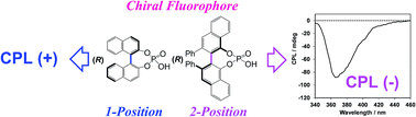
The signs of circularly polarised luminescence and circular dichroism of chiral biaryl fluorophores in the CHCl3-dissolved state (solution state) and the PMMA-film-dispersed state (solid state) were successfully controlled by modifying the internal axial chirality and the axial bonding position of the biaryl units.
To rationally invert the sign of the Cotton CPL signal of a chiral fluorophore, an organic fluorophore with opposite point chirality (stereogenic centers) is needed. However, this approach has practical limitations because enantiopure fluorophore pairs must be isolated. Thus, an elegant approach for fine controlling the CPL sign of chiral fluorophores without use of opposite point chirality is well acknowledged.3 In recent papers, we demonstrated several novel approaches for facile control of the CPL signs of binaphthyl-based fluorophores possessing the same axial chirality (stereogenic bonds) by changing the dihedral bond angle between two naphthyl units.4 This facile CPL sign inversion approach is also achievable by modifying steric demanding factors between adjacent binaphthyl units and by choosing external matrices (fluidic solutions, solid polymers, and inorganic salts).4
To gain a deeper understanding of how biaryl moiety affects the CPL sign, in this study we focused on a relationship between the internal axial bond position of biaryls with the same (R)- and (S)-axial chirality and the resulting CPL sign. To this end, we chose three pairs of optically active aryl fluorophores (Scheme 1): (i) (R)-(−)-3,3′-diphenyl-2,2′-bi-1-naphthol (VANOL) hydrogen phosphate [(R)-1] and its (S)-analog [(S)-1] as binaphthyls, (ii) (R)-(−)-2,2′-diphenyl-4-biphenanthrol (VAPOL) hydrogen phosphate [(R)-2] and its (S)-analog [(S)-2] as bisphenanthryls with an axial bonding position similar to (R)-1, and (iii) (R)-(−)-1,1′-binaphthyl-2,2′-diyl hydrogen phosphate [(R)-3] and its (S)-analog [(S)-3] as a different internal axial bonding position of these biaryls. Herein, we demonstrate for the first time that the CPL sign is determined solely by the internal axial bonding position of these biaryls.
The CPL spectra in CHCl3 solution and PMMA films were measured using a JASCO CPL-200 spectrofluoropolarimeter (Tokyo, Japan) at room temperature. The instrument used a scattering angle of 0° from the excitation of unpolarised, monochromated incident light with a bandwidth of 10 nm. The specimens in PMMA films were prepared analogously to the samples used for the solid-state fluorescence spectra. (R)-1 and (R)-2 at a concentration of 1.0 × 10⁻⁴ M in CHCl3 solutions were excited at 307 and 335 nm, respectively. (R)-1 and (R)-2 in PMMA films were excited at 320 and 330 nm, respectively. The CPL spectra were approximated using the simple moving average (SMA) method.
![]() | ||
Fig. 1 CPL (upper panel) and PL (lower panel) spectra of (R)-1 (black lines) and (S)-1 (grey lines) in CHCl3 (1.0 × 10⁻⁴ M). |
The fluorophore (R)-1 in CHCl3 has a PL maximum (λem) at 372 nm with an absolute value of PL quantum yield (ΦF) of 0.12 (indicated by black line). The differences in the λem and ΦF values between (R)-1 and (R)-3 [λem = 350 nm and ΦF = 0.29]4a,10 was calculated. Remarkably, the ΦF value of (R)-1 was almost half that of (R)-3. This may arise from thermal deactivation due to higher vibrational modes in (R)-1 than (R)-3.
As expected, (R)-1 can emit CPL in the order of |gem| ∼ 10−3, where the dimensionless Kuhn's anisotropy (gem) is defined as gem = 2(IL − IR)/(IL + IR). The CPL spectrum of (R)-1 in CHCl3 is shown in Fig. 1 (upper panel, black line). Surprisingly, although (R)-1 and (R)-3 commonly possess the same axially chiral binaphthyl units, they exhibit opposite signs in their CPL: a negative CPL sign for (R)-1 and a positive CPL sign for (R)-3 (Fig. 2, black lines for (R)-3).11
![]() | ||
Fig. 2 CPL (upper panel) and PL (lower panel) spectra of (R)-3 (black lines) and (S)-3 (grey lines) in CHCl3 (1.0 × 10⁻⁵ M).11 |
To verify whether (R)-1 introduced any artifacts in this CPL spectrum, the CPL spectrum of (S)-1 in CHCl3 was obtained in a similar way (Fig. 1, grey lines). The CPL spectra of (R)-1 and (S)-1 are mirror images of each other. The value of |gem| is ∼3.5 × 10⁻³ for 1, which is larger than that of 3 (|gem| ∼ 1.5 × 10⁻³).10 These results suggest that the CPL sign can be affected by the internal axial bonding position of binaphthyl units as well as by axial chirality.
To elucidate the origin of the CPL reversal, circular dichroism (CD) and UV-Vis absorption spectra of (R)-1 and (S)-1 in CHCl3 were measured and compared to those of (R)-3 and (S)-3. UV-Vis bands due to π–π* transitions characteristic of naphthyl groups in 1 are clearly observed between 250 and 350 nm (Fig. 3).
![]() | ||
Fig. 3 CD (upper panel) and UV-Vis (lower panel) spectra of (R)-1 (black lines) and (S)-1 (grey lines) in CHCl3 (1.0 × 10⁻⁵ M). |
The Cotton CD bands of (R)-1 and (S)-1 clearly demonstrate an almost mirror-image relationship due to π–π* transition moments. To quantitatively determine the CD amplitudes, we used the dimensionless Kuhn's anisotropy factor in the ground state, which is defined as gCD = Δε/ε. The |gCD| value at the first Cotton CD band (λCD = 343 nm) for 1 is ∼4.0 × 10⁻⁴. On the other hand, the |gCD| value of 3 at the first Cotton CD band (λCD = 324 nm) is ∼1.6 × 10−3.10 As in the case of CPL spectra, the CD spectra exhibit opposite signs: a negative sign for (R)-1 and a positive sign for (R)-3 (Fig. 4, black lines for (R)-3).11
![]() | ||
Fig. 4 CD (upper panel) and UV-Vis (lower panel) spectra of (R)-3 (black lines) and (S)-3 (grey lines) in CHCl3 (1.0 × 10⁻⁵ M).11 |
The signs of the CPL and the first Cotton CD band of (R)-1 are both negative, while those of (R)-3 are both positive. The sign reversal in the first Cotton CD bands is thus responsible for the reversal in the sign of the CPL bands observed for (R)-1 and (R)-3.
In the case of (R)-2, which is an extension of π-conjugation of (R)-1, (R)-2 in CHCl3 weakly emits at 373 nm (λem) with ΦF 0.09 (lower panel in Fig. 5, black line) and shows a negative CPL band (Fig. 5, upper panel, black line). To verify whether any artifacts existed in the CPL spectrum, we obtained the CPL spectrum of (S)-2 in CHCl3 for comparison (grey lines in Fig. 5). Because the CPL spectra of (R)-2 and (S)-2 exhibit an almost mirror-image relationship, the |gem| value for 2 is ∼1.3 × 10⁻³. Note that CPL signs for (R)-2 and (R)-3 are opposite: a negative for (R)-2 and a positive for (R)-3.
![]() | ||
Fig. 5 CPL (upper panel) and PL (lower panel) spectra of (R)-2 (black lines) and (S)-2 (grey lines) in CHCl3 (1.0 × 10⁻⁴ M). |
The CD and UV-Vis absorption spectra of (R)-2 in CHCl3 are shown in Fig. 6 (indicated by black lines). In contrast to (R)-1, the characteristic peaks originating from the π–π* transition of the biphenanthryl group in 2 are observed between 260 and 370 nm. To check if there are artefacts in the (R)-2 spectrum, we measured the CD spectrum of (S)-2 in CHCl3 (Fig. 6, grey lines). It is evident that (R)-2 and (S)-2 display an almost mirror-image relationship in their CD spectra. The |gCD| value of the first Cotton CD band (λCD = 361 nm) is ∼3.1 × 10⁻⁴. As expected, (R)-1 and (R)-2 exhibit the same signs in their CD spectra: a negative sign for both (R)-1 and (R)-2.
![]() | ||
Fig. 6 CD (upper panel) and UV-Vis (lower panel) spectra of (R)-2 (black lines) and (S)-2 (grey lines) in CHCl3 (1.0 × 10⁻⁵ M). |
To explain the origins of the opposite signs between (R)-1, (R)-2, and (R)-3 in their CPL and CD spectra, the equilibrium structures of (R)-1 and (R)-2 were examined theoretically (Fig. 7) and compared with the equilibrium structure of (R)-3 shown previously.10
The dihedral angles θ ((O)C–C–C–C(O)) of (R)-1, (R)-2, and (R)-3 are calculated as – 55°, – 61°, and – 53°,10 respectively. No significant difference in these θ angles between (R)-1, (R)-2, and (R)-3 is obtained theoretically, suggesting that the origin of the opposite CPL/CD signs cannot be simply ascribed to different conformations around the central C–C rotational bond.
The electronic excitation spectra of (R)-1 and (R)-2 are employed theoretically as that of (R)-3 was done previously.10 Fig. 8 shows calculated rotational strengths of (R)-1 and (R)-2 as a function of excitation wavelengths (λ) (Fig. 8a and b). These calculations suggest that (R)-1 and (R)-2 should have negative CD signals at the long-wavelength edge of the spectra (∼330 nm and ∼340 nm, respectively). The experimental CD spectra of (R)-1 and (R)-2 in CHCl3 are consistent with these theoretical results. The calculated CD intensities at these wavelengths can be explained as a result of non-coplanar monoaryl geometry of these molecules and reflected from the coupling of π–π* electronic transitions over each of the naphthalene and phenanthryl rings. In contrast, (R)-3 has a weak positive Cotton CD signal at the long-wavelength edge in the CD spectrum (∼340 nm).10 These CD calculations agree well with the experimental CD characteristics.
![]() | ||
Fig. 8 Calculated excitation wavelengths (l) and rotational strengths (R) for (a) (R)-1 and (b) (R)-2. Unit of rotational strength: 10−40 ergesucm G−1. |
For practical use as a CPL emitter, a detailed characterization of solid-state chiroptical properties is important. We recently studied the solid-state chiroptical properties of several chiral fluorophores doped into poly(methyl methacrylate) (PMMA) glassy matrices.4b,4d,10,12 The CPL spectra of (R)-1 and (R)-2 dispersed in spin-coated PMMA films clearly exhibit CPL and PL properties, as shown in Fig. 9 and 10 (black lines), respectively. (R)-1 and (R)-2 in PMMA films emit at λem 372 and 376 nm and have ΦF 0.16 and 0.19, respectively. The CPL spectra between (R)-1 and (S)-1, and between (R)-2 and (S)-2 are near mirror images of each other. (R)-1 and (R)-2 in PMMA films exhibit CPL in the order of |gem| ∼ 2.0 × 10⁻³ and ∼7.9 × 10⁻⁴, respectively.
![]() | ||
Fig. 9 CPL (upper panel) and PL (lower panel) spectra for (R)-1 (black lines) and (S)-1 (grey lines) in the PMMA films. |
![]() | ||
Fig. 10 CPL (upper panel) and PL (lower panel) spectra of (R)-2 (black lines) and (S)-2 (grey lines) in PMMA films. |
For 1 and 2, a subtle enhancement in the ΦF value in the PMMA films compared to ΦF in the CHCl3 solution was observed due to suppression of thermal deactivation modes because 1 and 2 are embedded into the glassy PMMA matrix. As reported previously, (R)-3 in PMMA has ΦF 0.14 and |gem| ∼ 1.4 × 10⁻³, which is similar to (R)-1 and (R)-2.4a,10 It is worth noting that relationships between (R)-1 and (R)-3 and between (R)-2 and (R)-3 maintain their opposite sign in their CPL spectra: a negative sign for (R)-1 and (R)-2, conversely, a positive sign for (R)-3.10
The CD and UV-Vis spectra of 1 and 2 dispersed in spin-coated PMMA films were measured (Fig. 11 and 12). CD and UV-Vis spectral profiles of (R)-1 and (R)-2 in PMMA are similar to those observed in the CHCl3 solution at longer wavelengths. The |gCD| values in the first Cotton CD bands of (R)-1 and (R)-2 are ∼2.1 × 10⁻³ at 336 nm and ∼5.2 × 10⁻⁴ at 361 nm, respectively. The CD spectra of (S)-1 and (S)-2 in the PMMA films are almost mirror images of those of the corresponding (R)-1 and (R)-2.
![]() | ||
Fig. 11 CD (upper panel) and UV-Vis (lower panel) spectra of (R)-1 (black lines) and (S)-1 (grey lines) in the PMMA films. |
![]() | ||
Fig. 12 CD (upper panel) and UV-Vis (lower panel) spectra of (R)-2 (black lines) and (S)-2 (grey lines) in the PMMA films. |
The solid-state chiroptical results indicate that the CPL and CD signals in PMMA films originate dominantly from the intramolecular interactions within the isolated molecule itself rather than from intermolecular interactions between multiple molecules. Additionally, the CPL sign even in PMMA can change in response to the internal axial bonding position of biaryl units.
This journal is © The Royal Society of Chemistry 2015 |