Yan Wanga,
Zhe-sheng Feng*a,
Lu-lin Wanga,
Le Yu†
b,
Jin-ju Chena,
Zi Liangc and
Rui Wangc
aState Key Laboratory of Electronic Thin Films and Integrated Devices, University of Electronic Science and Technology of China, Chengdu 610054, China. E-mail: fzs@uestc.edu.cn; Fax: +86 28 83202569; Tel: +86 28 83207590
bCatalysis Research Center, Hokkaido University, Sapporo, 001-0021, Japan
cAlternative Energy and Power Generation Technology Institute, DEC Central Academy, Dongfang Electric Corporation Limited, Chengdu 611731, China
First published on 6th October 2014
Mn doped LiFe1−xMnxPO4 (x = 0, 0.01, 0.03, 0.05, 0.07) cathode materials are synthesized by a carbothermal reduction method. The structure, morphology and electrochemical performance of the samples are characterized by X-ray diffraction (XRD), scanning electron microscopy (SEM), X-ray photoelectron spectroscopy (XPS), Raman, and charging–discharging tests. It is found that an appropriate amount of Mn doping did not affect the olivine structure and morphology of LiFePO4, but greatly improves its electrochemical performance. Especially, the LiFe0.97Mn0.03PO4 sample exhibits the best electrochemical performance, which shows the maximum discharge capacity of 148.2 mA h g−1 at 0.1 C and a capacity retention ratio of 98% after 60 cycles at various rates. Based on first-principles density functional theory (DFT), the lithium ion migration channels, energy band structures, densities of states, and charge density diffusion of LiFePO4 and LiFe15/16Mn1/16PO4 are calculated to further investigate the influence of Mn doping on the LiFePO4 lattice. The results prove that the higher lithium ion conduction and electronic conductivity of LiFe15/16Mn1/16PO4 are attributed to Mn doping.
Theoretical calculation has the advantage in supplementing the real experiments that one has full manipulate the relevant variables. The first-principles investigation has been shown to be a useful method to predict the properties of cathode materials in lithium-ion batteries.13–16 For example, Ceder et al. calculated activation barriers to Li ion motion for LiMPO4 (M = Mn, Fe, Co, Ni) olivine materials by using first-principles method, and demonstrated that Li ion diffuses through one-dimensional channels with high energy barriers to cross between the channels.13 In recent years, a lot of research works on metal ion doping of LiFePO4 by first-principle have been reported.17–21 Shi et al. investigated Mg doping LiFePO4 at Li and Fe sites using first-principles density-functional theory, and elucidated the dependence of the electrochemical properties on the concentration of Mg dopant.17
It is essential to study systematically the micro-structural changes and electrochemical performance of metal ion doped LiFePO4 for a better understanding on metal ion doping. Though some interesting experimental or theoretical results have been obtained, no joint experimental and theoretical investigations have been carried out to study the effect of metal ion doping on the physical and electrochemical characterization of LiFePO4, and the intrinsic reasons of electrochemical properties enhancing of metal ion doped LiFePO4 have not been well clarified.
In this paper, the Mn doped LiFe1−xMnxPO4 (x = 0, 0.01, 0.03, 0.05, 0.07) were prepared via a simple carbothermal reduction method. The effect of metal ion Mn doping with different amounts on the structure, morphology and electrochemical performance of LiFePO4 was studied in detail. In addition, by the first-principles, we calculated and compared the structural and electronic properties of LiFePO4 and LiFe15/16Mn1/16PO4. Combined with the experimental results, the mechanism that metal ion Mn doping improves the electrochemical performance of LiFePO4 is proposed theoretically.
The electrochemical properties of LiFe1−xMnxPO4 (x = 0, 0.01, 0.03, 0.05, 0.07) samples were measured by CR2032 button-type cell. The cathode electrodes were prepared by coating the slurry of a mixture composed of 80 wt% active material, 10 wt% acetylene black, and 10 wt% polyvinylidene fluoride (dissolved in N-methyl pyrrolidone) onto aluminum foil current-collector, and then dried at 120 °C for 10 h under vacuum. The electrolyte was 1 M LiPF6 in 1:
1
:
1 (v/v) ethylene carbonate, dimethyl carbonate and diethyl carbonate. A Celgard microporous polypropylene sheet was used as the separator, and a lithium metal foil was used as the anode electrode. The cells assembly was performed under an argon atmosphere in a glove box. The charge–discharge and cycling properties of the cells were evaluated between 2.2 and 4.2 V (vs. Li+/Li) using a EWARE BST-8 electrochemical test instrument at room temperature.
![]() | ||
Fig. 1 XRD patterns of LiFe1−xMnxPO4 (x = 0, 0.01, 0.03, 0.05, 0.07) samples (a). Magnification part in the range of 24.8–26.4° of XRD patterns (b). |
The detailed lattice parameters of LiFe1−xMnxPO4 (x = 0, 0.01, 0.03, 0.05, 0.07) samples are calculated and listed in Table 1. As being shown, the lattice parameters of Mn doped LiFePO4 are larger than those of undoped sample. Moreover, with increased x, the increase in the unit cell volume is almost linear. It is in good agreement with A. Yamada's report.26 That is because average value of the radius of Mn ion (0.84 Å) is larger than that of Fe ion (0.74 Å) in octahedral coordination.
Sample | a (Å) | b (Å) | c (Å) | V (Å3) |
---|---|---|---|---|
LiFePO4 | 6.0012 | 10.3176 | 4.6861 | 290.15 |
LiFe0.99Mn0.01PO4 | 6.0020 | 10.3189 | 4.6913 | 290.55 |
LiFe0.97Mn0.03PO4 | 6.0035 | 10.3213 | 4.6924 | 290.76 |
LiFe0.95Mn0.05PO4 | 6.0052 | 10.3216 | 4.6937 | 290.93 |
LiFe0.93Mn0.07PO4 | 6.0066 | 10.3288 | 4.6939 | 291.21 |
The morphological feature of the material is an extremely important factor for the electrochemical property of the cathode material, especially for its high-rate charge–discharge performance. The scanning electron microscopy images of LiFe1−xMnxPO4 (x = 0, 0.01, 0.03, 0.05, 0.07) samples are shown in Fig. 2. It can be seen that the LiFe1−xMnxPO4 is composed of irregular particles, which aggregate slightly with a broad particle size distribution of 100–600 nm. It indicates that a small amount of Mn doping does not have any influence on morphology of LiFe1−xMnxPO4. The nano-sized LiFe1−xMnxPO4 particles can shorten the diffusion path of lithium ions as well as providing rapid and easy Li+ transport.
In order to identify the oxidation state of Fe and Mn in the samples, X-ray photoelectron spectroscopy (XPS) characterization is carried out and reported in Fig. 3. The Fe 2p spectrum consists of two components (Fe 2p3/2 and Fe 2p1/2) because of spin–orbit coupling. The binding energy position of the main peak is related to the oxidation state of Fe. For the ferrous ion in LiFePO4, the main peaks of Fe 2p3/2 and Fe 2p1/2 are emerged at ∼710 and ∼724 eV, respectively. For the ferric iron in FePO4, the main peaks of Fe 2p3/2 and Fe 2p1/2 are centred at ∼713 and ∼726 eV, respectively. For the LiFe0.97Mn0.03PO4 sample, the peak position (710.4 and 723.8 eV) in Fig. 3(a) is consistent with Fe2+, and there is no peak assigned to Fe3+. The Mn 2p spectrum shape for LiFe0.97Mn0.03PO4 sample is similar with that of Fe 2p. It can be observed that a main peak centers at ∼641 eV which can be attributed to Mn 2p3/2 in the typical form of Mn2+. From the analysis of XPS, we confirm that Mn2+ has been incorporated into the lattice and remained the valence as in the starting material. Therefore, the oxidation states of Fe and Mn in the LiFe1−xMnxPO4 samples are +2.
Fig. 4 shows the Raman spectra of LiFe0.97Mn0.03PO4 and LiFePO4 samples. It can be seen that the peaks observed agree very well to the assignments of LiFePO4 reported in the literatures.28,29 The bands in the Raman spectra between 300 and 700 cm−1 ν2 and ν4 comprise bending modes of a PO43− anion. Three bands observed between 900 and 1200 cm−1 belong to intramolecular symmetric and asymmetric stretching modes of the PO43− anion (ν1 and ν3). The weak broad bands at ∼1350 and ∼1580 cm−1 can be assigned to the D band and G band of carbonaceous materials, respectively. This proves that a small amount of residual carbon from acetylene black may be present in the LiFe1−xMnxPO4 (x = 0, 0.01, 0.03, 0.05, 0.07) samples.
Fig. 5(a) shows the first charge–discharge profile of LiFe1−xMnxPO4 (x = 0, 0.01, 0.03, 0.05, 0.07) samples between 2.2 and 4.2 V at 0.1 C. All samples exhibit the typical electrochemical behavior of LiFePO4, with a long charge and discharge voltage plateau at ∼3.5 and ∼3.4 V. It indicates that the well-known crystal LiFePO4 is successfully synthesized and small amounts of Mn doping do not destroy the crystal structure of LiFePO4. It is difficult to observe voltage plateau of Mn2+/Mn3+ at ∼4.0 V due to very small concentration of Mn in LiFe1−xMnxPO4 (x = 0, 0.01, 0.03, 0.05, 0.07) samples. Pristine LiFePO4 shows the lowest charge–discharge capacity of 135.2 and 131.9 mA h g−1, followed by LiFe0.93Mn0.07PO4 of 142.4 and 140.7 mA h g−1, LiFe0.99Mn0.01PO4 of 144.7 and 142.4 mA h g−1, and then LiFe0.95Mn0.05PO4 of 146.5 and 145.1 mA h g−1. LiFe0.97Mn0.03PO4 has the highest charge–discharge capacity of 151.4 and 148.2 mA h g−1, which is nearly 90% of theoretical capacity. Moreover, the LiFe0.97Mn0.03PO4 has an almost 100% capacity retention after 20 charge–discharge cycles at 0.1 C (Fig. 5(b)). This means that LiFe0.97Mn0.03PO4 has a good electrochemical characteristic at low current rate.
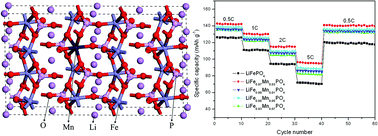
To study the rate capability of LiFe1−xMnxPO4 (x = 0, 0.01, 0.03, 0.05, 0.07), the electrodes are discharged at various current rates. Fig. 5(c) presents the discharge capability of LiFe0.97Mn0.03PO4 electrode at various current rates. The LiFe0.97Mn0.03PO4 sample exhibits discharge capacity of 142.5, 130.5, 115.0, and 96.3 mA h g−1 as it is discharged at 0.5, 1, 2, and 5 C, while that of LiFePO4 is 126.1, 111.0, 94.2, and 71.8 mA h g−1, respectively (Fig. 5(d)). Apparently, LiFe0.97Mn0.03PO4 has an outstanding rate performance at more than 0.5 C. From Fig. 5(d), as the current rate is reduced again to 0.1 C, the capacity of the LiFe0.97Mn0.03PO4 sample retains 98% after 60 cycles. It supports that the LiFe0.97Mn0.03PO4 displays a better rate capability at various rates compared with other LiFe1−xMnxPO4 electrodes, and enhances the rate capability and cycling performance effectively. Furthermore, it can be seen that all LiFe1−xMnxPO4 electrodes exhibit excellent rate capabilities. This indicates that proper content Mn doping plays a significant role in enhancing the electrochemical performances of LiFePO4.
In order to investigate the effect of Mn doping on electrochemical performance of LiFe1−xMnxPO4, the first principles calculations have been performed to calculate and analyze the structure and electronic characterizations of LiFePO4 and LiFe15/16Mn1/16PO4. (Only a maximum of 112-atom LiFePO4 supercell with 2 × 2 × 1 repetition can be built due to restriction of the computational resources.) Pure bulk olivine-type LiFePO4 has an orthorhombic unitcell, which accommodates four LiFePO4 formula-units with the space group of Pnma. Fig. 6 shows the three-dimensional framework of LiFe15/16Mn1/16PO4 supercell, which contains 16 LiFePO4 formula-units. It can be seen that the Mn doping model is built by replacing one Fe atom from the LiFePO4 supercell with 2 × 2 × 1 repetition. Table 2 summarizes the average bond lengths of Fe–O, P–O, and Li–O adjacent to Mn atom in LiFe15/16Mn1/16PO4 and LiFePO4 supercell. As we know, the wide [010] direction lithium ion channels will promote Li ion migration. It is clear from Table 2 that the LiFe15/16Mn1/16PO4 has shorter Fe–O bond lengths and longer Li–O distance, which implies broadening of the adjacent lithium ion channels. Therefore, it can be concluded that doping Mn atoms in LiFePO4 supercell may improve the lithium ion conduction.
M–O bond length (average) | LiFe15/16Mn1/16PO4 | LiFePO4 |
---|---|---|
Fe1–O | 2.109 | 2.119 |
Fe2–O | 2.120 | 2.124 |
Fe3–O | 2.108 | 2.124 |
P1–O | 1.562 | 1.554 |
P2–O | 1.568 | 1.557 |
P3–O | 1.565 | 1.554 |
Li1–O | 2.173 | 2.163 |
Li2–O | 2.214 | 2.179 |
To further analyze the impact of Mn doping on the characterizations of LiFePO4, we examine the electronic structures of LiFePO4 with and without Mn doping. Fig. 7 shows calculated electronic band structures of LiFePO4 and LiFe15/16Mn1/16PO4 along the high symmetry point across the first Brillouin zone. The Fermi level is at 0 eV on the energy axis. The calculated band gap of LiFePO4 displays an indirect band gap about 0.61 eV that is consistent with the previously reported ∼0.60 eV.30,31 The calculation results are reasonable for a comparison, although it is underestimated comparing with the experimental value of 3.75 eV32 due to the well-known drawback of DFT. According to Fig. 7(b), band gap of LiFe15/16Mn1/16PO4 is about 0.44 eV, which is much lower than that of LiFePO4. Some papers33–38 have demonstrated similar results by theoretical calculation. The narrower band gap indicates that the electronic conductivity of LiFePO4 could be enhanced by doping Mn within LiFePO4 lattice.
The density of states (DOS) of undoped and doped models are calculated to investigate how the band gap changes, and the results are shown in Fig. 8. Fig. 8(c) shows the partial density of states (PDOS) for Fe 3d states, P 3p states, O 2p states, and Li 2s and 2p states of LiFePO4. It can be seen that two major valence bands include the high one located between −0.63 eV and the Fermi level and the low one located between −9.12 eV and −3.06 eV. The former is formed primarily by the Fe 3d orbit with minor contribution from O 2p orbit, while the latter is formed by a bonding state of the hybridized P 3p and O 2p orbit. The PDOS around the Fermi level is concerned due to the importance of the electron orbits near the Fermi level in the electron transport processes. The dominant electronic character of LiFePO4 near the Fermi level is contributed by Fe 3d orbit. The DOS of Mn atom and its neighboring Fe, O, P, Li atoms are compared (Fig. 3(c)) to clarify the effect of Mn doping on the electronic structure of LiFePO4 near the Fermi level. It is clear that the PDOS at the Fermi level mainly come from the Mn-3d orbit, which hybrids with its neighboring Fe-3d and O-2p orbits. The results demonstrate that the PDOS of Mn atoms play a key role in band gap reduction of LiFe15/16Mn1/16PO4.
![]() | ||
Fig. 8 The calculated TDOS and PDOS of LiFePO4 (a and c) and LiFe15/16Mn1/16PO4 (b and d) obtained from DFT calculations. |
Charge density distribution of LiFePO4 and LiFe15/16Mn1/16PO4 are investigated and illustrated in Fig. 9 to give a clear description about bonding nature between Mn atoms and adjacent O atoms. It can be seen that no significant change in the charge density distribution is observed by introducing Mn doping. However, the electron clouds of Fe atoms overlapped with that of O atoms (Fig. 9(b)), while the corresponding covalent bond is weak in LiFePO4. It indicates that there should be more electrons transfer from Fe atoms to O atoms in LiFe15/16Mn1/16PO4. Furthermore, a covalent bond is also formed between Mn and its adjacent O atoms, although it is weaker than the corresponding Fe–O bond. These results demonstrate that a strong interaction of Fe–O bonding by introducing Mn doping in LiFe15/16Mn1/16PO4 supercell induces the narrowed band gap.
Footnote |
† Current address: Guangzhou Tinci Materials Technology Co. Ltd., Guangzhou 510760, China. |
This journal is © The Royal Society of Chemistry 2014 |