Efficient photoluminescent complexes of 400–1800 nm wavelength emitting lanthanides containing organic sensitizers for optoelectronic devices†
Zubair Ahmed and
K. Iftikhar*
Department of Chemistry, Jamia Millia Islamia, New Delhi-110 025, India. E-mail: kiftikhar@jmi.ac.in; Fax: +91-11-26980229/26982489; Tel: +91-11-26837297
First published on 13th November 2014
Abstract
An in situ solution processed reaction of a bidentate O,O′-chelating anionic hexafluoroacetylacetone (Hhfaa) and a bidentate N,N′-chelating 2,2′-bipyridine ligand with LnCl3·6H2O in the presence of a base afforded the UV-sensitised 400–1800 nm wavelength emitting lanthanide complexes, [Pr(hfaa)3(bpy)2] and [Ln(hfaa)3(bpy)](Ln = Tb, Dy, Tm and Lu). The single-crystal analysis indicates that the Pr complex is ten-coordinate with a distorted bicapped square antiprism while the Dy complex is eight-coordinate with a distorted square antiprism geometry, and the bpy units, in the complexes, are involved in π–π stacking interactions and hydrogen bonding, respectively. The assembly of the hfaa− (a low vibrational frequency ligand) and bpy ligand makes an efficient protective coordination environment (PrO6N4 or DyO6N2) around the Pr (red emission), Tb (green emission), Dy (yellow emission) and Tm (blue emission) ions which leads to high quantum yields and longer emission lifetimes. The quantum efficiency of the complexes is enhanced in the solid state. Furthermore, these volatile and luminescent complexes were used as emitting layers to fabricate red-, green- and yellow-light emitting devices and their electroluminescence performances were investigated. The best devices with the structure ITO/CuPc (20 nm)/[Pr(hfaa)3(bpy)2] or [Tb(hfaa)3(bpy)] or [Dy(hfaa)3(bpy)] (80 nm)/BCP (25 nm)/AlQ (30 nm)/LiF (1 nm)/Al (200 nm) exhibit a maximum brightness of 183, 1765 and 532 cd m−2 with a current efficiency of 0.58, 3.6 and 0.76 cd A−1, respectively, which indicates an improved EL performance over the devices based on Pr, Tb and Dy complexes in the literature.
Introduction
The chemistry of luminescent lanthanide complexes is a challenging and fruitful area due to both a variety of coordination geometries1 and potential applications in luminescent probes,2 laser systems,3 pressure/damage sensors4 and emitting materials in organic light emitting diodes (OLEDs).5 In the field of luminescence, lanthanide complexes have attracted intensive research because of narrow line like emissions of optically pure colours and long radiative lifetimes which results from the shielding of the 4f-orbitals by the 5s and 5p shells. The 4f–4f transitions that result in light emission from the lanthanide are spin- and parity-forbidden and have a very low absorption coefficient (ε ≤ 1–10 M−1 cm−1),6 thus requiring the use of organic chromophores or “antenna” that possess a reasonably large molar absorption cross section to indirectly excite the metal centre.The ligand, 2,2′-bipyridine (bpy) is one of the widest used ligand in the design of highly luminescent lanthanide-containing systems because of its (i) intense absorption band in the near-UV and (ii) ability to efficiently transfer energy onto the Ln(III) ion excited states (antenna effect),7 giving rise to molecular species that can be used as probes and labels for a variety of chemical and biological applications.8 Beside bpy ligand, β-diketones and its complexes with lanthanides are intensively studied because of their good solubility and good volatility.9 The β-diketones has strong absorption within a large wavelength range for its π–π* transition and consequently has been targeted for its ability to sensitize the luminescence of the Ln(III) ions. These β-diketones give thermodynamically stable and photoluminescent compounds.10–13 Moreover, the photophysical properties of lanthanide β-diketonates (β-DKs) complexes could be tuned by fluorination on β-diketone because fluorination affects the triplet state energy of the ligands.14 Since the lanthanide tris(β-DKs) are coordinatively unsaturated, mostly found as [Ln(β-DKs)3(H2O)2], they can rapidly react with one or more ancillary neutral N-/O-donor ligand or their derivatives to form coordinatively saturated complexes making themselves more luminescent by suppressing the radiationless transition via vibrational relaxation by removing the high-frequency O–H oscillators.15–19
In the field of OLEDs, the electroluminescence (EL) is an important phenomenon where an applied electric field generates an excited species which upon relaxation emits photons. For this purpose, the electroluminescent lanthanide tris(β-diketonates) are ideal candidates because (i) lanthanide-based materials can generate narrow emission due to f–f transitions originating from emitting state of Ln(III) ion and (ii) efficient excitation of the lanthanide which occurs through the triplet state of the surrounding ligands allowing the possibility of using both singlet and triplet excitons created by electron–hole recombination to be used in radiative processes. The use of both singlet and triplet state by the emissive material could theoretically allow device efficiency to reach 100%.5 The electroluminescence from the devices based on Eu(III) complexes are of great interest because this metal ion emits narrow emission bands in red region.5,20–23 While the electroluminescence from Tb(III) complexes are also of great significance because they emit in green region, but the reports are scanty.24,25 Moreover, the complexes of Pr(III), Dy(III) and Tm(III) which emits in red, blue and white, and blue regions, respectively, are also less reported.26–28 Lee et al.29 have reported a device based on Pr(III) complex, [Pr(DBM)3bath] with a brightness of 50 cd m−2 achieved at 15 V. Zhi-Yong Guo et al.30 designed and fabricated a number of devices based on a dysprosium complex, [Dy(PM)3(TP)2] (PM = 1-phenyl-3-methyl-4-isobutyryl-5-pyrazolone and TP = triphenyl phosphine oxide) with light output ranging from 98 cd m−2 at 11.9 V to 524 cd m−2 at 19 V. Zheng group25 have fabricated a number of devices based on Tb–tris(β-diketonates) complexes with a maximum brightness of 58 cd m−2 at 25 V. However, the devices based on the Pr(III), Tb(III) and Dy(III) complexes with good electroluminescence performances are still need to be explored.
In this paper, we report a modified, one step in situ, route for the synthesis of ternary β-diketonate complexes of Pr, Tb, Dy, Tm and Lu with hexafluoroacetylacetone (Hhfaa) and 2,2′-bipyridine (bpy) at room temperature (303 K). The photophysical properties of the paramagnetic Pr, Tb, Dy and Tm in visible and NIR regions in solution and solid state are investigated and being presented. Furthermore, these volatile and highly luminescent complexes, as emitting layer, were used to fabricate red-, green- and yellow-light emitting electroluminescence devices. The characteristics of the synthesized complexes which are in favor of fabrication of low temperature processed OLEDs are: (i) high luminescence efficiencies, (ii) high volatility, (iii) lightweight, (iv) good transparency (v) easily thermally evaporated (mp 160–215) and (v) high decomposition temperature than the temperature required for the thermal evaporation (ca. 75 °C).
Experimental section
Materials and methods
The commercially available chemicals that were used without further purification are: Ln2O3 (Ln = Tb, Dy, Tm and Lu; 99.9%) and Pr6O11. These oxides were purchased either from Aldrich or Leico Chem., USA. The oxides were converted to the corresponding chlorides LnCl3·6H2O, by the standard procedure.31 The hexafluoroacetylacetone (MTM Lancaster, England) and 2,2′-bipyridine (Merck, Germany) were used as received. The solvents used in this study were either AR or spectroscopic grade. 2,9-Dimethyl-4,7-diphenyl-1,10-phenanthroline (BCP, 98%) and tert-butylacetyl chloride (99%) were obtained from Acros; N,N′-diphenyl-N,N′-bis(3-methylphenyl)-1,1′-biphenyl-4,4′-diamine (TPD, 99%) was purchased from Aldrich. The electron transport material tris(8-hydroxyquinolinato)aluminium (AlQ) and the hole-injecting material copper phthalocyanine (CuPc, 99%) were obtained from the eLight Corporation.The elemental analyses were performed at the Chemistry Department, Banaras Hindu University, Varanasi. The melting points of the complexes were recorded by conventional capillary method as well as on a DSC analyzer in aluminum pans with a heating rate of 10 °C min−1. The electrospray ionization mass spectra of the complexes in positive ion mode were recorded on Waters Micromass Q-T mass spectrometer. The infrared spectra were recorded on a Perkin-Elmer spectrum RX I FT-IR spectrophotometer as KBr disc in the range 4000–400 cm−1. The thermal analysis of the complexes were carried out under dinitrogen atmosphere with a heating rate of 10 °C min−1 on Exstar 6000 TGA/DTA and DSC 6220 instruments from SIINT, Japan. The NMR spectra of the complexes in CDCl3 were recorded on a BRUKER AVANCE II 400 NMR Spectrometer using TMS as internal standard. The absorption spectra (200–1100 nm) were recorded on a Perkin-Elmer Lambda-40 spectrophotometer, with the samples contained in 1 cm3 stoppered quartz cell of 1 cm path length in the concentration range between 2 × 10⁻⁵ and 5 × 10⁻⁵ M.
The electroluminescence (EL) spectra were measured on a JY SPEX CCD3000 spectrometer. Current–luminance–voltage properties were measured by using a Keithley source measurement unit (Keithley 2400 and Keithley 2000) with a calibrated silicon photodiode. Steady state luminescence and excitation spectra were recorded on Horiba – Jobin Yvon Fluorolog 3-22 spectrofluorimeter with a 450 W xenon lamp as the excitation source and a R-928P Hamamatsu photomultiplier tube as detector. The photoluminescence spectra in NIR region and lifetime were recorded on an Edinburgh FLS920 fluorescence spectrometer equipped with a Hamamatsu R5509-72 super-cooled photomultiplier tube at 193 K and a TM300 emission monochromator with NIR grating blazed at 1000 nm. Corrected spectra were obtained via a calibration curve supplied with the instrument. A Voigt function was chosen, by using Peak Fit v 4.12 (Jandel Software, Inc.) to fit the peaks in order to determine the peak center maximum, full width at half-maximum (fwhm or peak width), and peak area. The instrument slits were set at emission/excitation = 2/2 nm. The relative quantum yield (Φr) of the sensitized Ln(III) emission of the complexes, in visible region, were measured in chloroform at room temperature and are cited relative to a reference solution of quinine bisulfate in 1 N H2SO4 (η = 1.338, Φr = 54.6%)32 with an experimental error of 10%. The compound, [Yb(tta)3(H2O)2] in toluene (η = 1.4964, Φr = 0.35%; tta = thenoyltrifluoroacetylacetonate)33 was used as standard for NIR emitting Pr(III), Tb(III), Dy(III) and Tm(III) ions (estimated error ±10%). The relative quantum yield was calculated using the equation.32
 | (1) |
 | (2) |
X-ray structure determination
Single crystals suitable for X-ray analysis were obtained by slow evaporation of chloroform solutions of [Pr(hfaa)3(bpy)2] and [Dy(hfaa)3(bpy)] complexes. X-ray diffraction studies of crystal mounted on a capillary were carried out on a BRUKER AXS SMARTAPEX diffractometer with a CCD area detector (KR) 0.71073 Å, monochromator: graphite.37 Frames were collected at T = 293 K by ω, φ and 2θ-rotation at 10 s per frame with SAINT.38 The measured intensities were reduced to F2 and corrected for absorption with SADABS.37 Structure solution, refinement, and data output were carried out with the SHELXTL program.39 Non-hydrogen atoms were refined anisotropically. C–H hydrogen atoms were placed in geometrically calculated positions by using a riding model. O–H hydrogen atoms were localized by difference Fourier maps and refined in subsequent refinement cycles. Images were created with the Diamond program.40 Crystallographic and refinement data are summarized in Table 1.†| Empirical formula | C35 H19 F18 N4 O6 Pr | C25 H11 Dy F18 N2 O6 |
| Formula weight | 1074.45 | 939.86 |
| Temperature | 293(2) K | 293(2) K |
| Wavelength | 0.71073 Å | 0.71073 Å |
| Crystal system | Monoclinic | Monoclinic |
| Space group | C2/c | P21/c |
| Unit cell dimensions | a = 21.9910(15) Å, α = 90° | a = 21.3765(4) Å, α = 90° |
| b = 11.9203(3) Å, β = 129.584(11)° | b = 18.9450(4) Å, β = 104.645(2)° | |
| c = 20.7629(15) Å, γ = 90° | c = 16.3980(4) Å, γ = 90° | |
| Volume | 4194.7(4) Å3 | 6425.1(2) Å3 |
| Z | 4 | 8 |
| Density (calculated) | 1.701 mg m−3 | 1.943 mg m−3 |
| Absorption coefficient | 1.289 mm−1 | 2.472 mm−1 |
| F(000) | 2104 | 3608 |
| Crystal size | 0.30 × 0.22 × 0.10 mm3 | 0.30 × 0.22 × 0.16 mm3 |
| Theta range for data collection | 2.09 to 28.84° | 2.10 to 28.98° |
| Index ranges | −28 ≤ h ≤ 28, −16 ≤ k ≤ 15, −27 ≤ l ≤ 24 | −21 ≤ h ≤ 28, −25 ≤ k ≤ 25, −21 ≤ l ≤ 20 |
| Reflections collected | 16![[thin space (1/6-em)]](https://www.rsc.org/images/entities/char_2009.gif) 366 366 |
49![[thin space (1/6-em)]](https://www.rsc.org/images/entities/char_2009.gif) 206 206 |
| Independent reflections | 4895 [R(int) = 0.0294] | 14![[thin space (1/6-em)]](https://www.rsc.org/images/entities/char_2009.gif) 669 [R(int) = 0.0281] 669 [R(int) = 0.0281] |
| Completeness to theta = 26.00° | 99.7% | 99.8% |
| Absorption correction | Semi-empirical from equivalents | Semi-empirical from equivalents |
| Max. and min. transmission | 0.8745 and 0.8187 | 0.8874 and 0.8145 |
| Refinement method | Full-matrix least-squares on F2 | Full-matrix least-squares on F2 |
| Data/restraints/parameters | 4895/1/290 | 14![[thin space (1/6-em)]](https://www.rsc.org/images/entities/char_2009.gif) 669/3/937 669/3/937 |
| Goodness-of-fit on F2 | 0.996 | 1.018 |
| Final R indices [I > 2sigma(I)] | R1 = 0.0529, wR2 = 0.1471 | R1 = 0.0541, wR2 = 0.1475 |
| R indices (all data) | R1 = 0.0659, wR2 = 0.1526 | R1 = 0.0902, wR2 = 0.1620 |
| Largest diff. peak and hole | 1.435 and −0.762 e Å−3 | 1.366 and −1.157 e Å−3 |
Preparation of EL devices
The layers (organic or emitting) in EL devices were evaporated onto a pre-cleaned ITO glass substrate with a speed of 0.1–0.3 nm s−1 under high vacuum (≤5.0 × 10⁻⁵ Pa). LiF and Al were evaporated with a different speed of 0.01 and 0.8 nm s−1, respectively under the same high vacuum. The thicknesses of the deposited layers and the evaporation speed of the individual materials were monitored in vacuum with quartz crystal monitors.Synthesis of the complexes
All the complexes were synthesized by a similar method. The synthesis of [Tb(hfaa)3(bpy)] given here is representative.A solution of Hhfaa (1.486 g, 7.1 mmol) in ethanol (5 mL) was added to 0.53 mL (0.1216 g, 7.1 mmol) of 25% ammonia solution in a 50 mL beaker and was kept covered for half an hour. Then bpy (0.3718 g, 2.37 mmol) and TbCl3·6H2O (0.8725 g, 2.37 mmol), each in 5 mL ethanol solution, were added to this NH4hfaa solution. The reaction mixture was stirred at room temperature (303 K) for 5 hours during which a white precipitate appeared. The precipitate was filtered off repeatedly. The filtrate, thus obtained, was covered and left for slow evaporation at room temperature. White crystals appeared after three days, which were filtered off and washed with CHCl3. The compound was recrystallized twice from carbon tetrachloride and dried in vacuum over P4O10.
Results and discussion
Synthesis and characterization
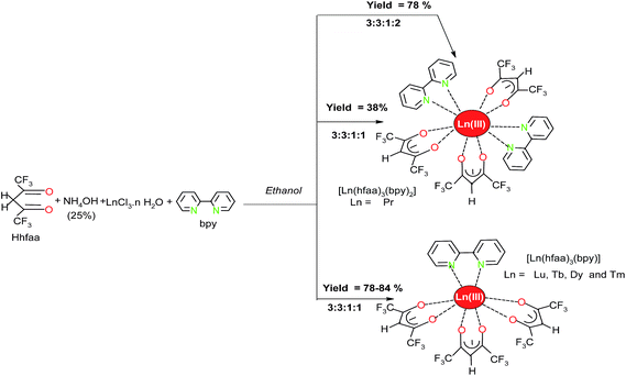
The praseodymium yielded ten-coordinate complex, [Pr(hfaa)3(bpy)2] although Pr : bpy stoichiometry was 1![[thin space (1/6-em)]](https://www.rsc.org/images/entities/char_2009.gif) :
:![[thin space (1/6-em)]](https://www.rsc.org/images/entities/char_2009.gif) 1. While the others are eight-coordinate, [Ln(hfaa)3bpy] (Ln = Tb, Dy, Tm and Lu) (Scheme 1). Initially, the yield of the Pr complex was much lower than those of Tb, Dy, Tm and Lu complexes since two bpy units per metal (Pr) were consumed. We have, therefore, also carried out synthesis of the Pr complex using the praseodymium chloride, Hhfaa, NH4OH and bpy in 1
1. While the others are eight-coordinate, [Ln(hfaa)3bpy] (Ln = Tb, Dy, Tm and Lu) (Scheme 1). Initially, the yield of the Pr complex was much lower than those of Tb, Dy, Tm and Lu complexes since two bpy units per metal (Pr) were consumed. We have, therefore, also carried out synthesis of the Pr complex using the praseodymium chloride, Hhfaa, NH4OH and bpy in 1![[thin space (1/6-em)]](https://www.rsc.org/images/entities/char_2009.gif) :
:![[thin space (1/6-em)]](https://www.rsc.org/images/entities/char_2009.gif) 3
3![[thin space (1/6-em)]](https://www.rsc.org/images/entities/char_2009.gif) :
:![[thin space (1/6-em)]](https://www.rsc.org/images/entities/char_2009.gif) 3
3![[thin space (1/6-em)]](https://www.rsc.org/images/entities/char_2009.gif) :
:![[thin space (1/6-em)]](https://www.rsc.org/images/entities/char_2009.gif) 2 mole ratio i.e. two bpy units per Pr atom. It improved the yield from 38 to 78%.
2 mole ratio i.e. two bpy units per Pr atom. It improved the yield from 38 to 78%.
TGA-DTA analysis
The thermograms of the complexes are similar in shape and show one step weight loss. The total weight loss for the complexes is 99.8% which is consistent with volatilization (Fig. S1 in ESI†). The DTA curve of the complexes displays two endothermic peaks; one peak at lower temperature corresponds to melting and other peak at higher temperature is consistent with the volatilization. The melting point of the eight-coordinate complexes is lower (between 166 and 170 °C) than that of the ten-coordinate Pr complex (206 °C). It indicates that the ten-coordinate Pr complex is thermally more stable than the eight-coordinate complexes. The reason for higher thermal stability could be related to π–π stacking interaction. A comparison of the thermal stability of the eight-coordinate complexes reveals that the thermal stability and volatility of the complexes increase with decreasing size of lanthanide. It is important to note that these complexes are stable at temperature as high as 220 °C and volatilize which is very important in view of the use of these complexes as molecular materials processed by thermal evaporation.Infrared spectroscopy
The IR spectra of the complexes are similar, exhibiting (C![[double bond, length as m-dash]](https://www.rsc.org/images/entities/char_e001.gif) O) and (C
O) and (C![[double bond, length as m-dash]](https://www.rsc.org/images/entities/char_e001.gif) C) stretching vibrations at 1659 and 1517 cm−1, respectively.41 The bands of free bpy are appreciably shifted to higher wave number in the complexes indicating coordination of heterocyclic ligand(s) through the nitrogen atoms of the bipyridine moiety. The strong absorption band appearing between 1257 and 1144 cm−1 is assigned to CF3 stretching mode.42
C) stretching vibrations at 1659 and 1517 cm−1, respectively.41 The bands of free bpy are appreciably shifted to higher wave number in the complexes indicating coordination of heterocyclic ligand(s) through the nitrogen atoms of the bipyridine moiety. The strong absorption band appearing between 1257 and 1144 cm−1 is assigned to CF3 stretching mode.42
Mass spectrometry
The ESI-MS spectra of the complexes were obtained in chloroform (Fig. S2–6 in ESI†). The intact molecular peak has been observed in all the complexes. The spectra show the peaks for species, [M + H]+. The [M + H]+ peak of Dy, Tm and Lu complexes are observed with 100%. It is important to note that none of the peak corresponding to chelate, [Ln(hfaa)3 + H]+ is observed. The other peaks observed in the mass spectra of the complexes may account for the diverse ionic species formed due to the fragmentation, ligand exchange and redistribution in the vapor phase and gets support from literature reports.43,44Single crystal X-ray crystallography
The X-ray crystal structures of the Pr and Dy complexes are shown in Fig. 1 and 2. The selected bond lengths and bond angles are given in Table 2, and Tables S1 and S2 in ESI.† The Pr(III) ion in the complex is ten-coordinated and consists of six O-atoms from three hfaa ligands and four N-atoms from two bidentate bpy ligands making the geometry around the Pr1 atom as a distorted bicapped square antiprismatic. Moreover, the bpy units are involved in π–π stacking interactions with an interplanar distance of 4.02 Å (Fig. S7 in ESI†), which is responsible for higher melting point of the complex. The crystal structure of the Pr complex is similar to [La(hfaa)3(bpy)2].11 However, the average bond lengths of Pr–O and Pr–N are 2.505(3) Å and 2.752(4) Å, respectively which are shorter than that reported for similar ten-coordinate [La(hfaa)3(bpy)2].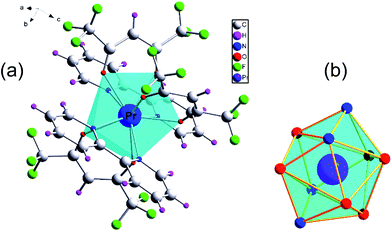 | ||
| Fig. 1 (a) Molecular structure of [Pr(hfaa)3(bpy)2] complex. (b) Coordination geometry around Pr(III) ion. Thermal ellipsoids are drown at the 30% probability level. | ||
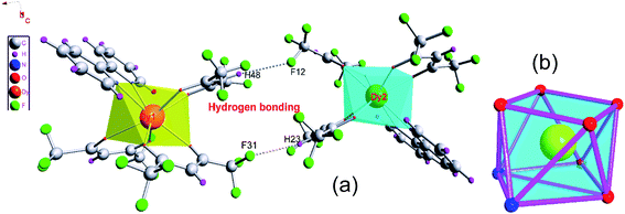 | ||
| Fig. 2 (a) Molecular structure of [Dy(hfaa)3(bpy)] complex. (b) Coordination geometry of Dy complex. Thermal ellipsoids are drown at the 30% probability level. | ||
| Pr complex | Dy complex | ||||
|---|---|---|---|---|---|
| Unit 1 | Unit 2 | ||||
| N(1)–Pr(1) | 2.750(4) | N(1)–Dy(1) | 2.520(5) | N(3)–Dy(2) | 2.520(5) |
| N(2)–Pr(1) | 2.754(4) | N(2)–Dy(1) | 2.520(5) | N(4)–Dy(2) | 2.515(5) |
| O(1)–Pr(1) | 2.525(3) | O(1)–Dy(1) | 2.369(4) | O(7)–Dy(2) | 2.344(4) |
| O(2)–Pr(1) | 2.495(4) | O(2)–Dy(1) | 2.354(4) | O(8)–Dy(2) | 2.343(4) |
| O(3)–Pr(1) | 2.495(3) | O(5)–Dy(1) | 2.318(4) | O(9)–Dy(2) | 2.348(4) |
| Pr(1)–O(2)#1 | 2.495(4) | O(6)–Dy(1) | 2.333(4) | O(10)–Dy(2) | 2.315(4) |
| Pr(1)–O(3)#1 | 2.495(3) | O(3)–Dy(1) | 2.346(4) | O(11)–Dy(2) | 2.330(4) |
| Pr(1)–O(1)#1 | 2.525(3) | O(4)–Dy(1) | 2.350(4) | O(12)–Dy(2) | 2.344(4) |
| Pr(1)–N(1)#1 | 2.750(4) | ||||
| Pr(1)–N(2)#1 | 2.754(4) | ||||
The unit cell of Dy complex contains two [Dy(hfaa)3(bpy)] complexes which are attached to each other via hydrogen bonding (Fig. 2). The complex consists of two square planes with negligible mean deviations of 0.007 and 0.101 Å from least square planes. In the ideal square antiprism these planes would be planar but here the square faces are creased about the diagonals N1–O5 and N3–O2 with angles of 3.17° and 4.41°. The results indicate that the Dy center consists of six O-atoms from three hfaa ligands and two N-atoms from one bpy unit arranged in a distorted square antiprismatic geometry.
The average bond lengths of Dy–O (hfaa) and Dy–N (bpy) bonds of the present complex are 2.345(4) and 2.520(5) Å, respectively. A comparative study shows that the average Dy–O(hfaa) bond length is 0.023 Å larger than the average Dy–O bond length of [Dy(bfa)3phen] (ref. 45) (bfa = 4,4,4-trifluoro-1-phenyl-1,3-butanedione; phen = 1,10-phenanthroline). The larger Dy–O bond lengths of hfaa complex than that of bfa (having one CF3 and one phenyl group) complex could be due to the larger negative inductive effect of the six fluorine atoms on two terminals of hfaa (two CF3 groups) which decreases more electron density on coordinating oxygen atoms and, in turn increase the residual acidity of the Dy(III) making it a better complexing site for incoming ancillary ligands. As a result, the Dy–N(bpy) bond length is 0.05 Å smaller than that of Dy–N bond length of bfa complex. The smaller Ln–N bond length of ancillary ligands in ternary lanthanide tris(β-diketonates) complexes could be an asset for the efficient energy transfer from ligands to metal ion.
In a recent paper, crystal structure of [Dy(hfaa)3(bpy)], based on the data obtained at low temperature (103 K) has been reported.13 It is important to point out that the reported crystal structure data obtained at 103 K is different from our data collected at room temperature (293 K). The following are the points of difference: (i) the bond lengths Dy1–N1, Dy1–N2, Dy1–O1 and Dy1–O2 obtained by us at room temperature are longer than those reported from X-ray diffraction data collected at 103 K; whereas the Dy1–O3, Dy1–O4, Dy1–O5 and Dy1–O6 are shorter in the present complex than the values obtained at 103 K. Furthermore for Dy2 unit, Dy–N and Dy–O bond lengths are longer at 293 K than those reported from data obtained at 103 K, (ii) the unit cell dimensions are shorter in the reported structure13 than those observed at 293 K in the present study and (iii) both the complexes crystallize in monoclinic P21/c with Z = 8, the angles, α and γ are of similar magnitude (α = γ = 90°) for both structures whereas third angle, β is shorter (β = 106.14) than that reported for 103 K structure. These results indicate that the degree of distortion in the geometry is more at room temperature (293 K) than that at lower temperature (103 K).
1H NMR spectra
The NMR spectra of the complexes were obtained in CDCl3 by dissolving sufficient amount of the samples (the solutions were sufficiently concentrated; 6–10 mg/0.50 mL). The chemical shifts, δ (ppm), and the paramagnetic shifts, Δδ (ppm) are given in the Table S3.† The spectrum of the diamagnetic Lu complex displays five signals (one for hfaa and four due to bpy) in the intensity ratio of 3![[thin space (1/6-em)]](https://www.rsc.org/images/entities/char_2009.gif) :
:![[thin space (1/6-em)]](https://www.rsc.org/images/entities/char_2009.gif) 2
2![[thin space (1/6-em)]](https://www.rsc.org/images/entities/char_2009.gif) :
:![[thin space (1/6-em)]](https://www.rsc.org/images/entities/char_2009.gif) 2
2![[thin space (1/6-em)]](https://www.rsc.org/images/entities/char_2009.gif) :
:![[thin space (1/6-em)]](https://www.rsc.org/images/entities/char_2009.gif) 2
2![[thin space (1/6-em)]](https://www.rsc.org/images/entities/char_2009.gif) :
:![[thin space (1/6-em)]](https://www.rsc.org/images/entities/char_2009.gif) 2 (Fig. S8†) and is consistent with the coordination of only one bpy to lutetium. The spectra of the paramagnetic complexes are very interesting and display huge paramagnetic shifts (Fig. 3 and S9–S11 in ESI†). The important features of the spectra of the complexes investigated are obvious: (i) only one set of signals are observed for the aromatic as well as β-diketone protons which substantiates presence of only one species in the solution, (ii) the spectra of the complexes show sizable downfield or up field shifts of bpy and diketonate resonances, (iii) the intensity ratio of bpy and methane protons resonances is 4
2 (Fig. S8†) and is consistent with the coordination of only one bpy to lutetium. The spectra of the paramagnetic complexes are very interesting and display huge paramagnetic shifts (Fig. 3 and S9–S11 in ESI†). The important features of the spectra of the complexes investigated are obvious: (i) only one set of signals are observed for the aromatic as well as β-diketone protons which substantiates presence of only one species in the solution, (ii) the spectra of the complexes show sizable downfield or up field shifts of bpy and diketonate resonances, (iii) the intensity ratio of bpy and methane protons resonances is 4![[thin space (1/6-em)]](https://www.rsc.org/images/entities/char_2009.gif) :
:![[thin space (1/6-em)]](https://www.rsc.org/images/entities/char_2009.gif) 4
4![[thin space (1/6-em)]](https://www.rsc.org/images/entities/char_2009.gif) :
:![[thin space (1/6-em)]](https://www.rsc.org/images/entities/char_2009.gif) 4
4![[thin space (1/6-em)]](https://www.rsc.org/images/entities/char_2009.gif) :
:![[thin space (1/6-em)]](https://www.rsc.org/images/entities/char_2009.gif) 4
4![[thin space (1/6-em)]](https://www.rsc.org/images/entities/char_2009.gif) :
:![[thin space (1/6-em)]](https://www.rsc.org/images/entities/char_2009.gif) 3 in the case of Pr (ten-coordinate) and 2
3 in the case of Pr (ten-coordinate) and 2![[thin space (1/6-em)]](https://www.rsc.org/images/entities/char_2009.gif) :
:![[thin space (1/6-em)]](https://www.rsc.org/images/entities/char_2009.gif) 2
2![[thin space (1/6-em)]](https://www.rsc.org/images/entities/char_2009.gif) :
:![[thin space (1/6-em)]](https://www.rsc.org/images/entities/char_2009.gif) 2
2![[thin space (1/6-em)]](https://www.rsc.org/images/entities/char_2009.gif) :
:![[thin space (1/6-em)]](https://www.rsc.org/images/entities/char_2009.gif) 2
2![[thin space (1/6-em)]](https://www.rsc.org/images/entities/char_2009.gif) :
:![[thin space (1/6-em)]](https://www.rsc.org/images/entities/char_2009.gif) 3 for eight-coordinate complexes. It substantiates presence of two bpy units in the cases of ten-coordinate complex, and (iv) in case of the Pr complex, the bpy protons shift are not uniformly directed and H-3 protons experience larger upfield displacement than the H-2 protons (Fig. S10 in ESI†). This suggests the important role of the geometric factor (3
3 for eight-coordinate complexes. It substantiates presence of two bpy units in the cases of ten-coordinate complex, and (iv) in case of the Pr complex, the bpy protons shift are not uniformly directed and H-3 protons experience larger upfield displacement than the H-2 protons (Fig. S10 in ESI†). This suggests the important role of the geometric factor (3![[thin space (1/6-em)]](https://www.rsc.org/images/entities/char_2009.gif) cos2
cos2![[thin space (1/6-em)]](https://www.rsc.org/images/entities/char_2009.gif) θ − 1) in deciding magnitude and direction of the shift. The room temperature paramagnetic shift obtained for methine protons of β-diketone moiety has its sign opposed to that of paramagnetic shifts of aromatic protons. The opposite direction shift of the methine resonance reflects importance of the geometric factor 3
θ − 1) in deciding magnitude and direction of the shift. The room temperature paramagnetic shift obtained for methine protons of β-diketone moiety has its sign opposed to that of paramagnetic shifts of aromatic protons. The opposite direction shift of the methine resonance reflects importance of the geometric factor 3![[thin space (1/6-em)]](https://www.rsc.org/images/entities/char_2009.gif) cos2
cos2![[thin space (1/6-em)]](https://www.rsc.org/images/entities/char_2009.gif) θ − 1 in changing the sign of the shift and that the paramagnetic shift in these complexes is dominated by dipolar interaction. This suggestion is strongly supported by the observations on [Ln(fod)3(bpy)] (ref. 46) and [Ln(hfaa)3(phen)] (ref. 47) complexes (fod = anion of 6,6,7,7,8,8,8-heptafluoro-2,2-dimethyl-3,5-octadione) and Ln(C5H5)3B (B = uncharged aprotic Lewis base), where the signal positions of the ligand B have their sign opposed to that of ring protons.48 Therefore, it seems likely that the average geometric factor of the aromatic and the methine protons have opposite sign and the paramagnetic shift is exclusively due to dipolar interaction.
θ − 1 in changing the sign of the shift and that the paramagnetic shift in these complexes is dominated by dipolar interaction. This suggestion is strongly supported by the observations on [Ln(fod)3(bpy)] (ref. 46) and [Ln(hfaa)3(phen)] (ref. 47) complexes (fod = anion of 6,6,7,7,8,8,8-heptafluoro-2,2-dimethyl-3,5-octadione) and Ln(C5H5)3B (B = uncharged aprotic Lewis base), where the signal positions of the ligand B have their sign opposed to that of ring protons.48 Therefore, it seems likely that the average geometric factor of the aromatic and the methine protons have opposite sign and the paramagnetic shift is exclusively due to dipolar interaction.
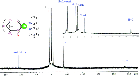 | ||
| Fig. 3 1H NMR spectrum of [Tb(hfaa)3(bpy)] in CDCl3. Inset: resolution of region from −40.0 to 10.0 ppm(δ). | ||
Absorption spectroscopy
The absorption spectra of free ligands (Hhfaa and bpy) (Fig. S12 in ESI†) and the complexes (Pr, Tb, Dy and Tm) were recorded as 5 × 10⁻⁵ M chloroform solutions (Fig. S13 in ESI†). The ligands, Hhfaa or bpy, absorb only in the UV range due to their singlet–singlet (1π–π*) electronic transitions. The bpy shows a strong band at 292 nm, whereas a strong absorption band is observed at 275 nm for Hhfaa. The spectrum of the complexes contain essentially the combined ligands absorption (λmax between 318 and 322 nm), this band in the spectrum of the complexes is shifted to longer wavelength (red-shift) as compared free bpy or Hhfaa due to coordination of the these ligands to metal. The determined molar absorption coefficient values from the spectra for free Hhfaa and bpy are 6.7 × 10³ M−1 cm−1 and 7.8 × 10³ M−1 cm−1, respectively whereas for the complexes Pr, Tb, Dy and Tm are 3.61 × 10⁴ M−1 cm−1, 2.83 × 10⁴ M−1 cm−1, 2.77 × 10⁴ M−1 cm−1 and 2.92 × 10⁴ M−1 cm−1, respectively. This result shows that the molar absorption coefficient values are more than three times higher than that of the free Hhfaa, indicating the presence of three β-diketone ligands together with one or two bpy ligand(s) in the corresponding complexes. Moreover, the higher molar absorption coefficient of Hhfaa or bpy reveals that the β-diketone or heterocyclic ligand (bpy) has a strong ability to absorb light.Photoluminescence (visible and NIR region)
![[thin space (1/6-em)]](https://www.rsc.org/images/entities/char_2009.gif) 500 cm−1) and bpy (22
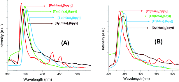
500 cm−1) and bpy (22![[thin space (1/6-em)]](https://www.rsc.org/images/entities/char_2009.gif) 900 cm−1) lie 6133 cm−1 and 6534 cm−1, respectively, above the 1D2 emitting level of Pr(III) ion which are well suited for efficient energy transfer from the ligands to the metal. The quantum yield (1.5%) of present Pr complex is higher as compared to those reported for other Pr(III) complexes.49 Moreover, only few reports describing the luminescence behaviour of Pr(III) complexes in the visible region are available in the literature.50,51
900 cm−1) lie 6133 cm−1 and 6534 cm−1, respectively, above the 1D2 emitting level of Pr(III) ion which are well suited for efficient energy transfer from the ligands to the metal. The quantum yield (1.5%) of present Pr complex is higher as compared to those reported for other Pr(III) complexes.49 Moreover, only few reports describing the luminescence behaviour of Pr(III) complexes in the visible region are available in the literature.50,51
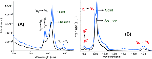 | ||
| Fig. 5 Emission spectra of [Pr(hfaa)3(bpy)2] in solution (black) and solid state (blue) in visible (A, λex = 340 nm) and NIR region (B, λex = 400 nm). Concentration = 5 × 10⁻⁵ M. | ||
For energy transfer mechanism, it appears that the both the ligands transfer energy to the Pr(III) and populate the higher energy levels, 1I6 and 3P1. These excited levels may relax by transferring the energy to the lower lying levels, 3P0 and 1D2 which results in strong emission in the visible region. At the same time, it is also believed that 1I6, 3P1, and 3P0 excited states after receiving energy from the ligand centered triplet state rapidly transfer the energy to the 1D2 emissive level of Pr(III) via non-radiative relaxation which leads to NIR emission. The intensity of the emission transitions of the lanthanide complexes depends on the effective overlap between the ligand triplet state and the emitting level of the Ln(III) centre. Therefore, the strong luminescence observed for the praseodymium complex, either in the visible or NIR regions, reflects a good match between the ligand centred triplet state and 1D2 emissive state of the Pr(III). In addition, the suitable energy difference between the triplet state of bpy and emitting level (1D2) of Pr(III) leads to an efficient energy transfer which establishes that the bpy is a good sensitizers for Pr(III) ion. The quantum yield of the Pr complex in NIR region is determined to be 0.0042% which is higher than that reported for other Pr complex.50 Moreover, the examples of sensitized emission from Pr(III) complexes in the NIR region are rarely reported.51
The emission spectrum of the Tb complex (Fig. 6) is dominated by 5D4 → 7F5 transition (544 nm) followed by the 5D4 → 7F6 transition (485 nm). The transition, 5D4 → 7F6 is relatively more sensitive to the chemical environment and symmetry of the coordination polyhedron. While the transition, 5D4 → 7F5 is considered an internal reference to account for the differences in emission transition ratios induced by the ligands.52,53 For the chelate, [Tb(hfaa)3(H2O)2],54 the intensity ratios for different emission transitions are reported as f4–6(0.22), f4–5(1.00), f4–4(0.11), f4–3(0.08) and f4–2(0.08). For present [Tb(hfaa)3(bpy)], these ratios are calculated as f4–6(0.33), f4–5(1.00), f4–4(0.13), f4–3(0.06) and f4–2(0.07). This result indicates that the presence of bpy as ancillary ligand increases the luminescent intensity (∼1.5-fold) of the 5D4 → 7F6 transition and is responsible for more induced nephelauxetic effect. Moreover, the energy difference between the triplet state of the organic ligands and 5D4 emitting level of Tb(III) ion should be 2400 ± 300 cm−1 for efficient energy transfer from ligands to Tb(III) ion.55 The energy difference between the triplet state of the ligands (hfaa and bpy) and 5D4 (20![[thin space (1/6-em)]](https://www.rsc.org/images/entities/char_2009.gif) 430 cm−1) level are ∼1500 and ∼2470 cm−1, respectively. This indicates that energy gap is not suitable for hfaa which results in less efficient energy transfer and in addition there will be a back energy transfer from Tb(III) ion to hfaa, which has negative effect on the luminescence of the Tb(III) complex. The back energy transfer from Tb (5D4) to ligands is confirmed by increased emission lifetime from 414 μs at room temperature to 776 μs at 77 K (Fig. S14 in ESI†). The quantum yield of the hydrated chelate, [Tb(hfaa)3(H2O)2] (ref. 52) is reported to be 27% which is increased to 29%, in the present complex, upon binding of bpy ligand to Tb(III). Moreover, the quantum yield of the present Tb(III) complex is much higher than the those reported for [Tb(hfaa)3(bpyO2)] (0.75%) (ref. 52) and [Tb(tta)2(terpyridine-carboxylate)] (13%).56 The energy gap reported between the triplet state of bpyO2 and 5D4 level of Tb is very small (∼40 cm−1) which lead to back energy transfer and resulted in low quantum yield. The high quantum yield of the present Tb(III) complex could be due to suitable energy gap of ∼2470 cm−1 between triplet state of bpy and emitting level of Tb(III) ion.
430 cm−1) level are ∼1500 and ∼2470 cm−1, respectively. This indicates that energy gap is not suitable for hfaa which results in less efficient energy transfer and in addition there will be a back energy transfer from Tb(III) ion to hfaa, which has negative effect on the luminescence of the Tb(III) complex. The back energy transfer from Tb (5D4) to ligands is confirmed by increased emission lifetime from 414 μs at room temperature to 776 μs at 77 K (Fig. S14 in ESI†). The quantum yield of the hydrated chelate, [Tb(hfaa)3(H2O)2] (ref. 52) is reported to be 27% which is increased to 29%, in the present complex, upon binding of bpy ligand to Tb(III). Moreover, the quantum yield of the present Tb(III) complex is much higher than the those reported for [Tb(hfaa)3(bpyO2)] (0.75%) (ref. 52) and [Tb(tta)2(terpyridine-carboxylate)] (13%).56 The energy gap reported between the triplet state of bpyO2 and 5D4 level of Tb is very small (∼40 cm−1) which lead to back energy transfer and resulted in low quantum yield. The high quantum yield of the present Tb(III) complex could be due to suitable energy gap of ∼2470 cm−1 between triplet state of bpy and emitting level of Tb(III) ion.
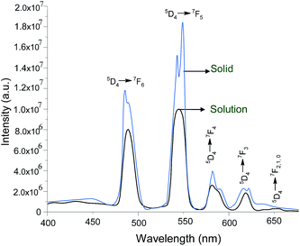 | ||
| Fig. 6 Emission spectra of [Tb(hfaa)3(bpy)] in solution (black, concentration = 5 × 10⁻⁵ M; (λex = 340 nm) and solid state (blue) at room temperature in visible region. | ||
It is interesting to mention that the quantum yield of [Tb(hfaa)3bpy] complex reported13 from the solid state emission spectrum in visible region, upon exciting the complexes at 355 nm is only 3.4% which is much lower than what we observed (29%). The higher quantum yield of the complex in our case could be due excitation of the complex at correct wavelength (340 nm) which lead to highest emission intensity of the transitions.
The emission spectra of Dy complex in the visible and NIR regions are shown in Fig. 7. In the visible region, the complex shows two transitions, 4F9/2 → 6H15/2 (blue) and 4F9/2 → 6H13/2 (yellow) in which 4F9/2 → 6H15/2 is magnetically allowed and does not vary with the local field around the Dy(III) ion while 4F9/2 → 6H13/2 is a forced electric-dipole transition and is hypersensitive (Fig. 7A). The emission intensity ratio of 4F9/2 → 6H13/2 and 4F9/2 → 6H15/2 transitions is 3.7. Such high value is expected for systems with no inversion center57 and is responsible for the intense yellow-green emission colour of the present complex. Moreover, the 4F9/2 → 6H13/2 transition is prominent only when Dy(III) ion is located at a low symmetry site which allows the intensification of this transition. Therefore, this transition is used as a probe for the site symmetry in Dy(III) systems.58,59 The intensification of the 4F9/2 → 6H13/2 transition in the present Dy(III) complex could be due to the distorted square antiprismatic geometry of the complex. The quantum yield (1.7%) of present Dy complex is higher than that reported for [Dy(bfa)3phen] (0.2%).45 In NIR region, the Dy(III) has several important NIR emission bands (Fig. 7B) which are of interest for optical communications. The band at 1303 nm offers the opportunity to develop new materials suitable for optical amplifiers operating at 1.3 μm while the emission band at 1532 nm matches well with the third working window of telecommunications. Thus, the NIR emission of the Dy complex could be the basis for the future telecommunication network. Moreover, only few reports are available about the visible and NIR luminescence of Dy(III) in inorganic systems. However, visible and NIR luminescence of Dy(III) in organic complexes are rarely reported.45,51 Furthermore, no definite data are available regarding optimum energy gap between the triplet state of the organic ligands and 4F9/2 emitting level of Dy(III) ion for an efficient energy transfer.57 The energy difference between the emitting level of Dy(III) ion (21![[thin space (1/6-em)]](https://www.rsc.org/images/entities/char_2009.gif) 000 cm−1) and triplet state of bpy ligand in present Dy complex seems suitable since the measured quantum yield is higher than those reported in the literature.45,51 Moreover, it is believed that the major part of the excitation energy is transferred from the ligands to Dy(III) ion by populating the 4F9/2 level which results in visible as well as NIR emission. At the same time, some part of the excitation energy is relaxed to the lower lying states which are responsible for weak emission transitions observed in the NIR spectrum. The determined quantum yield of present complex in NIR region is 0.0036% which is higher than that reported in the literature.51
000 cm−1) and triplet state of bpy ligand in present Dy complex seems suitable since the measured quantum yield is higher than those reported in the literature.45,51 Moreover, it is believed that the major part of the excitation energy is transferred from the ligands to Dy(III) ion by populating the 4F9/2 level which results in visible as well as NIR emission. At the same time, some part of the excitation energy is relaxed to the lower lying states which are responsible for weak emission transitions observed in the NIR spectrum. The determined quantum yield of present complex in NIR region is 0.0036% which is higher than that reported in the literature.51
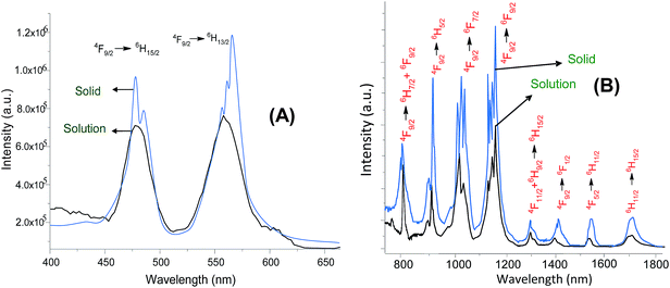 | ||
| Fig. 7 Emission spectra of [Dy(hfaa)3(bpy)] in solution (black) and solid state (blue) in visible (A, λex = 340 nm) and NIR region (B, λex = 400 nm). Concentration = 5 × 10⁻⁵ M. | ||
The luminescence of Tm(III) ion, either in visible or NIR region, is less reported. The emission spectrum of the Tm complex consists of a peak at 475 nm (1G4 → 3H6) and does not show any emission due to the ligands (Fig. 8A). This means that the energy difference (ΔE ∼ 1625 cm−1) between the triplet state of bpy and emitting level, 1G4 (21![[thin space (1/6-em)]](https://www.rsc.org/images/entities/char_2009.gif) 275 cm−1) of Tm(III) is suitable for efficient energy transfer from bpy to Tm(III) ion. Moreover, the quantum yield of present Tm complex is determined to be 0.060% which is higher than that reported for the complex, [Tm(hfaa)(TOPO)] (TOPO = trioctylphosphine oxide).60 In the NIR region, two emission bands, 3H4 → 3H6 (803 nm) and 3H4 → 3F4 (1472 nm) are observed (Fig. 8B). The band at 1472 nm consists of three peaks, which could be ascribed to the Stark splitting of 4f electronic levels.61 Moreover, the fwhm of the 1472 nm band is 95 nm which is very broad and such a broad spectrum enables a wide gain bandwidth for optical amplification.62 Therefore, the 1472 nm band is the potential candidate of broadening amplification band from C band (1530–1560 nm) to S+ band (1450–1500 nm) in optical telecommunication. The quantum yield of Tm complex in NIR region is determined to be 0.0019%. For the energy transfer mechanism, it is assumed that the part of excitation energy is transferred to 3F2 and 3F3 levels which, in turn, may relax the excited electrons to the 3H4 level and then radiatively decay to the lower lying levels results in NIR emission. At the same time, the remaining part of excitation energy is transferred to the 1G4 level which results in visible emission at 475 nm. For better understanding of the energy transfer between metal ion and the triplet states of the ligands, the energy level diagram is shown in Fig. S15 in ESI.†
275 cm−1) of Tm(III) is suitable for efficient energy transfer from bpy to Tm(III) ion. Moreover, the quantum yield of present Tm complex is determined to be 0.060% which is higher than that reported for the complex, [Tm(hfaa)(TOPO)] (TOPO = trioctylphosphine oxide).60 In the NIR region, two emission bands, 3H4 → 3H6 (803 nm) and 3H4 → 3F4 (1472 nm) are observed (Fig. 8B). The band at 1472 nm consists of three peaks, which could be ascribed to the Stark splitting of 4f electronic levels.61 Moreover, the fwhm of the 1472 nm band is 95 nm which is very broad and such a broad spectrum enables a wide gain bandwidth for optical amplification.62 Therefore, the 1472 nm band is the potential candidate of broadening amplification band from C band (1530–1560 nm) to S+ band (1450–1500 nm) in optical telecommunication. The quantum yield of Tm complex in NIR region is determined to be 0.0019%. For the energy transfer mechanism, it is assumed that the part of excitation energy is transferred to 3F2 and 3F3 levels which, in turn, may relax the excited electrons to the 3H4 level and then radiatively decay to the lower lying levels results in NIR emission. At the same time, the remaining part of excitation energy is transferred to the 1G4 level which results in visible emission at 475 nm. For better understanding of the energy transfer between metal ion and the triplet states of the ligands, the energy level diagram is shown in Fig. S15 in ESI.†
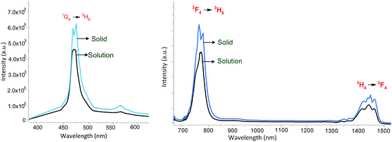 | ||
| Fig. 8 Emission spectra of [Tm(hfaa)3(bpy)] in solution (black) and solid state (blue) in visible (A, λex = 340 nm) and NIR region (B, λex = 400 nm). Concentration = 5 × 10⁻⁵ M. | ||
In order to see the effect on photophysical parameters, the spectra of the complexes were also recorded in the solid state and are compared with the spectra in solution (Fig. 5–8). It is interesting to note that the emission transitions (visible and NIR region) of the complexes are slightly shifted from their position in solution spectra and appear at higher wavelength with increased intensity and greater stark splitting. The emission transitions: 1D2 → 3H4 of Pr, 5D4 → 7F6 of Tb, 4F9/2 → 6H13/2 of Dy and 1G4 → 3H6 of Tm in visible region and 1D2 → 3F4 (Pr), 4F9/2 → 6F7/2 and 6F9/2 (Dy) and 3H4 → 3H6 and 3H4 → 3F4 (Tm) in the NIR region are splitted into many components with enhanced emission intensity. The higher emission intensity could be related to absence of solvent-based quenching in the solid state. Since in the solution, the C–H bonds of the chloroform could provide a quenching effect which would be detrimental on the luminescence intensity. Moreover, the higher emission intensity and Stark splitting of the transitions could also be attributed to low molecular symmetry of the complexes in the solid state. The Stark splitting has disappeared from the solution spectra, in some cases, which could be attributed to the changes in symmetry of the field around Ln(III) ions. This decrease in asymmetry of the ligand field around the Ln(III) ion and presence of C–H bonds of the chloroform consequently decrease the radiative transition probability and as a result the quantum yield is lowered in solution. This indicates that highly monochromatic clear emission is observed in solid state.
| ΦLn = τobs/τrad | (3) |
| Compound | Transition (cm−1) | Fwhma (nm) | KRADb (s−1) | KNRb (s−1) | τobs (μs) | QLnLnc (%) | Qreld (%) |
|---|---|---|---|---|---|---|---|
| a Full width at half maximum of emission peak {Fwhm (nm)}.b Radiative rate (KRAD) and non-radiative rate (KNR) were determined using equations; (KRAD = Qrel/τobs and KNR = 1/τobs − KRAD.c Intrisic quantum yield calculated by using the eqn (3).d Relative quantum yield calculated using the eqn (1) and (2).e Solution (chloroform).f Solid state. | |||||||
| [Pr(hfaa)3(bpy)2]e | 1D2 → 3H4 (16![[thin space (1/6-em)]](https://www.rsc.org/images/entities/char_2009.gif) 287) 287) |
9.11 | 1.36 × 10³ | 8.95 × 10⁴ | 11 | 21.69 | 1.5 |
| [Tb(hfaa)3(bpy)2]e | 5D4 → 7F6 (18![[thin space (1/6-em)]](https://www.rsc.org/images/entities/char_2009.gif) 382) 382) |
10.12 | 7.00 × 10² | 1.72 × 10³ | 414 | 13.80 | 29 |
| [Dy(hfaa)3(bpy)]e | 4F9/2 → 6H13/2 (17![[thin space (1/6-em)]](https://www.rsc.org/images/entities/char_2009.gif) 761) 761) |
5.6 | 1.30 × 10³ | 4.58 × 10⁴ | 13 | — | 1.7 |
| [Tm(hfaa)3(bpy)]e | 1G4 → 3H6 (21![[thin space (1/6-em)]](https://www.rsc.org/images/entities/char_2009.gif) 052) 052) |
— | 2.0 × 10³ | 3.31 × 10⁵ | 3 | 1.11 | 0.60 |
| [Pr(hfaa)3(bpy)2]f | 1D2 → 3H4 (16![[thin space (1/6-em)]](https://www.rsc.org/images/entities/char_2009.gif) 221) 221) |
9.17 | 1.56 × 10³ | 7.25 × 10⁴ | 13.5 | 26.62 | 1.8 |
| [Tb(hfaa)3(bpy)2]f | 5D4 → 7F6 (18![[thin space (1/6-em)]](https://www.rsc.org/images/entities/char_2009.gif) 388) 388) |
10.12 | 7.24 × 10² | 1.67 × 10³ | 418 | 13.60 | 30.3 |
| [Dy(hfaa)3(bpy)]f | 4F9/2 → 6H13/2 (17![[thin space (1/6-em)]](https://www.rsc.org/images/entities/char_2009.gif) 732) 732) |
5.8 | 1.36 × 10³ | 7.00 × 10⁴ | 14 | — | 1.9 |
| [Tm(hfaa)3(bpy)]f | 1G4 → 3H6 (21![[thin space (1/6-em)]](https://www.rsc.org/images/entities/char_2009.gif) 011) 011) |
— | 3.9 × 10³ | 4.96 × 10⁵ | 2.6 | 3.94 | 0.78 |
| Compound | Transition (cm−1) | Fwhma (nm) | KRADb (s−1) | KNRb (s−1) | τobs (μs) | Qrelc (%) |
|---|---|---|---|---|---|---|
| a Full width at half maximum of emission peak {Fwhm (nm)}.b Radiative rate (KRAD) and non-radiative rate (KNR) were determined using equations; KRAD = Qrel/τobs and KNR = 1/τobs − KRAD.c Relative quantum yield calculated using the eqn (1).d Solution (chloroform).e Solid State. | ||||||
| [Pr(hfaa)3(bpy)2]d | 1D2 → 3F4 (9541) | — | 7.77 × 10² | 1.85 × 10⁷ | 0.054 | 4.2 × 10⁻³ |
| [Dy(hfaa)3(bpy)]d | 4F9/2 → 6F9/2 (8462) | — | 8.78 × 10² | 2.44 × 10⁷ | 0.041 | 3.6 × 10⁻³ |
| [Tm(hfaa)3(bpy)]d | 3H4 → 3F4 (6520) | 95 | 4.52 × 10² | 2.38 × 10⁷ | 0.042 | 1.9 × 10⁻³ |
| [Pr(hfaa)3(bpy)2]e | 1D2 → 3F4 (9533) | — | — | — | 0.053 | — |
| [Dy(hfaa)3(bpy)]e | 4F9/2 → 6F9/2 (8423) | — | — | — | 0.044 | — |
| [Tm(hfaa)3(bpy)]e | 1D2 → 3F4 (9512) | 94 | — | — | 0.046 | — |
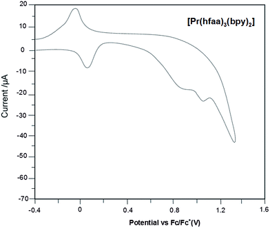
To investigate the electroluminescence properties of Pr, Tb and Dy complexes, twelve devices (3 single layered and 9 four layered) were fabricated in which these complexes were used as emitting layer (Fig. 9). The devices consist of TPD or CuPc as hole transport layer, AlQ as electron transfer layer and BCP (2,9-dimethyl-4,7-diphenyl-1,10-phenylanthroline) is used as the hole-blocking layer. In order to understand the charge injection and transport processes in devices, the HOMO (highest occupied molecular orbital) and the LUMO (lowest unoccupied molecular orbital) energy level of the complexes were estimated by cyclic voltammetry. The ferrocene/ferricenium redox couple ion (Fc/Fc+) was used as an internal standard. Two adjoining oxidation peaks were detected during the oxidation scan in chloroform. The onset potential was determined from the intersection of two tangents drawn at the rising and background currents of the CV. The CV of the Pr complex is shown in Fig. 10. The HOMO and LUMO energy levels were calculated by using the empirical relations.68
| EHOMO = −(1.4 ± 0.1) × (qVCV) − (4.6 ± 0.08) eV |
ELUMO = EHOMO − Eg (22![[thin space (1/6-em)]](https://www.rsc.org/images/entities/char_2009.gif) 460 cm−1) 460 cm−1) |
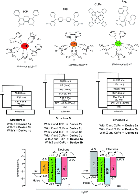 | ||
| Fig. 9 Molecular structures, configuration of device (with structures A, B and C) and energy band diagrams. | ||
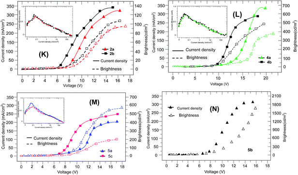
In terms of EL performance, no emission was observed from the single layered devices 1a and 1c while the EL spectrum of the device 1b is similar to PL spectrum and shows main emission peak at 544 nm. This device has a turn-on voltage of 12 V and gave a maximum brightness of 40 cd m−2 at 18 V. The turn-on voltage of device 2a was 7.5 V at 1 cd m−2 and a maximum brightness of 95 cd m−2 was achieved at 17.3 V with a current density of 251 mA cm−2 (Fig. 11K). Device 2b has a turn-on voltage at 6 V. Its maximum brightness was 106 cd m−2 at 16.2 V with a current density of 223 mA cm−2. It is well known that the turn-on voltage is related to the carrier injection and transport ability of the layer.69 The lower turn-on voltage of device 2b than that of 2a indicates that carrier injection and transport is more appropriate for device 2b. As a result, the brightness and efficiencies are higher for the 2b. Moreover, the Pr complex show intermolecular π–π interaction which facilitates the transport of electrons or holes in the devices and hence leads to good EL efficiencies. On the other hand, the turn-on voltage of device 3a was 10 V at 1 cd m−2 and a maximum brightness of 855 cd m−2 was achieved at 19 V. Device 3b had a turn-on voltage at 8 V and gave maximum brightness of 1198 cd m−2 at 16 V. The turn-on voltage of device 4a was 12 V at 1 cd m−2 and a maximum brightness of 354 cd m−2 was achieved at 21 V with a current density of 238 mA cm−2 (Fig. 11L). Device 4b had a turn-on voltage at 9 V. Its maximum brightness was 401 cd m−2 at 18 V with a current density of 209 mA cm−2. In order to improve the EL performance of the device, the thickness of the complex layer and BCP layer were changed from 50 nm (devices 2b, 3b and 4b) to 80 nm (devices 5a, 5b and 5c) and 20 (devices 2b, 3b and 4b) to 25 nm (devices 5a, 5b and 5c), respectively, since the thickness of the emitting layer and the BCP layer affect the performance of device.30 For the devices 5a, 5b and 5c, the maximum brightness of 183, 1765 and 532 cd m−2 with current densities of 202, 295 and 193 mA cm−2 were achieved at 16, 19 and 18 V, respectively (Fig. 11M and N). Moreover, the current efficiencies found for the devices 4a, 4b and 4c are as 0.58, 3.6 and 0.76 cd A−1, respectively.
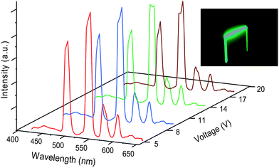
The EL spectra of the device 2a and 2b consist of three peaks at 586 (3P0 → 3H5), 615 nm (1D2 → 3H4) and 645 nm (3P0 → 3F2) (Fig. 12). The emission peak, 1D2 → 3H4 at 615 nm is dominating the spectra. While the EL spectra of 3b show four transitions as 5D4 → 7F6, 5D4 → 7F5, 5D4 → 7F4 and 5D4 → 7F3 (Fig. 13). For the devices 4a and 4b, the two transitions, 4F9/2 → 6H15/2 and 4F9/2 → 6H13/2 are observed in which 4F9/2 → 6H15/2 transition is dominant (Fig. 13). Besides the EL from the complexes in 2a, 3a and 4a, a strong emission peak at 500–510 nm due to BCP is observed at low voltage and become weakened as the voltage increases (Fig. 12 and 13). At the same time, the emission at 400 nm due to TPD which is very weak at low voltage become stronger at high voltages. This gradual decrease or increase in the emission of BCP or TPD with varying applied voltages could be due to the shifting in electron–hole recombination i.e. the charge carriers are not properly balanced in recombination zone. At low operating voltages, the mobility rate of holes is higher than that of electrons which moves the remnant holes to BCP layer and get coupled with electrons there which leads to formation of some excitons at BCP and, therefore, a weak emission is observed from this layer. In contrast, the electron mobility rate is proportional to an increasing applied field. As the operating voltages is increased, the recombination zone shifts gradually to the interface of the TPD and complex layers which results in emission from Ln(III) and TPD. The EL spectra of 2b, 3b and 4b (obtained by replacement of TPD by CuPc, the hole-inject and -transport layer) show the emission only from the complexes, no other emission is observed (Fig. 12–14). This result indicates that charge carriers are more balanced and well-confined in the emitting layer in presence of CuPc that could be due to the lower HOMO value of CuPc (5.2 eV) than that of TPD (5.4 eV) which leads to difficulty in holes transportation in CuPc than in TPD (Fig. 9). As a result, the transportation of the holes to BCP layer become smaller. Thus, the complexes with relatively balanced carrier injection and transporting ability are superior in the formation of excitons and leads to improve the device efficiency. As a result, the devices 2b, 3b and 4b are superior over the devices 2a, 3a and 4a, respectively. The 5a, 5b and 5c are found much more efficient devices as compared to 2b, 3b and 4b. The reason could be the increased thickness of emitting layer and BCP layer which leads to extension of recombination zone and thus decrease the quenching probability. Secondly, the reduction in the total current density which makes the device stable and perform better. The results show an improved electroluminescence performance of the present complexes based OLEDs over the devices based on Pr, Tb and Dy complexes reported in literature.25,29,30
 | ||
| Fig. 12 EL spectra of devices 2a and 2b at different operating voltages. Inset: image of Pr complex based OLED. | ||
Conclusion
An in situ solution processed reaction of an anionic hexafluoroacetylacetone and a bidentate 2,2′-bipyridine ligand with LnCl3·nH2O (n = 6) in the presence of a base, in 3![[thin space (1/6-em)]](https://www.rsc.org/images/entities/char_2009.gif) :
:![[thin space (1/6-em)]](https://www.rsc.org/images/entities/char_2009.gif) 3
3![[thin space (1/6-em)]](https://www.rsc.org/images/entities/char_2009.gif) :
:![[thin space (1/6-em)]](https://www.rsc.org/images/entities/char_2009.gif) 1
1![[thin space (1/6-em)]](https://www.rsc.org/images/entities/char_2009.gif) :
:![[thin space (1/6-em)]](https://www.rsc.org/images/entities/char_2009.gif) 1 or 3
1 or 3![[thin space (1/6-em)]](https://www.rsc.org/images/entities/char_2009.gif) :
:![[thin space (1/6-em)]](https://www.rsc.org/images/entities/char_2009.gif) 3
3![[thin space (1/6-em)]](https://www.rsc.org/images/entities/char_2009.gif) :
:![[thin space (1/6-em)]](https://www.rsc.org/images/entities/char_2009.gif) 2
2![[thin space (1/6-em)]](https://www.rsc.org/images/entities/char_2009.gif) :
:![[thin space (1/6-em)]](https://www.rsc.org/images/entities/char_2009.gif) 1 molar ratio, afforded the UV-sensitized lanthanide complexes, [Pr(hfaa)3(bpy)2] and [Ln(hfaa)3(bpy)] (Ln = Tb, Dy, Tm and Lu). The single crystal structure of Pr complex shows that the bpy units are involved in π–π stacking interactions while in the case of the Dy complex, the N-/H-atoms of β-diketone moieties are involved hydrogen bonding. The assembly of hfaa− and bipyridine ligands makes an efficient protective coordination environment around the Pr (red emission), Tb (green emission), Dy (yellow emission) and Tm (blue emission) ions which leads to high quantum yields and longer emission lifetimes. The electroluminescence (EL) studies indicates that the devices with CuPc as hole transporting layer are superior over the devices with TPD layer. Moreover, the best devices were obtained by increasing thickness of emitting layer and BCP layers with structure: ITO/CuPc (20 nm)/[Pr(hfaa)3(bpy)2] or [Tb(hfaa)3(bpy)] or [Dy(hfaa)3(bpy)] (80 nm)/BCP (25 nm)/AlQ (30 nm)/LiF (1 nm)/Al (200 nm), which indicate an highly improved EL performances over the devices reported in literature.
1 molar ratio, afforded the UV-sensitized lanthanide complexes, [Pr(hfaa)3(bpy)2] and [Ln(hfaa)3(bpy)] (Ln = Tb, Dy, Tm and Lu). The single crystal structure of Pr complex shows that the bpy units are involved in π–π stacking interactions while in the case of the Dy complex, the N-/H-atoms of β-diketone moieties are involved hydrogen bonding. The assembly of hfaa− and bipyridine ligands makes an efficient protective coordination environment around the Pr (red emission), Tb (green emission), Dy (yellow emission) and Tm (blue emission) ions which leads to high quantum yields and longer emission lifetimes. The electroluminescence (EL) studies indicates that the devices with CuPc as hole transporting layer are superior over the devices with TPD layer. Moreover, the best devices were obtained by increasing thickness of emitting layer and BCP layers with structure: ITO/CuPc (20 nm)/[Pr(hfaa)3(bpy)2] or [Tb(hfaa)3(bpy)] or [Dy(hfaa)3(bpy)] (80 nm)/BCP (25 nm)/AlQ (30 nm)/LiF (1 nm)/Al (200 nm), which indicate an highly improved EL performances over the devices reported in literature.
Acknowledgements
We thank Prof. Bachcha Singh, Department of Chemistry, Banaras Hindu University, Varanasi for his help in getting the micro analysis and Mr Avatar Singh, SAIF, Panjab University, Chandigarh for recording NMR of the complexes. The authors are thankful to the Department of Chemistry, Banaras Hindu University, Varanasi for X-ray diffraction data. Part of this work is supported by Special Assistance Programme (DRS-II) of the University Grant Commission (no. F.540/8/DRS/2013 (SAP-I)).References
- D.-L. Long, A. J. Blake, N. R. Champness, C. Wilson and M. Schroder, J. Am. Chem. Soc., 2001, 123, 3401–3402 CrossRef CAS.
- D. Parker and J. A. G. Williams, J. Chem. Soc., Dalton Trans., 1996, 3613–3628 RSC.
- J. D. B. Bradley and M. Pollnau, Laser Photonics Rev., 2011, 5, 368–403 CrossRef CAS.
- I. Sage and G. Bourhill, J. Mater. Chem., 2001, 11, 231–245 RSC.
- J. Kido and Y. Okamoto, Chem. Rev., 2002, 102, 2357–2368 CrossRef CAS PubMed.
- S. Quici, G. Marzanni, A. Forni, G. Accorsi and F. Barigelletti, Inorg. Chem., 2004, 43, 1294–1301 CrossRef CAS PubMed.
- S. I. Weissman, J. Chem. Phys., 1942, 10, 214–217 CrossRef CAS PubMed.
- J.-C. G. Bunzli and C. Piguet, Chem. Soc. Rev., 2005, 34, 1048–1077 RSC.
- Y. Zheng, J. Lin, Y. Liang, Y. Yu, Y. Zhou, C. Guo, S. Wang and H. Zhang, J. Alloys Compd., 2002, 336, 114–118 CrossRef CAS.
- K. Iftikhar, M. Sayeed and N. Ahmad, Inorg. Chem., 1982, 21, 80–84 CrossRef CAS.
- D. R. Van Staveren, G. A. van Albada, J. G. Haasnoot, H. Kooijman, A. M. M. Lanfredi, P. J. Nieuwenhuizen, A. L. Spek, F. Ugozzoli, T. Weyhermüller and J. Reedijk, Inorg. Chim. Acta, 2001, 315, 163–171 CrossRef CAS.
- (a) A. Bellusci, G. Barberio, A. G. Crispini, M. Ghedini, M. La Deda and D. Pucci, Inorg. Chem., 2005, 44, 1818–1825 CrossRef CAS PubMed; (b) C. G. Gameiro, E. F. da Silva Jr, S. Alves Jr, G. F. de Sa and P. A. Santa-Cruz, J. Alloys Compd., 2001, 323–324, 820–823 CrossRef CAS.
- Y.-L. Wang, Y. Ma, X. Yang, J. Tang, P. Cheng, Q.-L. Wang, L.-C. Li and D.-Z. Liao, Inorg. Chem., 2013, 52, 7380–7386 CrossRef CAS PubMed.
- Y. Zheng, J. Lin, Y. Liang, Q. Lin, Y. Yu, Q. Meng, Y. Zhou, S. Wang, H. Wang and H. Zhang, J. Mater. Chem., 2001, 11, 2615–2619 RSC.
- Y. Li, F.-K. Zheng, X. Liu, W.-Q. Zou, G.-C. Guo, C.-Z. Lu and J.-S. Huang, Inorg. Chem., 2006, 45, 6308–6316 CrossRef CAS PubMed.
- O. Moudam, B. C. Rowan, M. Alamiry, P. Richardson, B. S. Richards, A. C. Jones and N. Robertson, Chem. Commun., 2009, 6649–6651 RSC.
- C. Freund, W. Porzio, U. Giovanella, F. Vignali, M. Pasini, S. Destri, A. Mech, S. Di Pietro, L. Di Bari and P. Mineo, Inorg. Chem., 2011, 50, 5417–5429 CrossRef CAS PubMed.
- S. Biju, D. B. A. Raj, M. L. P. Reddy and B. M. Kariuki, Inorg. Chem., 2006, 45, 10651–10660 CrossRef CAS PubMed.
- G. Zucchi, V. Murugesan, D. Tondelier, D. Aldakov, T. Jeon, F. Yang, P. Thuery, M. Ephritikhine and B. Geffroy, Inorg. Chem., 2011, 50, 4851–4856 CrossRef CAS PubMed.
- F. Liang, Q. Zhou, Y. Cheng, L. Wang, D. Ma, X. Jing and F. Wang, Chem. Mater., 2003, 15, 1935–1937 CrossRef CAS.
- J. Yu, L. Zhou, H. Zhang, Y. Zheng, H. Li, R. Deng, Z. Peng and Z. Li, Inorg. Chem., 2005, 44, 1611–1618 CrossRef CAS PubMed.
- L. Huang, K.-Z. Wang, C.-H. Huang, F.-Y. Li and Y.-Y. Huang, J. Mater. Chem., 2001, 11, 790–793 RSC.
- Z. Q. Bian, D. Q. Gao, K. Z. Wang, L. P. Jin and C. H. Huang, Thin Solid Films, 2004, 460, 237–241 CrossRef CAS PubMed.
- R. Wang, D. Song, C. Seward, Y. Tao and S. Wang, Inorg. Chem., 2002, 41, 5187–5192 CrossRef CAS PubMed.
- Y. Zheng, J. Lin, Y. Liang, Q. Lin, Y. Yu, Q. Meng, Y. Zhou, S. Wang, H. Wang and H. Zhang, J. Mater. Chem., 2001, 11, 2615–2619 RSC.
- Y. Zheng, L. Fu and H. Zhang, J. Mater. Chem., 2002, 12, 919–923 RSC.
- J. Yu, H. Zhang, L. Fu, R. Deng, L. Zhou, H. Li, F. Liu and H. Fu, Inorg. Chem. Commun., 2003, 6, 852–854 CrossRef CAS.
- Z. Hong, W. Li and D. Zhao, Synth. Met., 1999, 104, 165–168 CrossRef CAS.
- Z. Hong, C. Liang, R. Li, F. Zang, D. Fan, W. Li, L. S. Hung and S. T. Lee, Appl. Phys. Lett., 2001, 79, 1942–1944 CrossRef CAS PubMed.
- Z.-F. Li, L. Zhou, J.-B. Yu, H.-J. Zhang, R.-P. Deng, Z.-P. Peng and Z.-Y. Guo, J. Phys. Chem. C, 2007, 111, 2295–2300 CAS.
- Z. Ahmed, W. A. Dar and K. Iftikhar, J. Coord. Chem., 2012, 65, 3932–3948 CrossRef CAS.
- W. H. Melhuish, J. Phys. Chem., 1961, 65, 229–235 CrossRef CAS.
- S. B. Meshkova, Z. M. Topilova, D. V. Bolshoy, S. V. Beltyukova, M. P. Tsvirko and V. Y. Venchikov, Acta Phys. Pol., 1999, 95, 983–990 CAS.
- A. Bril and A. W. J. De Jager-Veenis, J. Electrochem. Soc., 1976, 123, 396–398 CrossRef CAS PubMed.
- W. H. Melhuish, J. Opt. Soc. Am., 1964, 54, 183–186 CrossRef CAS.
- C. D. Mello Donega, S. Alves Jr and G. F. de Sa, Chem. Commun., 1996, 1199–1200 RSC.
- Bruker Analytical X-ray Systems, SMART: Bruker Molecular Analysis Research Tool, Version 5.618, Bruker AXS, Madison, WI, 2000 Search PubMed.
- Bruker Analytical X-ray Systems, SAINT-NT, Version 6.04, Bruker AXS, Madison, WI, 2001 Search PubMed.
- Bruker Analytical X-ray Systems, SHELXTL-NT, Version 6.10, Bruker AXS, Madison, WI, 2000 Search PubMed.
- B. Klaus, DIAMOND, Version 1.2c, University of Bonn, Germany, 1999 Search PubMed.
- K. Nakanishi, IR spectroscopy and Structure of Organic Compounds, Mir, Moscow, 1965, in Russian Search PubMed.
- F. S. Richardson, W. F. Wagner and D. E. Sands, J. Inorg. Nucl. Chem., 1968, 30, 1275–1284 CrossRef.
- A. P. Hunter, A. M. J. Lees and A. W. G. Platt, Polyhedron, 2007, 26, 4865–4876 CrossRef CAS PubMed.
- J. Zhang, P. D. Badger, S. J. Geib and S. Petoud, Inorg. Chem., 2007, 46, 6473–6482 CrossRef CAS PubMed.
- J. Feng, H.-J. Zhang, S.-Y. Song, Z.-F. Li, L.-N. Sun, Y. Xing and X.-M. Guob, J. Lumin., 2008, 128, 1957–1964 CrossRef CAS PubMed.
- K. Iftikhar, Polyhedron, 1996, 15, 1113–1120 CrossRef CAS.
- Z. Ahmed and K. Iftikhar, Inorg. Chim. Acta, 2010, 363, 2606–2615 CrossRef CAS PubMed.
- R. D. Fischer, in NMR of Paramagnetic Molecule: Principles and Applications, ed. G. N. La Mar, W. D. Horrocks Jr and R. H. Holm, Academic Press, N.Y., 1973, p. 521 Search PubMed.
- M. D. Regulacio, M. H. Pablico, J. A. Vasquez, P. N. Myers, S. Gentry, M. Prushan, S.-W. Tam-Chang and S. L. Stoll, Inorg. Chem., 2008, 47, 1512–1523 CrossRef CAS PubMed.
- W.-Y. Yang, L. Chen and S. Wang, Inorg. Chem., 2001, 40, 507–515 CrossRef CAS.
- S. Quici, M. Cavazzini, G. Marzanni, G. Accorsi, N. Armaroli, B. Ventura and F. Barigelletti, Inorg. Chem., 2005, 44(3), 529–537 CrossRef CAS PubMed.
- S. V. Eliseeva, V. Oxana, O. V. Kotova, F. Gumy, S. N. Semenov, V. G. Kessler, L. S. Lepnev, J.-C. G. Bunzli and N. P. Kuzmina, J. Phys. Chem. A, 2008, 112, 3614–3626 CrossRef CAS PubMed.
- S. Biju, M. L. P. Reddy, A. H. Cowley and K. V. Vasudevan, J. Mater. Chem., 2009, 19, 5179–5187 RSC.
- S. V. Eliseeva, D. N. Pleshkov, K. A. Lyssenko, L. S. Lepnev, J.-C. G. Bunzli and N. P. Kuzmina, Inorg. Chem., 2011, 50, 5137–5144 CrossRef CAS PubMed.
- M. Latva, H. Takalo, V. M. Mukkala, C. Matachescu, U. J. C. Rodríguez-Ubis and J. Kankare, J. Lumin., 1997, 75, 149–169 CrossRef CAS.
- S. A. Eugen, N. Gauthier, D. Imbert, R. Demadrille, J. Pécaut and M. Mazzanti, Inorg. Chem., 2013, 52, 14382–14390 CrossRef PubMed.
- G. Seeta Rama Raju, J. Y. Park, H. C. Jung, H. K. Yang, B. K. Moon, J. H. Jeong and J. H. Kim, Opt. Mater., 2009, 31, 1210–1214 CrossRef CAS PubMed.
- Q. Su, Z. Pei, L. Chi, H. Zhang, Z. Zhang and F. Zou, J. Alloys Compd., 1993, 192, 25–27 CrossRef CAS.
- F. Gu, S. F. Wang, M. K. Lu, G. J. Zhou, D. Xu and D. R. Yuan, Langmuir, 2004, 20, 3528–3531 CrossRef CAS.
- S. B. Meshkova, Z. M. Topilova, D. V. Bol'shoi and N. Nazarenko, J. Appl. Spectrosc., 2000, 67, 893–897 CrossRef CAS.
- Y. S. Yang, M. L. Gong, Y. Y. Li, H. Y. Lei and S. L. Wu, J. Alloys Compd., 1994, 207, 112–114 CrossRef.
- F. X. Zang, Z. R. Hong, W. L. Li, M. T. Li and X. Y. Sun, Appl. Phys. Lett., 2004, 84, 2679–2681 CrossRef CAS PubMed.
- M. J. Weber, Phys. Rev., 1968, 171, 283–291 CrossRef CAS.
- M. H. V. Werts, R. T. F. Jukes and J. W. Verhoeven, Phys. Chem. Chem. Phys., 2002, 4, 1542 RSC.
- G. M. Davies, R. J. Aarons, G. R. Motson, J. C. Jeffery, H. Adams, S. Faulkner and M. D. Ward, Dalton Trans., 2004, 1136–1144 RSC.
- S. V. Eliseeva, D. N. Pleshkov, K. A. Lyssenko, L. S. Lepnev, J. C. G. Bünzli and N. P. Kuzmina, Inorg. Chem., 2011, 50, 5137–5144 CrossRef CAS PubMed.
- D. B. A. Raj, B. Francis, M. L. P. Reddy, R. R. Butorac, V. M. Lynch and A. H. Cowley, Inorg. Chem., 2010, 49, 9055–9063 CrossRef CAS PubMed.
- B. Dandrade, S. Datta, S. Forrest, P. Djurovich, E. Polikarpov and M. Thompson, Org. Electron., 2005, 6, 11–20 CrossRef CAS PubMed.
- H. Xu, K. Yin and W. Huang, J. Phys. Chem. C, 2010, 114, 1674–1683 CAS.
Footnote |
| † Electronic supplementary information (ESI) available: Further details are given in Fig. S1–S17. CCDC 844575 and 844576. For ESI and crystallographic data in CIF or other electronic format see DOI: 10.1039/c4ra11330f |
| This journal is © The Royal Society of Chemistry 2014 |

