Rapid synthesis of NADPH responsive CdSe quantum dots from selenium nanoparticles

Mengcheng Zhoua, Bingxin Liub, Changli Lvb, Zhijun Chen*a and Jiacong Shena
aState Key Laboratory of Supramolecular Structure and Materials, Jilin University, 2699 Qianjin Street, 130012 Changchun, P. R. China. E-mail: zchen@jlu.edu.cn; Fax: +86-431-8519-3421; Tel: +86-186-8663-6807
bFaculty of Chemistry, Northeast Normal University, Changchun 130024, P. R. China

Received 26th September 2014 , Accepted 6th November 2014

First published on 7th November 2014


Abstract

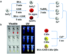
Herein we developed a new approach for CdSe quantum dots (QDs) synthesis. These bovine serum albumin (BSA) and glutathione (GSH) conjugated CdSe QDs (BSA–GSH–CdSe QDs) are of low cytotoxicity and NADPH responsive. The whole synthetic process can be completed within 8 minutes at room temperature.


Quantum dots (QDs) play an important role in various fields such as sensing,1 catalysis,2 solar cells,3 and bioimaging studies4 due to their unique electrooptical properties.5,6 However, several factors have greatly limited the application of QDs in broader areas. First of all, QD synthesis is normally carried out in an oil phase by heating under nitrogen protection and prolonged incubation.7–9 On the other hand, it remains a challenge to design and synthesize QDs that can respond to various cellular metabolites with high selectivity and low cytotoxicity. To overcome these limitations, it is critical to find a new synthetic method for QD production. Indeed, during the last few years several methods were developed concerning the synthesis of QDs under relatively milder conditions in contrast to the classical approach.10–19 Nevertheless, most of these approachs require multiple pH adjustments, prolonged incubation, high temperature, and are frequently only suitable for a small volume of QD synthesis. For comparison, methods of synthesizing QDs under mild conditions are summarized (Table S1).

Crystalline CdSe is among the best known QDs. Therefore, CdSe QDs were chosen as a model system to explore the possibility of modification and improvement of the QD synthetic protocol. It is known that a stable supply of the active selenium group (Se2−) is a critical and time-consuming step in the classical CdSe QD synthetic approach.20,21 Se2− ions can be easily oxidized by the trace amount of oxygen in the aqueous solution. Thus, finding of alternative ways to produce and stabilize Se2− might be the key to green synthesis of QDs. To this end, we reasoned that a powerful ROS scavenging system may be introduced into the reaction solution of QD synthetic system to stabilize Se2−. It was known that selenium nanoparticles (SeNPs) contain ROS removal function which is partially attributed to their large surface area and small size. Very recently, a green synthetic route for SeNP synthesis was established by using glutathione (GSH) as a reducing agent and bovine serum albumin (BSA) as a stabilizer.22,23 Notably, GSH is a free radical eliminating compound, widely distributed in almost all the organisms. This sulphur group containing molecule can interact with various metal ions and thus was frequently used for QD synthesis.24–27 Similarly, BSA was also sometimes used for HgS, ZnS and CdSe QD synthesis,28–30 which is likely due to its capability of interaction with variety of inorganic molecules and stabilize them.31,32

Herein this study, we developed an alternative route by using SeNPs as selenium source to initiate CdSe QD synthesis. This new system was exempted from N2 protection, low temperature and longtime incubation at oil phase, as compared to classical QD synthetic approach. The process can be divided into two phases: SeNP and CdSe QD synthesis (Fig. 1).


image file: c4ra11312h-f1.tif
Fig. 1 Synthetic route for BSA–GSH–CdSe QDs. (a) A, B, C, and D stand for SeO32−, SeO32−/Se aggregates/SeNPs, Se2−, and CdSe aggregate/CdSe QDs, respectively. 1, 2, and 3 refer to BSA, GSH, and BSA–GSH based synthetic systems. (b) Samples correlated to a under either visible or UV lights.

The first part of this synthetic process mainly concerns the production of SeNPs and this step was completed in 5 minutes. When selenite (Fig. 1A) and BSA was mixed, there is no colour change observed, suggesting no SeNP was formed (Fig. 1B1). While GSH was added into selenite solution, the mixture was changed from clear colourless to turbid red solution, indicating the formation of selenium aggregates (Fig. 1B2). SeNP synthesis was initiated by mixing of selenite, GSH and BSA. The colour of this mixture was rapidly switched from colourless to red, indicating the formation of SeNPs (Fig. 1B3). Sodium borohydride was then introduced into the SeNP solution followed by 3 minute-incubation. The solution was switched back to colourless again within 3 minutes, hinting the Se2− formation (Fig. 1C3). Cd2+ was then added into this Se2− containing solution. The colour of the solution was shifted to light yellow-green within one second, suggesting the formation of CdSe QDs, as also shown in the images taken under UV irradiation (360 nm) (Fig. 1D3). This rapid synthetic process can be completed within approximately 8 minutes. In the absence of BSA, this process somehow can also produce relatively unstable CdSe QDs (Fig. 1D2). Synthesis of CdSe QDs at RT using SeNPs showed advantages over other selenium sources tested so far (Fig. S1).

This SeNPs based approach for QD synthesis was summarized in Fig. 2a. SeNPs via this BSA–GSH assisted route was approximately 70 nm in size, as shown in scanning electron microscopy (SEM) images and dynamic light scattering (DLS) measurements (Fig. 2b). The core size of the BSA–GSH–CdSe QDs was approximately 4.5 nm with clear visible lattice fringes, as shown in transmission electron microscope (TEM) images (Fig. 2c). UV-vis spectra showed a strong shoulder peak centered at 425 nm (Fig. 2d). Fluorescence spectra showed that the main excitation and emission peaks of BSA–GSH–CdSe QDs were localized at 398 and 535 nm, respectively (Fig. 2e). As expected, BSA–GSH–CdSe QDs emitted yellow-green fluorescence under UV irradiation (Fig. 2f).


image file: c4ra11312h-f2.tif
Fig. 2 Schematic synthetic route and spectroscopic characterization of SeNPs and BSA–GSH–CdSe QDs. (a) Schematic presentation of BSA–GSH–CdSe QD synthesis. (b) Representative SEM image and DLS analysis of SeNPs. (c) Representative TEM image and a size histogram of BSA–GSH–CdSe QDs. Inset: enlarged images with visible lattice fringes. (d) UV-visible spectra of GSH (black), BSA (red), BSA–GSH–CdSe QDs (blue), and SeNPs (green). (e) Fluorescent excitation (red) and emission (black) curves of BSA–GSH–CdSe QDs. (f) Substrates, intermediate products and synthesized BSA–GSH–CdSe QDs under visible (upper) and UV (lower) lights.

These CdSe QDs were conjugated with BSA and GSH, as confirmed by FT-IR spectra (Fig. S2). Based on literatures, the size of SeNPs was partially dependent on the ratio of BSA/GSH/SeO32−.22,23 As a consequence, a mild blue shift of BSA–GSH–CdSe QDs was observed along with the increase of BSA concentration in the reaction mixture (Fig. S3), suggesting that BSA might act as a “size regulator” of CdSe QDs. Compared to other proteins such as mucin and collagen, BSA appeared to be an excellent candidate for synthesizing CdSe QDs in that BSA conjugated QDs show much higher fluorescence under the same synthetic condition (Fig. S4). BSA–GSH–CdSe QDs are fairly stable in different buffers (Fig. S5a), pH (Fig. S5b) and resistant to high salt condition (Fig. S5c) and can tolerate longtime incubation at RT (Fig. S5d).

Recent years the design of biosensors for the recognition of biological active small molecules has received considerable attention,33 especially for nucleotides and their derivatives such as adenosine triphosphate (ATP) and nicotinamide adenine dinucleotide phosphate (NADP(H)). NADP(H) are structurally similar to ATP and are cofactors for hundreds of cellular enzymes for nearly all the living organisms, where NADPH is the reduced form and NADP+ is the oxidized form. Cellular redox condition is largely associated with the status of these cofactors, for instance, the ratio of[NADP+]/[NADPH]. Therefore, it is highly important to generate sensors that can detect the content of these cofactors in aqueous solution. Several excellent methods have been developed for ATP recognition,34 whereas the detection method for NADP(H) are relatively less and suffer from laborious and low selectivity, such as electroanalysis35–37 and spectrum detection.38,39 It is highly valuable to find new approaches that can improve the simplicity, sensitivity and selectivity of the NAD(P)(H) probing system.

Interestingly, BSA–GSH–CdSe QDs can response to NADPH in aqueous solutions likely through forming a supramolecular complex (Fig. 3a). In contrast to most of currently available NADPH nano-sensors that detection mechanism lies in fluorescent quenching,38,39 a clear change of fluorescence emission wavelength was observed when BSA–GSH–CdSe QDs were mixed with NADPH (Fig. 3b). The blue shift of emission of BSA–GSH–CdSe QDs was accompanied by an increase of fluorescence intensity along with the raise of NADPH concentration (Fig. 3b and S6). There is a linear relationship between the fluorescence intensity and NADPH concentration at the range of 0–13 mM with a detection limit of 250 μM (Fig. 3c). In contrast, when MPA–CdSe QDs synthesized using classical method40 was incubated with NADPH, there is no blue shift event observed, instead the emission peaks of MPA–CdSe QDs and NADPH were separated, indicating the absence of physical interaction between MPA–CdSe QDs and NADPH (Fig. S7).


image file: c4ra11312h-f3.tif
Fig. 3 BSA–GSH–CdSe QDs acted as a NADPH sensor. (a) Schematic presentation of putative mechanism of BSA–GSH–CdSe QDs based NADPH sensor. (b) The blue shift and fluorescent increase of BSA–GSH–CdSe QDs toward NADPH. (c) The linear relationship between the fluorescence intensity of BSA–GSH–CdSe QDs and NADPH concentration (I482 and I0 refer to the fluorescent intensity at 482 and 535 nm, respectively). (d) The selectivity of BSA–GSH–CdSe QDs toward NADPH and other related compounds and biomolecules (5 mM). (e) There was no clear cytotoxicity of BSA–GSH–CdSe QDs observed toward E. coli cells. OD600 stands for the optical density (OD) at 600 nm of the bacterial cells.

To investigate the selectivity of BSA–GSH–CdSe QDs based nano-sensor, the fluorescence changes of this nanomaterial were carefully analysed upon adding of NADPH and its derivatives and several other biomolecules or compounds as follows: NADP, ATP, adenosine diphosphate (ADP), adenosine monophosphate (AMP), guanosine triphosphate (GTP), cytidine triphosphate (CTP), uridine triphosphate (UTP), 2′-deoxyguanosine-5′-triphosphate (d-GTP), 2′-deoxyadenosine-5′-triphosphate (d-ATP), 2′-deoxythymidine-5′-triphosphate (d-TTP), 2′-deoxycytidine-5′-triphosphate (d-CTP), and D-galactose, glycine, GSH, mucin, Na+, and H2O (Fig. 3d and S8). Except for NADPH, all the other reagents tested so far did not show obvious blue shift and fluorescence enhancement at 482 nm, suggesting a high selectivity of BSA–GSH–CdSe QDs toward NADPH. The zeta potential of CdSe QDs and NADPH was −14 and −12.3 mV, respectively, which was shifted to −18.7 mV when they were mixed, hinting a stable interaction between them. The cytotoxicity of this material was evaluated by using bacteria Escherichia coli cells as a model system. The growth of E. coli cells was not affected by adding of 2 μM of BSA–GSH–CdSe QDs, as measured at OD600 (Fig. 3e).

Human serum is a complex mixture containing large amount of ions and biological molecules. Therefore, it provides a good platform to evaluate sensor function of BSA–GSH–CdSe QDs with special reference to their NADPH recognition properties. Human blood sera from volunteers were diluted 10 times using phosphate buffered saline (PBS buffer, pH 7.4) before use. These sera containing buffers and plain PBS buffer were used as media to measure NADPH content using the proposed method. The serum showed hardly any interference effect on the NADPH concentration determination (Table S1 and Fig. S9), suggesting BSA–GSH–CdSe QDs can be a promising biocompatible NADPH sensor.

In summary, we have developed a new approach for facile and rapid synthesis of CdSe QD at RT using eco-friendly molecule BSA and GSH. This visual process can be easily monitored under visible and UV lights by naked eyes. Notably, the whole reaction is completed within 8 minutes. Further, there is no sophisticated equipment or skills required during the synthetic process. Moreover, these nanomaterials prepared in this study are NADPH responsive and can potentially be used in probing NADPH in biotic environment. Finally, this new CdSe QD green synthesis method is expected to be extended to other QDs or nanoparticles.

Acknowledgements

This work was supported by the National Natural Science Foundation of China (NSF)(no. 21372097), Open Fund of State Key Laboratory of Reproductive Biology at Institute of Zoology of Chinese Academy of Sciences, and Center of International Mobility (CIMO) grant from Finland.

Notes and references

  1. R. Gui, H. Jin, X. Liu, Z. Wang, F. Zhang, J. Xia, M. Yang and S. Bi, Chem. Commun., 2014, 50, 14847 RSC.
  2. R. Freeman, X. Liu and I. Willner, Nano Lett., 2011, 11, 4456 CrossRef CAS PubMed.
  3. Z. Tachan, M. Shalom, I. Hod, S. Rühle, S. Tirosh and A. Zaban, J. Phys. Chem. C, 2011, 115, 6162 CAS.
  4. P. Zrazhevskiy, M. Sena and X. Gao, Chem. Soc. Rev., 2010, 39, 4326 RSC.
  5. M. Bruchez, M. Moronne, P. Gin, S. Weiss and A. P. Alivisatos, Science, 1998, 281, 2013 CrossRef CAS.
  6. W. C. W. Chan and S. Nie, Science, 1998, 281, 2016 CrossRef CAS.
  7. Z. A. Peng and X. G. Peng, J. Am. Chem. Soc., 2001, 123, 183 CrossRef CAS.
  8. M. A. Hines and P. Guyot-Sionnest, J. Phys. Chem., 1996, 100, 468 CrossRef CAS.
  9. S. H. Yu, Y. S. Wu, J. Yang, Z. H. Han, Y. Xie, Y. T. Qian and X. M. Liu, Chem. Mater., 1998, 10, 2309 CrossRef CAS.
  10. J. H. Li, C. L. Ren, X. Y. Liu, Z. D. Hu and D. S. Xue, Mater. Sci. Eng., A, 2007, 458, 319 CrossRef PubMed.
  11. O. M. Primera-Pedrozo, Z. Arslan, B. Rasulev and J. Leszczynski, Nanoscale, 2012, 4, 1312 RSC.
  12. W. E. Mahmoud, A. M. Al-Amri and S. J. Yaghmour, Opt. Mater., 2012, 34, 1082 CrossRef CAS PubMed.
  13. Y. Wang, Y. Zhang, F. Wang, D. E. Giblin, J. Hoy, H. W. Rohrs, R. A. Loomis and W. E. Buhro, Chem. Mater., 2014, 26, 2233 CrossRef CAS PubMed.
  14. M. P. Reddy, W. Madhuri, I. S. S. K. Rao, I. G. Kim and R. R. Reddy, Int. J. ChemTech Res., 2014, 6, 2048 CAS.
  15. Q. Sun, S. Fu, T. Dong, S. Liu and C. Huang, Molecules, 2012, 17, 8430 CrossRef CAS PubMed.
  16. Q. Wang, S. Li, P. Liu and X. Min, Mater. Chem. Phys., 2012, 137, 580 CrossRef CAS PubMed.
  17. A. M. Al-Amri, S. J. Yaghmour and W. E. Mahmoud, J. Cryst. Growth, 2011, 334, 76 CrossRef CAS PubMed.
  18. J. C. Newton, K. Ramasamy, M. Mandal, G. K. Joshi, A. Kumbhar and R. Sardar, J. Phys. Chem. C, 2012, 116, 4380 CAS.
  19. X. He, L. Gao and N. Ma, Sci. Rep., 2013, 3, 2825 Search PubMed.
  20. A. L. Rogach and A. Kornowski, J. Phys. Chem. B, 1999, 103, 3065 CrossRef CAS.
  21. A. L. Rogach, D. Nagesha, J. W. Ostrander, M. Giersig and N. A. Kotov, Chem. Mater., 2000, 12, 2676 CrossRef CAS.
  22. J. S. Zhang, X. Y. Gao, L. D. Zhang and Y. P. Bao, BioFactors, 2001, 15, 27 CrossRef CAS.
  23. B. Huang, J. Zhang, J. Hou and C. Chen, Free Radical Biol. Med., 2003, 35, 805 CrossRef CAS.
  24. H. Qian, C. Dong, J. Weng and J. Ren, Small, 2006, 2, 747 CrossRef CAS PubMed.
  25. Y. Zheng, S. Gao and J. Y. Ying, Adv. Mater., 2007, 19, 376 CrossRef CAS.
  26. Y. Nakane, Y. Tsukasaki, T. Sakata, H. Yasuda and T. Jin, Chem. Commun., 2013, 49, 7584 RSC.
  27. Y. Li, Y. Zhou, H. Y. Wang, S. Perrett, Y. Zhao, Z. Tang and G. Nie, Angew. Chem., Int. Ed., 2011, 50, 5860 CrossRef CAS PubMed.
  28. N. Goswami, A. Giri, S. Kar, M. S. Bootharaju, R. John, P. L. Xavier, T. Pradeep and S. K. Pal, Small, 2012, 8, 3175 CrossRef CAS PubMed.
  29. W. Zhou and F. Baneyx, ACS Nano, 2011, 5, 8013 CrossRef CAS PubMed.
  30. Q. Wang, F. Ye, T. Fang, W. Niu, P. Liu, X. Min and X. Li, J. Colloid Interface Sci., 2011, 355, 9 CrossRef CAS PubMed.
  31. J. Xie, Y. Zheng and J. Y. Ying, J. Am. Chem. Soc., 2009, 131, 888 CrossRef CAS PubMed.
  32. L. Yang, R. Xing, Q. Shen, K. Jiang, F. Ye, J. Wang and Q. Ren, J. Phys. Chem. B, 2006, 110, 10534 CrossRef CAS PubMed.
  33. N. Hildebrandt, ACS Nano, 2011, 5, 5286 CrossRef CAS PubMed.
  34. H. Huang, Y. Tan, J. Shi, G. Liang and J. J. Zhu, Nanoscale, 2010, 2, 606 RSC.
  35. S. P. Pogorelova, M. Zayats, T. Bourenko, A. B. Kharitonov, O. Lioubashevski, E. Katz and I. Willner, Anal. Chem., 2003, 75, 509 CrossRef CAS.
  36. K. Schubert, W. Khalid, Z. Yue, W. J. Parak and F. Lisdat, Langmuir, 2010, 26, 1395 CrossRef CAS PubMed.
  37. Z. Dai, G. Lu, J. Bao, X. Huang and H. Ju, Electroanalysis, 2007, 19, 604 CrossRef CAS.
  38. M. Y. Kim, Y. S Kim, J. Kim, S. S. Hah, T. J. Kim and Y. D. Kim, Biotechnol. Lett., 2011, 33, 623 CrossRef CAS PubMed.
  39. G. Garai-Ibabe, L. Saa and V. Pavlov, Anal. Chem., 2013, 85, 5542 CrossRef CAS PubMed.
  40. Z. Deng, F. L. Lie, S. Shen, I. Ghosh, M. Mansuripur and A. J. Muscat, Langmuir, 2009, 25, 434 CrossRef CAS PubMed.

Footnote

Electronic supplementary information (ESI) available: Materials and methods, FT-IR spectra, fluorescence spectra, stability measurements, and several other experiments. See DOI: 10.1039/c4ra11312h

This journal is © The Royal Society of Chemistry 2014