
Tunable blue-green-emitting Ca3Si2O4N2:Ce3+, Eu2+ phosphor with energy transfer for light-emitting diodes
Yanyan Li,
Quansheng Wu,
Xicheng Wang,
Jianyan Ding,
Qiang Long and
Yuhua Wang*
Key Laboratory for Special Function Materials and Structural Design of the Ministry of Education, School of Physical Science and Technology, Lanzhou University, Lanzhou, 730000, China. E-mail: wyh@lzu.edu.cn; Fax: +86-931-8913554; Tel: +86-931-8912772
First published on 18th November 2014
Abstract
A series of Ce3+, Eu2+ codoped Ca3Si2O4N2 phosphors have been synthesized by traditional solid state reactions, and the crystal structures and luminescence properties were investigated in detail. Rietveld structure refinement indicates that Ca3Si2O4N2 crystallizes in a cubic unit cell with space group Pa![[3 with combining macron]](https://www.rsc.org/images/entities/char_0033_0304.gif) (205) and lattice constant a = 15.0712 Å. Tunable blue-green emitting Ca3Si2O4N2:Ce3+, Eu2+ phosphors have been obtained by codoping Ce3+ and Eu2+ into the host and varying their relative ratios. Compared with the Eu2+ ion singly doped phosphor, the codoped phosphors have wider absorption in the ultraviolet (UV) range and stronger emission of Eu2+, which are attributed to the effective energy transfer from Ce3+ to Eu2+. The energy transfer from Ce3+ to Eu2+ is demonstrated to be a dipole–dipole mechanism. The Ce3+, Eu2+ codoped phosphors might be candidates for blue-green components in UV white light-emitting diodes (WLEDs).
(205) and lattice constant a = 15.0712 Å. Tunable blue-green emitting Ca3Si2O4N2:Ce3+, Eu2+ phosphors have been obtained by codoping Ce3+ and Eu2+ into the host and varying their relative ratios. Compared with the Eu2+ ion singly doped phosphor, the codoped phosphors have wider absorption in the ultraviolet (UV) range and stronger emission of Eu2+, which are attributed to the effective energy transfer from Ce3+ to Eu2+. The energy transfer from Ce3+ to Eu2+ is demonstrated to be a dipole–dipole mechanism. The Ce3+, Eu2+ codoped phosphors might be candidates for blue-green components in UV white light-emitting diodes (WLEDs).
1 Introduction
White light-emitting diodes (WLEDs), the so-called next-generation solid-state lighting, offer benefits in terms of reliability, energy-saving, maintenance and safety, and therefore are gaining much attention.1 Hence, WLEDs are expected to be promising candidates to replace conventional incandescent and fluorescent lamps. Generally, commercial phosphor converted WLEDs (pc-WLEDs) are mainly fabricated through the combination of a 450–470 nm blue InGaN chip and a yellowish phosphor (typically YAG:Ce3+) coating. However, such methods suffer from a poor color-rendering index (CRI) and high correlated color temperature (CCT = 7756 K), owing to the absence of red emissions in the long wavelength region.2,3 Accordingly, during the past few years, WLEDs fabricated with tricolor phosphors and ultraviolet (UV) LEDs are considered to be very promising for the realization of high-quality light sources because of their higher CRI and homogeneous color distribution in comparison to current commercial WLEDs.4 LED phosphors are generally required to have a high conversion efficiency, excellent chemical stability and high thermal quenching temperatures.5 In addition, they are also expected to have a tunable emission color,6 allowing WLED designers to have greater flexibility in choosing the emission wavelength and gain an improvement either in the CCT or CRI of WLEDs and for phosphor applications in UV LEDs. In certain Eu2+ doped matrices, the green to orange emission and UV to blue-light excitation are commonly observed through the strong crystal-field splitting and the lower energy of the centroid of the 5d level.7 Hence, Eu2+ can be sensitized by a well-known blue-emitting Ce3+ in such lattices because of the spectral overlap between the emission band of Ce3+ and the excitation band of Eu2+. Consequently, tunable emissions can be produced in this kind of Ce3+, Eu2+ codoped single composition host.8–10Nitride/oxynitride hosts are good candidates for host lattices for phosphors due to several advantages, such as high chemical stability and good thermal quenching ability, and exhibit intense luminescence for application in WLEDs when activated with Ce3+/Eu2+, such as in M2Si5N8,11,12 MSiN2,13 Ca-α-SiAlON,14 CaAlSiN3,15,16 MSi2O2N2 (M = Ca, Sr, Ba),17 MSiAl2O3N2 (M = Sr, Ba)18 and so on. Some of them have already been put to practical use.
Eu2+ activated Ca3Si2O4N2 was first reported by Liu's group.19 Although the excitation spectrum of Eu2+ doped Ca3Si2O4N2 covers the region from about 290 to 410 nm, the optimal excitation wavelength is about 328 nm, which doesn't match quite well with UV LED chips. Wang et al.20 studied the O/N ordering in the structure of Ca3Si2O4N2 and simply showed the excitation and emission spectra of Ce3+ doped Ca3Si2O4N2 phosphors, which can be excited at about 350 nm and give a blue emission. Subsequently, C. H. Huang21 studied the photoluminescence properties of Ce3+ doped Ca3Si2O4N2 phosphors in detail. A systematic study on the energy transfer between Eu2+ and Ce3+ in Ca3Si2O4N2 host, however, was not reported, to the best of our knowledge. In this paper, we report the luminescence properties of Ce3+, Eu2+ codoped Ca3Si2O4N2 phosphors. After codoping with Ce3+, the emission intensity of Eu2+ was enhanced and a series of tunable blue-green colors can be obtained by varying the relative ratio of Ce3+/Eu2+ under the irradiation of 350 nm. Moreover, the energy transfer mechanism between Ce3+ and Eu2+ has been investigated systematically.
2 Experimental section
Samples of Ca3−xSi2O4N2:xCe3+ (0.005 ≤ x ≤ 0.03), Ca2.995Si2O4N2:0.005Eu2+ and Ca2.98−ySi2O4N2:0.02Ce3+, yEu2+ (0.0025 ≤ y ≤ 0.05) investigated in this work were synthesized through solid state reactions. CaCO3 (A. R.), Si3N4 (Sigma-Aldrich 99.9%), CeO2 (99.99%) and Eu2O3 (99.999%) were employed as raw materials. Briefly, the raw materials were weighed in appropriate proportions and finely ground, and then filled into boron nitride (BN) crucibles. Subsequently, the powder mixtures were preheated at 900 °C (the temperature was increased at a rate of 10 °C min−1) for 2 h to ensure the decomposition of CaCO3, and then the temperature was increased to 1450 °C (at a rate of 5 °C min−1) and maintained for 8 h. After firing, the samples were cooled to 500 °C (at a rate of 5 °C min−1) and finally quenched to room temperature. In order to control the oxygen content in the reaction, the whole heating processes for both Ce3+ doped and Ce3+, Eu2+ co-doped samples are conducted in a reductive atmosphere of NH3/N2.All measurements were made on finely ground powder. The phase purity of samples was analyzed by X-ray diffraction (XRD) using a D2 PHASER X-ray Diffractometer with Ni-filtered Cu Kα radiation. Structure refinement was performed using the Rietveld method,22 using the Materials Studio (MS) program.23 Photoluminescence (PL) and PL excitation (PLE) spectra were measured at room temperature using a FLS-920T fluorescence spectrophotometer equipped with a 450 W Xe light source and double excitation monochromators. The PL decay curves were measured by a FLS-920T fluorescence spectrophotometer with a nF900 nanosecond flashlamp as the light source. High temperature luminescence intensity measurements were carried out by using an aluminum plaque with cartridge heaters; the temperature was measured by thermocouples inside the plaque and controlled by using a standard TAP-02 high temperature fluorescence controller. The powder morphology was investigated by using a scanning electron microscope (SEM; S-3400, Hitachi, Japan). The elemental compositions were determined using an energy-dispersive X-ray spectroscope (EDX), which was attached to the transmission electron microscope (TEM, FEI Tecnai F30, operated at 300 kV).
3 Results and discussion
Phase identification and crystal structure
Fig. 1(a) plots the experimental, calculated, background and difference results of the XRD profiles for the Rietveld refinement of Ca3Si2O4N2 host. The resulting crystallographic data are summarized in Table 1, which indicate that the powder sample crystallizes in a cubic unit cell with space group Pa(205) and lattice constant a = 15.0712 Å and Z = 24. The refinement finally converged to Rwp = 13.08%, Rp = 9.03%. Selected XRD patterns of Ce3+ doped and Ce3+, Eu2+ co-doped phosphors with different doping contents are illustrated in Fig. 1(b), which match well the calculated XRD patterns, indicating that the obtained samples are all of single phase.
 | ||
| Fig. 1 (a) Experimental (crosses) and calculated (red solid line) XRD patterns of the Ca3Si2O4N2 host. The blue solid lines represent the difference between experimental and calculated data and the pink sticks mark the Bragg reflection positions. The inset shows the corrugated 12-membered ring structure viewed from [111]. (b) XRD patterns of Ca2.995Si2O4N2:0.005Eu2+, Ca3Si2O4N2:0.02Ce3+, and Ca2.98−ySi2O4N2:0.02Ce3+, yEu2+ (0.0025 ≤ y ≤ 0.05) samples. (c) Unit cell structure and (d) EDX spectrum of Ca3Si2O4N2 host. (e) and (f) SEM images of Ca3Si2O4N2:0.02Ce3+, 0.005Eu2+. | ||
| Crystallographic data | |
| Formula | Ca3Si2O4N2 |
| Crystal system | Cubic |
| Space group | Pa(205) |
| a (Å) | 15.0712 |
| Cell volume (Å3) | 3423.288 |
| Z | 24 |
| Rietveld data | |
| Program | Materials studio |
| Range | 5°–110° |
| Step | 0.02 |
| Rwp | 13.08% |
| Rp | 9.03% |
Fig. 1(c) shows the refined crystal structure of Ca3Si2O4N2. In this structure, [SiO2N2] tetrahedral construct highly corrugated 12-membered rings rather than the [Si(O,N)4−m]n layers. The tetrahedral connect each other by sharing N atoms on bridging positions and the O atoms are located at terminal positions, which can be seen from the inset of Fig. 1(a). In the structure of Ca3Si2O4N2, the Ca2+ ions have seven different coordination environments, and only the Ca(6) ion is eight-coordinated, while the other Ca2+ ions are six-coordinated. The ionic radii of the six- and eight-coordinated Ca2+ are 1.00 and 1.12 Å, respectively. Similarly, the ionic radii of the six- and eight-coordinated Ce3+ are 1.01 and 1.14 Å, while 1.17 and 1.25 for six- and eight-coordinated Eu2+, respectively.24 Because of the similarities of their ionic radii, the Ce3+ and Eu2+ are expected to randomly occupy Ca2+ sites in the Ca3Si2O4N2 crystal structure. The corresponding EDX spectrum analysis which is shown in Fig. 1(d) indicates that the sample has a chemical composition of Ca, Si, N and O and the Cu peak is ascribed to the copper grid supporting the TEM sample.
The SEM images of Ca3Si2O4N2:0.005Eu2+, 0.02Ce3+ are shown in Fig. 1(e) and (f). It can be observed that the grains exhibit ellipsoid shapes and they have a particle size ranging from about 2–7 μm.
Photoluminescence properties and energy transfer
The PL and PLE spectra of Ca3Si2O4N2:0.02Ce3+ are shown in Fig. 2. The excitation spectrum consists of three absorption bands that peaking at about 290, 320 and 350 nm, which correspond to the transitions from the ground state of the Ce3+ ions to the field splitting levels of the 5d state.21 Upon excitation at 350 nm, the emission spectrum shows an asymmetric broad band peaking at 436 nm. This asymmetric emission was attributed to the spin–orbit coupling of the Ce3+ 4f1 ground state, which splits into two levels (2F5/2 and 2F7/2),25 and the occupation of Ce3+ ions in independent six- and eight-coordinate Ca2+ ion sites. | ||
| Fig. 2 PL and PLE spectra of Ca3Si2O4N2:0.02Ce3+. | ||
The dependence of the peak position and emission intensity of Ca3Si2O4N2:Ce3+ on the Ce3+ content is shown in Fig. 3. With increasing the content of Ce3+, the emission intensity is maximized at x = 0.02, and then decreases due to the concentration quenching effect. In addition, the peak positions of the emission bands shift toward a longer wavelength for about 10 nm as the content of Ce3+ dopant increases. This phenomenon can be explained in terms of energy transfer from the Ce3+ ions at higher 5d level to those at the lower levels, which decreases the emission energy from the 5d excited state to the 4f ground state and results in a redshift of the emission.21
 | ||
| Fig. 3 The dependence of the peak position and the emission intensity on the Ce3+ content. | ||
As depicted in Fig. 4(b), the PL spectrum of the Eu2+ singly doped phosphor shows a broad green emission band centering at 510 nm attributed to the typical 4f65d1–4f7 (8S7/2) transition of Eu2+, and the PLE spectrum shows a broad absorption from 280 to 420 nm with a maximum at 337 nm. The PL and PLE spectra of Ca3Si2O4N2:0.02Ce3+ are also shown in Fig. 4(a) for comparison. A significant spectral overlap between the PL spectrum of Ca3Si2O4N2:0.02Ce3+ and the PLE spectrum of Ca3Si2O4N2:0.005Eu2+ is observed, which indicates that the energy transfer from the Ce3+ to Eu2+ ions can be expected in Ce3+ to Eu2+ codoped host. Fig. 4(c) depicts the PL and PLE spectra of the Ca3Si2O4N2:0.02Ce3+, 0.005Eu2+ phosphor. The codoped phosphor shows a blue emission band of Ce3+ ions and a green emission band of Eu2+ ions at the irradiation of 350 nm. When monitoring at 500 nm and 436 nm, the peak positions of the PLE spectra are all located at about 350 nm, which is more suitable for the UV chip compared to the Eu2+ singly doped phosphor.
 | ||
| Fig. 4 PL and PLE spectra of (a) Ca3Si2O4N2:0.02Ce3+, (b) Ca3Si2O4N2:0.005Eu2+, and (c) Ca3Si2O4N2:0.02Ce3+, 0.005Eu2+ phosphors. | ||
Fig. 5 displays the PL spectra of Ca3Si2O4N2:0.02Ce3+, yEu2+ samples with a fixed Ce3+ content of 0.02 and a varying Eu2+ content y in the range of 0.0025–0.05, as well as the PL spectrum of Ca3Si2O4N2:0.005Eu2+. One can see that the PL intensity for Ce3+ decreases monotonically with an increase in doping Eu2+ content. Meanwhile, the emission of Eu2+ increases gradually until the Eu2+ content is above 0.005 and concentration quenching occurs. Subsequently, the emission of the Eu2+ falls with a further increase in y. The observed variations in the emission of the Ce3+ and Eu2+ ions further support the occurrence of the effective energy transfer from the Ce3+ to Eu2+ ions. Comparing the PL spectrum of Ca3Si2O4N2:0.005Eu2+ with that of Ca3Si2O4N2:0.02Ce3+, 0.005Eu2+, we can find that after codoping with Ce3+, the emission of Eu2+ in Ca3Si2O4N2 is obviously enhanced.
 | ||
| Fig. 5 PL spectra of Ca3Si2O4N2:0.02Ce3+, yEu2+ (0.0025 ≤ y ≤ 0.05) and Ca3Si2O4N2:0.005Eu2+. | ||
Additionally, it is noticeable that the emission peaks of the Eu2+ ions gradually shift to longer wavelength for about 10 nm with an increase of the content of Eu2+ from 0.0025 to 0.05. This red-shift behavior is consistent with the phenomenon that reported by Liu,19 which is ascribed to the change in the crystal-field splitting of Eu2+. Consequently, the phenomenon can be explained in terms of energy transfer from Eu2+ ions at the higher 5d levels to those at the lower levels and this causes the emission energy from the 5d excited state to the 4f ground state to become lower, and therefore, the emission shifts to longer wavelength.
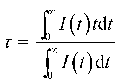
To further study the energy transfer process, the PL decay curves of the Ce3+ ions in Ca3Si2O4N2:0.02Ce3+, yEu2+ (0 ≤ y ≤ 0.03) phosphors were measured by monitoring at 436 nm with an irradiation of 350 nm, and depicted in a logarithmic intensity in Fig. 6. The decay curves for Ce3+ emission in Ca3Si2O4N2:0.02Ce3+, yEu2+ (0 ≤ y ≤ 0.03) phosphors deviate slightly from the single exponential function, which is also consistent with the structure analysis that more than one Ce3+ emitting center exists in the phosphor. An increase in Eu2+ content will enhance this deviation, which can further verify the energy transfer between Ce3+ and Eu2+. The decay process of these samples are characterized by an average lifetime, τ, which can be calculated using eqn (1) as follows,26–28
 | (1) |
| η = 1 − τ/τ0 | (2) |
 | ||
| Fig. 6 The decay curves of the Ce3+ ion in Ca3Si2O4N2:0.02Ce3+, yEu2+ (0 ≤ y ≤ 0.03) phosphors demonstrated on a logarithmic intensity scale. | ||
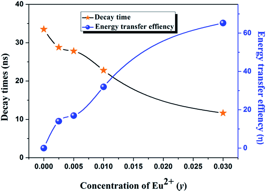 | ||
| Fig. 7 Dependence of the decay time of Ce3+ ions and energy transfer efficiency on the Eu2+ concentration in Ca3Si2O4N2:0.02Ce3+, yEu2+ (0 ≤ y ≤ 0.05) phosphors. | ||
In general, energy transfer from the sensitizer to activator in a phosphor may take place via a multipolar interaction29 or an exchange interaction at higher concentration. Based on Dexter's energy transfer formula of multipolar interaction and Reisfeld's approximation, the following relation can be obtained:30,31
 | (3) |
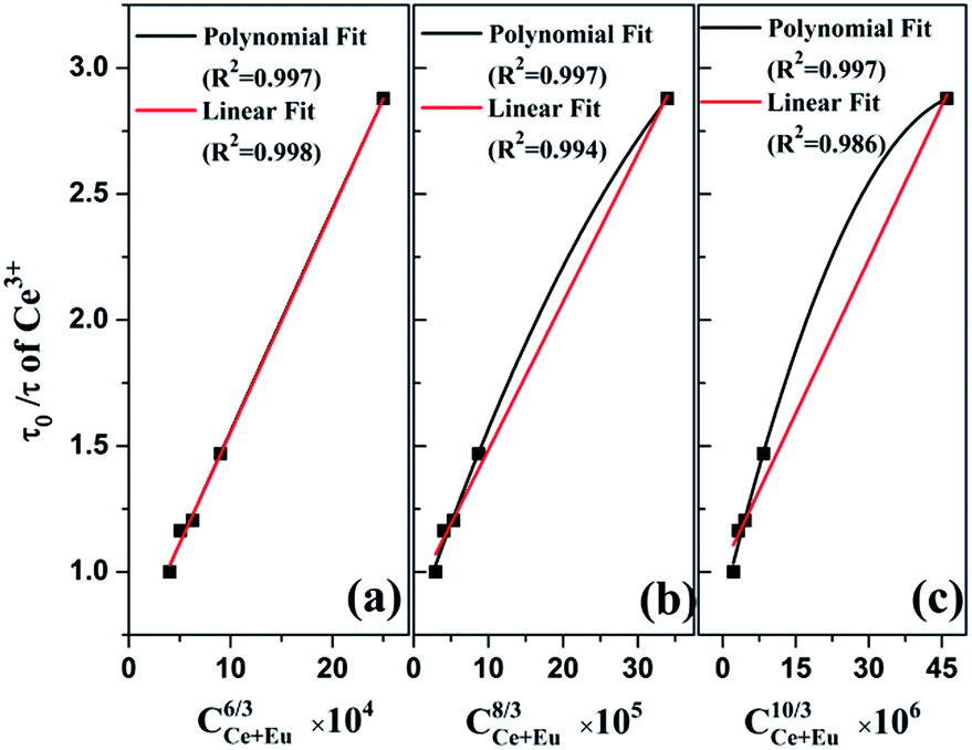 | ||
| Fig. 8 Dependence of τ0/τ of Ce3+ on (a) C6/3, (b) C8/3, and (c) C10/3. | ||
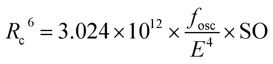
For electric dipole–dipole interaction, the critical distance Rc between Ce3+ and Eu2+ can be expressed by32
 | (4) |
The x and y values of the CIE chromaticity coordinates for the Ca3Si2O4N2:0.02Ce3+, yEu2+ (0.0025 ≤ y ≤ 0.05) phosphors are calculated and presented in Fig. 9. It can be seen that the emitting color of the phosphors can be easily modulated from blue (0.245, 0.285) to green (0.283, 0.515) by simply varying the value of y from 0.0025 to 0.05 due to the different emission composition of the Ce3+ and Eu2+ ions. Thus, blue-green emitting phosphors which can be efficiently excited by the UV chips are obtained via the energy transfer from the Ce3+ to Eu2+ ions and the Ca3Si2O4N2:Ce3+, Eu2+ phosphors could have potential value as blue-green phosphors used for UV WLEDs.
 | ||
| Fig. 9 CIE chromaticity coordinates for the Ca3Si2O4N2:0.02Ce3+, yEu2+ (0.0025 ≤ y ≤ 0.05) phosphors. | ||
Thermal quenching properties
For application in high power LEDs, the thermal stability of phosphors is one of the important issues to be considered. The temperature dependence of the emission spectra of Ca3Si2O4N2:0.02Ce3+, 0.005Eu2+ and Ca3Si2O4N2:0.005Eu2+ are shown in Fig. 10(a) and (b), respectively, which show a relatively poor thermal stability. At 150 °C, the emission intensity of the Ca3Si2O4N2:0.02Ce3+, 0.005Eu2+ phosphor is about 27% of that measured at room temperature while 47% for Ca3Si2O4N2:0.005Eu2+, which can be seen from Fig. 10(c). Compared with Eu2+ singly doped Ca3Si2O4N2, more rare earths ions were doped into Ca3Si2O4N2:0.02Ce3+, 0.005Eu2+ and then more deficiencies formed due to the radii mismatch of rare earth ions and Ca2+ ions. As a result, the probability of non-radiative process increased and thus leading to the worse thermal stability. The temperature quenching mechanism could be explained by the configuration coordinate diagram as shown in Fig. 10(d). With the increase of the temperature, the phonon vibration is strengthened and more electrons located at the lowest excited state C can absorb the phonon energy and are excited to state E. Electrons at state E can go back to the ground state A by relaxation and give no emission, making the number of electrons which can go back to the ground state through radiation decrease. Thus, the emission intensity of the phosphor will decrease with the increment of temperature. | ||
| Fig. 10 The temperature dependence of the emission spectra of (a) Ca3Si2O4N2:0.02Ce3+, 0.005Eu2+ and (b) Ca3Si2O4N2:0.005Eu2+, and (c) the dependence of their normalized PL intensity on working temperature. (d) The schematic configuration coordinate diagram for the explanation of temperature quenching. | ||
4 Conclusions
In summary, a series of Ce3+, Eu2+ codoped Ca3Si2O4N2 phosphors have been synthesized and the photoluminescence properties have been investigated in detail. Rietveld structure refinement indicates that Ca3Si2O4N2 crystallizes in a cubic unit cell with space group Pa(205) and lattice constant of a = 15.0712 Å and Z = 24. When Ce3+ and Eu2+ were codoped in Ca3Si2O4N2, the photoluminescence spectra displayed tunable blue-green emission by varying their relative ratios. Besides, the emission intensity of Eu2+ was enhanced via codoping with Ce3+ and the codoped phosphors have wider absorption in the UV range due to the effective energy transfer from the Ce3+ to Eu2+. The dipole–dipole interaction mechanism should be mainly responsible for the energy transfer from Ce3+ to Eu2+ in Ca3Si2O4N2. The experimental results indicate that the Ca3Si2O4N2:Ce3+, Eu2+ phosphor might have promising applications in UV WLEDs.
Acknowledgements
This work was supported by the National Natural Science Funds of China (Grant no. 51372105) and Specialized Research Fund for the Doctoral Program of Higher Education (no. 20120211130003).Notes and references
- N. Hirosaki, R.-J. Xie, K. Kimoto, T. Sekiguchi, Y. Yamamoto, T. Suehiro and M. Mitomo, Appl. Phys. Lett., 2005, 86, 211905 CrossRef PubMed.
- H. Jang, Y.-H. Won and D. Jeon, Appl. Phys. B, 2009, 95, 715–720 CrossRef CAS.
- A. A. Setlur, W. J. Heward, Y. Gao, A. M. Srivastava, R. G. Chandran and M. V. Shankar, Chem. Mater., 2006, 18, 3314–3322 CrossRef CAS.
- T. Taguchi, IEEJ Trans. Electr. Electron. Eng., 2008, 3, 21–26 CrossRef CAS.
- P. Dai, X. Zhang, L. Bian, S. Lu, Y. Liu and X. Wang, J. Mater. Chem. C, 2013, 1, 4570 RSC.
- P. F. Smet, A. B. Parmentier and D. Poelman, J. Electrochem. Soc., 2011, 158, R37–R54 CrossRef CAS PubMed.
- C.-H. Huang, Y.-T. Lai, T.-S. Chan, Y.-T. Yeh and W.-R. Liu, RSC Adv., 2014, 4, 7811–7817 RSC.
- P. Li, Z. Wang, Z. Yang and Q. Guo, RSC Adv., 2014, 4, 27708–27713 RSC.
- Q. Wu, Y. Li, X. Wang, Z. Zhao, C. Wang, H. Li, A. Mao and Y. Wang, RSC Adv., 2014, 4, 39030–39036 RSC.
- M. Zhang, Y. Liang, R. Tang, D. Yu, M. Tong, Q. Wang, Y. Zhu, X. Wu and G. Li, RSC Adv., 2014, 4, 40626–40637 RSC.
- Y. Q. Li, G. De With and H. Hintzen, J. Lumin., 2006, 116, 107–116 CrossRef CAS PubMed.
- M. Zeuner, F. Hintze and W. Schnick, Chem. Mater., 2008, 21, 336–342 CrossRef.
- C. Duan, X. Wang, W. Otten, A. Delsing, J. Zhao and H. Hintzen, Chem. Mater., 2008, 20, 1597–1605 CrossRef CAS.
- R.-J. Xie, N. Hirosaki, K. Sakuma, Y. Yamamoto and M. Mitomo, Appl. Phys. Lett., 2004, 84, 5404–5406 CrossRef CAS PubMed.
- Y. Li, N. Hirosaki, R. Xie, T. Takeda and M. Mitomo, Chem. Mater., 2008, 20, 6704–6714 CrossRef CAS.
- X. Piao, K.-i. Machida, T. Horikawa, H. Hanzawa, Y. Shimomura and N. Kijima, Chem. Mater., 2007, 19, 4592–4599 CrossRef CAS.
- Y. Q. Li, A. Delsing, G. De With and H. Hintzen, Chem. Mater., 2005, 17, 3242–3248 CrossRef CAS.
- W.-Y. Huang, F. Yoshimura, K. Ueda, Y. Shimomura, H.-S. Sheu, T.-S. Chan, C.-Y. Chiang, W. Zhou and R.-S. Liu, Chem. Mater., 2014, 26, 2075–2085 CrossRef CAS.
- Y.-C. Chiu, C.-H. Huang, T.-J. Lee, W.-R. Liu, Y.-T. Yeh, S.-M. Jang and R.-S. Liu, Opt. Express, 2011, 19, A331–A339 CrossRef PubMed.
- X.-M. Wang, C.-H. Wang, M. M. Wu, Y. X. Wang and X.-P. Jing, J. Mater. Chem., 2012, 22, 3388–3394 RSC.
- C. H. Huang, Y. C. Chiu and W. R. Liu, Eur. J. Inorg. Chem., 2014, 2014, 3674–3680 CrossRef CAS.
- H. Rietveld, J. Appl. Crystallogr., 1969, 2, 65–71 CrossRef CAS.
- H. Miura, T. Ushio, K. Nagai, D. Fujimoto, Z. Lepp, H. Takahashi and R. Tamura, Cryst. Growth Des., 2003, 3, 959–965 CAS.
- R. D. Shannon, Acta Crystallogr., Sect. A: Cryst. Phys., Diffr., Theor. Gen. Crystallogr., 1976, 32, 751–767 CrossRef.
- C.-H. Huang, T.-W. Kuo and T.-M. Chen, ACS Appl. Mater. Interfaces, 2010, 2, 1395–1399 CAS.
- N. Guo, Y. Song, H. You, G. Jia, M. Yang, K. Liu, Y. Zheng, Y. Huang and H. Zhang, Eur. J. Inorg. Chem., 2010, 2010, 4636–4642 CrossRef.
- F. Lahoz, I. R. Martín, J. Méndez-Ramos and P. Núñez, J. Chem. Phys., 2004, 120, 6180–6190 CrossRef CAS PubMed.
- G. Blasse, Phys. Lett. A, 1968, 28, 444–445 CrossRef CAS.
- G. Blasse and B. Grabmaier, Luminescent materials, Springer-Verlag, Berlin, German, 1994, p. 91 Search PubMed.
- G. Blasse, Philips Res. Rep., 1969, 24, 131–144 CAS.
- D. L. Dexter and J. H. Schulman, J. Chem. Phys., 1954, 22, 1063–1070 CrossRef CAS PubMed.
- Y. Song, G. Jia, M. Yang, Y. Huang, H. You and H. Zhang, Appl. Phys. Lett., 2009, 94, 091902 CrossRef PubMed.
| This journal is © The Royal Society of Chemistry 2014 |