DOI:
10.1039/C4RA10562A
(Paper)
RSC Adv., 2014,
4, 60953-60958
Reduction of ferrylmyoglobin by cysteine as affected by pH†
Received
16th September 2014
, Accepted 3rd November 2014
First published on 5th November 2014
Abstract
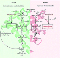
Reduction of the hypervalent meat pigment ferrylmyoglobin, MbFe(IV)
O, by cysteine is enhanced by acid due to protonation of ferrylmyoglobin to yield sulfmyoglobin as the main product, while at alkaline conditions, the rate decreases with the cysteine dianion as the reactant forming oxymyoglobin, MbFe(II)O2. The second-order rate constant for cysteine reacting with protonated ferrylmyoglobin is 5.1 ± 0.4 L mol−1 s−1 at 25 °C in 0.16 M aqueous sodium chloride and for the cysteine dianion reacting with ferrylmyoglobin 0.31 ± 0.15 L mol−1 s−1. For pH = 7.4 the activation parameters for sulfmyoglobin formation is ΔH‡ = 75 ± 2 kJ mol−1 and ΔS‡ = −250 ± 7 J mol−1 K−1 with similar values for homocysteine and glutathione. The difference in product is indicative of a shift from an electron-transfer/radical addition mechanism at low pH as in the stomach to a two-step electron-transfer mechanism at higher pH as in the intestine, and is discussed in relation to protection against the formation of radicals by sulphurous compounds during the digestion of red meat.
Introduction
Hypervalent iron is formed in the digestive tract during the digestion of meat by activation of metmyoglobin (MbFe(III)) by reactive oxygen species in the gut e.g. fatty acid hydroperoxide1 and also in vitro by hydrogen peroxide2 forming the very short lived perferryl compound (˙MbFe(IV)
O) and subsequently the more long-lived ferrylmyoglobin (MbFe(IV)
O).1–4 Both ˙MbFe(IV)
O and MbFe(IV)
O are known to be radical initiators of lipid oxidation and protein oxidation in meat and meat products during meat processing and storage affecting product quality negatively.5,6 Such radical mechanisms may also operate during digestion of red meat, and besides being radical initiators these hypervalent heme pigment may also liberate free iron forming reactive oxygen species in the gut. Red meat has been suggested to be one of a main causes in the development of colon cancer,3,7 and research are actively carried out to understand the toxic effects of reactive oxygen species (ROS) generated by hypervalent iron and how this can be counteracted by combining the intake of red meat with foods rich in natural antioxidants.8
Some thiols and hydrogen sulfide are known to react with MbFe(IV)
O and to form sulfmyoglobin (SulfMb) which is a protein with a modified chlorine type tetrapyrrole structure with a sulphur atom added to the pyrrole B ring of the porphyrin.10,11 SulfMb can, like myoglobin, exist as SulfMbFe(II) and SulfMbFe(III) with iron in different oxidation states.12,13 Cysteine is among the most reducing amino acids and may play a role in the protection of epithelial cells against radical attack, both as a free amino acid and incorporated into peptides like glutathione and larger proteins. Cysteine and glutathione are thus known to have cytoprotective effects against acute renal failure due to myoglobin toxicity, a condition that depends on iron catalyzed peroxidation.14 As previously reported, the products formed by reduction of MbFe(IV)
O by cysteine depends on pH.9,15 However, the complex structure of the data obtained by UV-vis spectroscopy invited for the use of multivariate curve resolution (MCR) in order to identify intermediate and reaction products for different conditions of pH.
Based on the extracted component spectra and the time concentration profiles of the absorbing components in the reactions mixture several reactions mechanisms were suggested.9 Accordingly, a more focused kinetic study has been initiated in order to identify reaction pathways under different pH-conditions and to determine rate constants for the dominating reactions for discussion of reaction mechanisms behind protection against hypervalent heme pigments by cysteine and cysteine derivatives and homologues.
Materials and methods
Chemicals
L-Cysteine was obtained from Merck (Darmstadt, Germany). Glutathione, L-homocysteine, and hydrogen peroxide (30% v/v) were obtained from Sigma-Aldrich (Steinheim, Germany) and used without further treatment. Catalase from bovine liver and myoglobin from horse heart (purity > 90%) was obtained from Sigma-Aldrich (Steinheim, Germany). Myoglobin was purified according to procedures previously described.9,11 Acetic acid was from AppliChem GmbH (Darmstadt, Germany). K2HPO4 and KH2PO4 were of analytical grade and supplied by J. T. Baker (Phillipsburg, NJ, US). Deionized water was obtained using a Milli-Q system Millipore Co. (Billerica, MA, US).
Reaction kinetics
Aqueous L-cysteine stock solutions (1.5 × 10⁻² mol L−1) were prepared in appropriate buffers. Ferrylmyoglobin was prepared from reaction of metmyoglobin (1 × 10⁻⁴ mol L−1) with H2O2 in a proportion of 1
:
2. Hydrogen peroxide excess was consumed by addition of catalase as previous described.9,11 Kinetic measurements at pH 6.0, 6.5, 7.0, 7.4, and 8.2 were performed using appropriate aqueous buffer using a two-compartment 1 cm × 1 cm quartz cell for fast mixture (Hellma Analytics, Müllheim, Germany) and UV-vis spectra recorded from 200 nm to 800 nm at different time intervals in an Multiskan GO Microplate Spectrophotometer (ThermoScientific, Bremen, Germany) thermostatted at 20.0, 25.0, or 30.0 °C. Kinetic measurements at pH 4.5, 5.0, 5.6, and 6.2 were performed using appropriate aqueous buffer using a SX 20 sequential mixing stopped-flow spectrophotometer (Applied Photophysics Ltd, Leatherhead, UK). Ferrylmyoglobin is unstable under acid conditions (pH 4.5–6.2), and kinetic data were obtained using a pH-jump technique. In acidic medium, the reaction was initiated by mixing a ferrylmyoglobin solution in a pH 7.4 phosphate buffer with low ionic strength with an acidic cysteine solution in acetate buffer at pH 4.5, 5.0, and 5.6 or phosphate buffer at 6.2 and 6.5 with an ionic strength of 0.32 M yielding a final ionic strength of 0.16 M at the desired pH. pH was measured in the reaction mixture using a combined glass electrode pH-meter model PHM240 (Radiometer Analytical, Copenhagen, Denmark). The formation of sulfmyoglobin was monitored at 625 nm and the decay of ferrylmyoglobin was monitored at 580 nm under pseudo-first order conditions for increasing concentration of cysteine. The auto-decay rate constant for ferrylmyoglobin was determined by mixing equal volumes of ferrylmyoglobin in low ionic strength buffer solution at pH 7.4 and an acid buffer at ionic strength 0.32 M resulting in a final 0.16 M ionic strength at the desired pH. The auto-decay followed first-order kinetics. The first-order rate constants were obtained by non-linear regression (Matlab R2012a, Mathworks, Natick MA, US). Concentration/time profiles were modelled as previously reported9 in agreement with different reactions scheme using Levenberg–Marquardt algorithm in an optimized toolbox at Matlab R2012a.
Results and discussion
Ferrylmyoglobin, MbFe(IV)
O, is known to be reduced by cysteine,15 and both metmyoglobin, metMbFe(III), and sulfmyoglobin, sulfMbFe(II), have been detected as the heme reaction product using UV-vis spectroscopy to monitor the reactions, see Fig. 1. Using multivariate curve resolution (MCR) of UV-vis spectra for reaction mixture of MbFe(IV)
O and cysteine for conditions of excess of cysteine in varying concentrations, it was previously detected that the product formed for low pH conditions was sulfMbFe(II) with an increasing contribution from metMbFe(III) for higher pH.9 The spectral profile and product assignment obtained by MCR analysis provide the proposed pathways for the reduction of MbFe(IV)
O by cysteine under different pH conditions (pH 6, 7, 8 and 9) as illustrated in Scheme 1. For pH 6, 7, and 8, MbFe(IV)
O reacted with cysteine forming mainly sulfMbFe(II) accordingly to the following rate equations: |
−d[MbFe(IV) O]/dt = −k1[MbFe(IV) O][cys] − k2[MbFe(IV) O][cys]
| (1) |
|
d[metMbFe(III)]/dt = k2[MbFe(IV) O][cys] − k3[metMbFe(III)][cys]
| (2) |
|
d[sulfMbFe(II)]/dt = k1[MbFe(IV) O][cys] + k3[metMbFe(III)][cys]
| (3) |
 |
| Fig. 1 Spectral changes for the reaction between MbFe(IV) O (5 × 10⁻⁵ mol L−1) and cysteine (1 × 10⁻² mol L−1) in aqueous solution at pH 7.4 and 25.0 °C. Absorption spectra were recorded with intervals of 10 seconds. | |
 |
| Scheme 1 Possible reaction pathways for the reduction of MbFe(IV) O by cysteine (RSH) under acidic, neutral, and alkaline yielding SulfMbFe(II), MbFe(III), and MbFe(II)O2. The proposed pathways are the basis for writing the differential rate laws 1–6. kautoreduction is related to the autoreduction of ferrylmyoglobin yielding metmyoglobin in which the protein backbone becomes modified.16 kautoxidation is related to the auto decay of oxymyoglobin producing superoxide and the oxidized metal center.17 | |
The rate constants as obtained from the MCR analysis and subsequent fitting of concentrations of eqn (1)–(3) for experiments at pH 6, 7, and 8 separately showed that only the reaction of MbFe(IV)
O with cysteine to yield sulfMbFe(II) significantly contribute to the reduction of MbFe(IV)
O, since k1 ⋙ k2 > k3, see Fig. 2A–C, which show the observed pseudo-first order rate constants k′1, k′2, and k′3 as a function of increasing cysteine concentration. For pH 6, 7 and 8 only the reaction corresponding to the first term in eqn (1) contributes to the over-all reduction with k′1 (s−1) equal to:
|
k′1 = kautoreduction + k1[cys]
| (4) |
where
kautoreduction is the natural decay of ferrylmyoglobin yielding metmyoglobin with the polypeptide chain slightly modified by oxidation.
16
 |
| Fig. 2 Derived first-order rate constants in s−1 for the reaction of MbFe(IV) O and metMbFe(III) with cysteine at 25.0 °C as function of excess [cysteine] for pH = 6 (A: k′1 = ■, k′2 = ●, k′3 = ▲), for pH = 7 (B: k′1 = ■, k′2 = ●, k′3 = ▲), for pH = 8 (C: k′1 = ■, k′2 = ●, k′3 = ▲), for pH = 9 (D: k′4 = ■, k′5 = ●, k′6 = ▲). The quality of the non-linear fitting was evaluated by the residuals; see Table 1 of ESI.† | |
Any formation of metMbFe(III) under these conditions seems to results from auto-reduction of MbFe(IV)
O and since k3 may be considered insignificant compared to k1, metMbFe(III) could be considered a spectator in the reaction. Notably, the second-order rate constant k1 (L mol−1 s−1) determined as the slope of k′1 as a linear function of [cys], depends on pH in a complex way which was analysed in separate experiments. As a example of concentration/time profile, concentrations of sulfMbFe(II) and metMbFe(III) as function of time in the reaction of MbFe(IV)
O with cysteine at pH 7 for 9.43 mM of cysteine is seen in Fig. 3. The concentration of metMbFe(III) present in the reaction mixture prior to reaction with cysteine was not changing during the progress of reaction in agreement with the insignificance of other reactions than the reaction corresponding to the first term in eqn (1) under these conditions.
 |
| Fig. 3 Non-linear fitting of rate equations of eqn (1)–(3) to concentration profiles of MbFe(IV) O (▼) as reactant and sulfMbFe(II) (■) and metMbFe(III) (♦) as products in the reaction of MbFe(IV) O with cysteine at pH = 7 and 25 °C. | |
For pH 9, MbFe(IV)
O reacts with cysteine forming metMbFe(III) and subsequent MbFe(II)O2 rather than sulfMbFe(II) according to the spectral analysis.9 The concentration/time profile of MbFe(IV)
O, metMbFe(III), MbFe(II)O2 (and sulfMbFe(II)) corresponds to contributions from the following rate equations:
|
−d[MbFe(IV) O]/dt = k4[MbFe(IV) O][cys−]
| (5) |
|
d[metMbFe(III)]/dt = k4[MbFe(IV) O][cys−] − k5[metMbFe(III)][cys−] + k6[MbFe(II)O2]
| (6) |
|
d[MbFe(II)O2]/dt = k5[metMbFe(III)][cys−] − k6[MbFe(II)O2][cys−]
| (7) |
The rate equations as obtained from the MCR analysis and subsequently fitting of concentrations to the eqn (5)–(7) for experiments at pH 9 showed that the reaction corresponding to the first term in eqn (7) determine the rate of formation of MbFe(II)O2 under the present conditions with an excess of cysteine, which at this high pH is deprotonated to the thiolate anion, since pKa for the thiol of cysteine is 8.3 and 10.7 for the amine group.18
In order to analyse the pH-dependence of cysteine reduction of MbFe(IV)
O at conditions of relevance for meat products and for digestion of meats, formation of sulfmyoglobin(II) was followed for 4.5 < pH < 8.2 using absorption at 625 nm, see Fig. 4A, at which wavelength sulfMbFe(II) has an absorption maximum, and metMbFe(III) and MbFe(IV)
O are in the isobestic point.9 The observed pseudo-first order rate constant corresponds to k′1 in eqn (1) and (2) and in Fig. 2A–C and k′1 was found to increase linearly on the concentration of excess cysteine as shown for pH 7.4 and 25 °C in Fig. 4B. The intercept (1.2 × 10⁻² s−1) in Fig. 4B is in agreement with the reported auto-reduction of ferrylmyoglobin, kautoreduction = 1.8 × 10⁻² s−1, at pH 7.5 and 25 °C.16
 |
| Fig. 4 (A): Absorbance time profile at 625 nm for the formation of sulfMbFe(II) during the reduction of 5 × 10⁻⁵ mol L−1 of MbFe(IV) O at pH = 7.4 and 25 °C with increasing concentrations of cysteine as indicated. (B): Observed first-order rate constant for formation of sulfMbFe(II) as dependent on excess of cysteine concentration. | |
SulfMbFe(II) is the main product for reduction of MbFe(IV)
O by cysteine for acidic and neutral conditions, eqn (8).
|
 | (8) |
The second-order rate constant, k1, for reduction of MbFe(IV)
O by cysteine to yield sulfMbFe(II) and an aldehyde form of oxidized cysteine19 depends on pH similarly to the dependence seen for ascorbate and chlorogenate,20 Fig. 5. The increase in rate for decreasing pH depends on protonation of MbFe(IV)
O
|
MbFe(IV) O + H+ ⇌ MbFe(IV) O,H+
| (9) |
with a p
Ka = 4.9 (
ref. 20) rather than on specific acid catalysis as seen for autoreduction of MbFe(
IV)
![[double bond, length as m-dash]](https://www.rsc.org/images/entities/char_e001.gif)
O and for auto-oxidation of MbFe(
II)O
2.
16,21 For neutral conditions, protonization of His
48 and His
119 in globin seems to enhance or hamper electron-transfer, respectively, as has been found for electron-transfer between cytochrome-C and oxymyoglobin.
22 Considering all three protolytic active groups, as in
Scheme 2, the observed pseudo-first order rate constant,
kobs (s
−1) for the formation of sulfmyoglobin as function of pH can be described as follow:
|
 | (10) |
where
Ka1 is the acid dissociation constant of MbFe(
IV)
![[double bond, length as m-dash]](https://www.rsc.org/images/entities/char_e001.gif)
O,H
+,
Ka2 is the acid dissociation constant of His
48 is ferrylmyoglobin and
Ka3 is the acid dissociation constant of His
119 in ferrylmyoglobin, see
Scheme 2. The pH-profile as seen in
Fig. 5 is based on a non-linear fitting procedure including the values p
Ka1 = 4.9, p
Ka2 ⋍ 5.6,
23 p
Ka3 ⋍ 6.6,
23 to yield

= 5.1 ± 0.4 L mol
−1 s
−1,

= 2.1 ± 0.4 L mol
−1 s
−1, and
kN1 = 0.31 ± 0.15 L mol
−1 s
−1. The value for

could not be satisfactory determined based on the non-linear fitting of the experimental data in
Fig. 5 to
eqn (10) from the non-linear fitting

= 0.29 ± 0.5 L mol
−1 s
−1.
 |
| Fig. 5 pH dependence at 25 °C for second-order rate constant for reduction of ferrylmyoglobin by cysteine in appropriate buffer. Solid line is obtained by non-linear regression to eqn (10) goodness-of-fit r2 = 0.9658. | |
 |
| Scheme 2 Suggested reaction scheme for reduction of MbFe(IV) O by cysteine under acidic and neutral conditions. From the reaction pH dependence (Fig. 5) the involvement of two histidine (His48/His119) residues in an acid-base equilibrium is proposed. | |
For higher pH, where the reductant is the thiolate dianion of cysteine with pKa of 8.3 for the thiol and 10.4 for the amino group, the product is oxymyoglobin (MbFe(II)O2):
For which a value of k5 = 0.12 ± 0.01 L mol−1 s−1 was found at pH 9.0 and 25 °C (see Fig. 2D).
Homocysteine together with glutathione were compared with cysteine at an intermediate pH of 7.4, where sulfMbFe(II) was the product of reduction. The reaction rates were very similar as were the activation parameters derived from the temperature dependence for the three reductants, see Table 1.
Table 1 Second-order rate constant for reduction of MbFe(IV)
O by cysteine, homocysteine, and glutathione in 0.16 M NaCl at pH 7.4 and calculated enthalpy and entropy of activation
|
k1 (L mol−1 s−1) |
ΔH‡ (kJ mol−1) |
ΔS‡ (J mol−1 K−1) |
20.0 °C |
25.0 °C |
35.0 °C |
Cysteine |
0.30 |
0.72 |
1.49 |
75 ± 2 |
−250 ± 7 |
Homocysteine |
0.23 |
0.70 |
1.39 |
89 ± 4 |
−251 ± 13 |
Glutathione |
0.32 |
0.79 |
1.56 |
77 ± 3 |
−248 ± 9 |
Discussion
Cysteine is concluded to reduce MbFe(IV)
O less efficiently than hydrogen sulfide as studied previously.11 For hydrogen sulfide the reaction with protonated ferrylmyoglobin has a rate constant of 2.5 × 10⁶ L mol−1 s−1 at 25 °C to be compared with the rate constant for cysteine
= 5.1 ± 0.4 L mol−1 s−1. For ferrylmyoglobin at higher pH, the reaction of HS− has a rate constant of 1.0 × 10⁴ L mol−1 s−1 at 25 °C while the thiolate dianion reduction of ferrylmyoglobin has a rate constant of 0.31 ± 0.15 L mol−1 s−1 at 25 °C. Notably, the product of the reduction of ferrylmyoglobin by hydrogen sulfide is sulfMbFe(II) independent of the pH, while sulfMbFe(II) is formed by cysteine reduction only at pH lower than the pKa for the thiol and amino group in cysteine, and MbFe(II)O2 is formed at higher pH.
One-electron oxidation of H2S results in the sulfur radicals HS˙ and S˙−, which will add to the porphyrin in metMbFe(III) forming sulfMbFe(II). For cysteine only the protonated form is supposed to form HS˙ following a 1,2-hydrogen shift as shown in Scheme 3, while the dianion form of cysteine will favor a 1,3-hydrogen shift yielding a less reactive tertiary carbon-centered radical.19 For higher pH conditions, it is accordingly suggested reduction of the initially formed metMbFe(III) yields oxymyoglobin. The two different reaction pathways for MbFe(IV)
O (and the protonated form MbFe(IV)
O,H+) reacting with cysteine and the cysteine dianion are outlined in Scheme 3.
 |
| Scheme 3 Proposed reaction mechanisms for the reduction of MbFe(IV) O by cysteine under acidic and alkaline conditions.19 | |
Cysteine is known to protect kidneys against myoglobin toxicity under myoglobinuric tubular injury.14 The mechanism is not obvious, since glutathione and homocysteine apparently worsen this pathology condition, in which hydrogen peroxide seems to be involved. Hypervalent myoglobin may be the damaging reactant, and cysteine may under the slightly acidic conditions reduce the hypervalent iron pigment preventing damaging lipid and especially protein oxidation otherwise harming sensitive membranes.
Conclusions
It is often recommended to add vegetables and fruits to meals rich in red meat in order to protect the digestive tract from radicals formed by the meat pigments during digestion. Fruits are rich in ascorbate and ascorbate is known to reduce protonated ferrylmyoglobin with a second-order rate constant of 2.0 × 10³ L mol−1 s−1, while chlorogenate, a common antioxidant in vegetables, is even faster with 3.0 × 10⁶ L mol−1 s−1 at 25 °C.20 Cysteine is far less efficient, but may still be of relevance, since meat proteins like myosin are rich in cysteine.24 Structural meat proteins like myosin are closely associated with myoglobin during meat processing and during digestion and may thus for steric reasons contribute to reduction of any hypervalent meat pigment formed during digestion.
From a mechanistic point of view, the difference between hydrogen sulfide and cysteine is interesting, since hydrogen sulfide react so much faster and since hydrogen sulfide, which may be formed in the intestine, form sulfMbFe(II) also under alkaline conditions. SulfMbFe(II) may serve as a dead-end for myoglobin reactions, while MbFe(II)O2 formed by cysteine reduction in alkaline conditions or by ascorbate and chlorogenate reduction may still be catalytic for radical formation. The formation of sulfmyoglobin may also impair iron absorption since heme iron account to approximately 40% of total iron bioavailability and inorganic iron absorption in the gut is very inefficient.25
Meat proteins like myosin may accordingly serve as an internal protection against hypervalent iron formed by peroxide oxidation of myoglobin and hemoglobin, since sulfmyoglobin as the product of reduction by cysteine during digestion prevents catalytic activity of the heme, in effect serving as a dead-end. This is in contrast to plant-derived antioxidants like ascorbate and chlorogenate, which although reacting faster form reduced heme pigment still with catalytic activity.
Acknowledgements
This research is part of the bilateral Brazilian/Danish Food Science Research Program “BEAM – Bread and Meat for the Future” supported by FAPESP (Grant 2011/51555-7) and by the Danish Research Council for Strategic Research (Grant 11-116064). D.R.C. thanks the Brazilian National Research Council – CNPq for the research Grant (305385/2009-7). H.P.K. was as a PhD student partly supported partly by Arla Foods amba, Denmark.
Notes and references
- J. P. F. Angeli, C. C. M. Garcia, F. Sena, F. P. Freitas, S. Miyamoto, M. H. G. Medeiros and P. D. Mascio, Free Radical Biol. Med., 2011, 51, 503 CrossRef CAS PubMed.
- S. Yara, J. Lavoie, J. Beaulieu, J. E. Delvin, D. Amre, V. Marc, E. Seidman and E. Levy, PLoS One, 2013, 8, 1 Search PubMed.
- D. J. Kelman, J. A. Degray and R. P. Mason, J. Biol. Chem., 1994, 269, 7458 CAS.
- C. U. Carlsen, J. K. S. Møller and L. H. Skibsted, Coord. Chem. Rev., 2005, 249, 485 CrossRef CAS PubMed.
- C. P. Baron and H. J. Andersen, J. Agric. Food Chem., 2002, 50, 3887 CrossRef CAS PubMed.
- M. V. Kroger-Ohlsen, H. Ostdal and M. L. Andersen, Arch. Biochem. Biophys., 2003, 416, 202 CrossRef CAS.
- S. Ishikawa, S. Tamaki, M. Ohata, K. Arihara and M. Itoh, Mol. Nutr. Food Res., 2010, 54, 1182 CAS.
- B. Lorrain, O. Dangles, C. Genot and C. Dufour, J. Agric. Food Chem., 2010, 54, 1182 Search PubMed.
- H. Pindstrup, C. Fernandez, J. M. Amigo and L. H. Skibsted, Chemom. Intell. Lab. Syst., 2013, 122, 78 CrossRef CAS PubMed.
- E. Cadenas, Biothiols Part A, 1995, 251, 106 CAS.
- S. H. Libardi, H. Pindstrup, D. R. Cardoso and L. H. Skibsted, J. Agric. Food Chem., 2013, 61, 2883 CrossRef CAS PubMed.
- P. Nicholls, Biochem. J., 1961, 81, 374 CAS.
- M. J. Chatfield, G. N. Lamar and R. J. Kauten, Biochemistry, 1987, 26, 6939 CrossRef CAS.
- R. A. Zager and K. M. Burkhart, Kidney Int., 1998, 53, 1661 CrossRef CAS PubMed.
- F. J. Romero, I. Ordonez, A. Arduini and E. Cadenas, J. Biol. Chem., 1992, 267, 1680 CAS.
- A. Mikkelsen and L. H. Skibsted, Z. Lebensm.–Unters. Forsch., 1995, 200, 171 CrossRef CAS.
- H. J. Andersen, G. Bertelsen and L. H. Skibsted, Acta Chem. Scand., Ser. A, 1988, 42, 226 CrossRef PubMed.
- D. R. Cardoso, D. W. Franco, K. Olsen, M. L. Andersen and L. H. Skibsted, J. Agric. Food Chem., 2004, 52, 6602 CrossRef CAS PubMed.
- T. Nauser, W. H. Koppenol and C. Schoneich, J. Phys. Chem. B, 2012, 116, 5329 CrossRef CAS PubMed.
- C. E. Carlsen, M. V. Kroger-Ohlsen, R. Bellio and L. H. Skibsted, J. Agric. Food Chem., 2000, 48, 204 CrossRef CAS PubMed.
- H. J. Andersen, G. Bertelsen and L. H. Skibsted, Acta Chem. Scand., Ser. A, 1988, 42, 226 CrossRef PubMed.
- G. B. Postnikova, V. S. Sivozhelezov and S. V. Tselikova, Mol. Eng., 1995, 4, 431 CrossRef CAS.
- Y. Kao, C. A. Fitch, S. Bhattacharya and C. J. Sarkisian, Biophys. J., 2000, 79, 1637 CrossRef CAS.
- R. Lamestch, A. Karlsson, K. Rosenvold, H. J. Andersen, P. Roepstorff and E. Bendixen, J. Agric. Food Chem., 2003, 51, 6992 CrossRef PubMed.
- R. Hurrell and I. Egli, Am. J. Clin. Nutr., 2010, 91, 1461S CrossRef CAS PubMed.
Footnote |
† Electronic supplementary information (ESI) available: Quality of the non-linear fitting evaluated by residual values and total residual and individual electronic absorption spectra for ferrylmyoglobin, metmyoglobin, sulfmyoglobin, and cysteine. See DOI: 10.1039/c4ra10562a |
|
This journal is © The Royal Society of Chemistry 2014 |
Click here to see how this site uses Cookies. View our privacy policy here.