
Single-molecule force spectroscopic study on chiral recognition of cysteine derivatives immobilized on a gold substrate by using AFM tips chemically modified with optically active crown ethers†
Yoshio Nakahara,
Hitoshi Mitani,
Shinpei Kado and
Keiichi Kimura*
Department of Applied Chemistry, Faculty of Systems Engineering, Wakayama University, Sakae-dani 930, Wakayama 640-8510, Japan. E-mail: kkimura@center.wakayama-u.ac.jp; Tel: +81 73 457 8254
First published on 22nd October 2014
Abstract
The chiral recognition of cysteine derivatives immobilized on a gold substrate using atomic force microscopy (AFM) tips chemically modified with optically active crown ethers was quantitatively investigated with single-molecule force spectroscopy (SMFS). Interestingly, the chiral recognition ability of the optically active crown ether-modified tip was entirely opposite to that of its optical isomer-modified tip. The difference of rupture forces for the chiral recognition was determined to be about 40 pN under our experimental conditions.
Introduction
Solid surfaces chemically modified with chiral host molecules have attracted considerable attention because they can be used for important applications such as enantiospecific sensors,1 separation of chiral compounds2,3 and asymmetric catalysts.4–6 Despite this importance, the mechanism of chiral recognition on the solid surface is not well understood.7 In order to elucidate the properties of chiral solid surfaces, a few analytical methods such as scanning tunnelling microscopy,8–10 atomic force microscopy (AFM)11 and the quartz crystal microbalance method12 have been developed but there still remain many subjects for research.13,14Until now, host–guest interactions on solid surfaces have often been investigated using AFM-based single-molecule force spectroscopy (SMFS).15–21 In these studies, host and guest molecules were immobilized on a tip and a substrate individually, and their host–guest binding events were revealed at the single-molecule level by analysing the force–extension curves reflecting the interaction between the tip and the substrate. Since SMFS can provide insights into the kinetics and thermodynamics of intermolecular association, this analytical method is expected to be a highly useful tool for clarifying the detailed mechanism of host–guest interactions on the solid surface.
Since optically active crown ethers were first reported by Cram et al. in the 1970s,22,23 various kinds of chiral host molecule have been synthesized.24,25 Among them, optically active (18-crown-6)-2,3,11,12-tetracarboxylic acid 1 (ref. 26) and its derivatives have been used most frequently for enantiomeric recognition.27–30 In this study, the chiral recognition of cysteine derivatives on a solid surface using AFM tips chemically modified with 1 was quantitatively investigated using AFM-based SMFS. In addition, the results of the chiral recognition on the solid surface obtained from the AFM study were compared with those in solution evaluated by NMR study. Fig. 1 shows the chemical structures of chiral compounds used in this study. In the AFM study, 1 and cysteine methyl ester perchlorate salt 3 were immobilized on an AFM tip and a gold substrate, respectively. In the NMR study, optically active (18-crown-6)-2,3,11,12-tetrakis(N,N-dimethylcarboxamide) 2 was used instead of 1 because the four carboxyl groups of 1 were capped with nonpolar functional groups in the AFM study. No report on AFM-based SMFS for the chiral recognition of optically active crown ethers has yet been published, to the best of our knowledge.
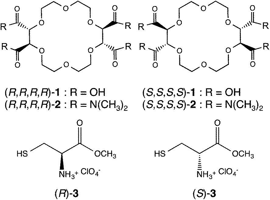 | ||
| Fig. 1 Chemical structures of optically active crown ethers and cysteine derivatives used in this study. | ||
Experimental
Materials
All chemicals for the preparation of optically active crown ether-modified tips, except for 2 (ref. 31) and 1-{[5-(1,2-dithiolan-3-yl)pentanoyl]oxy}-2,5-pyrrolidinedione,32 were of commercially available purity, and used without further purification. Also, 3 was obtained from cysteine methyl ester chloride salt by anion exchange. Organic solvents for the synthesis were purified by conventional methods, such as distillation. Deionized water (resistivity: 18 MΩ cm) was prepared using a Milli-Q system. Ethanol for AFM measurements was obtained from Sigma-Aldrich (Japan) and used without further purification.Chemical modification
The tips and the gold-coated substrates used for AFM force measurements were chemically modified with thiol and disulfide derivatives.33 The V-shaped AFM cantilevers were commercially available: Si3N4 type coated with Au/Cr on both sides (k = 0.02 N m−1, OMCL-TR400PB-1, Olympus, Japan). The cantilevers were pretreated by immersion into a piranha solution (concentrated H2SO4/28% H2O2, 7/3, v/v) at room temperature for 30 min to clean their surface (CAUTION: Piranha solution is extremely dangerous and should be handled with great care). The cantilevers were then thoroughly rinsed with deionized water.The AFM tips chemically modified with optically active crown ethers were prepared according to Fig. 2. The cleaned tips were treated by immersion into an ethanol solution containing 1.0 mM 1-{[5-(1,2-dithiolan-3-yl)pentanoyl]oxy}-2,5-pyrrolidinedione at room temperature for 24 h. After being rinsed with ethanol, the tips were fully dried in the atmosphere. The tips were treated by immersion into a DMF solution containing 0.20 mM α-aminopropyl-ω-aminopropoxy-polyoxyethylene (Mw = 3500, Mw/Mn = 1.4) and 5.0 mM N,N-dimethylaminopyridine (DMAP) at room temperature for 2 h. After being rinsed with DMF, the tips were fully dried in the atmosphere. Next, the tips were treated by immersion into a 1.0 wt% NaHCO3 aqueous solution containing 1.0 mM 1 and 10 mM 1-ethyl-3-(3-dimethylaminopropyl)carbodiimide hydrochloride (WSC) at room temperature for 24 h. After being rinsed with ion-exchange water, the tips were fully dried in the atmosphere. Furthermore, the tips were immersed into acetic anhydride at room temperature for 2 h for the remaining amino groups to be nonpolarized. After being rinsed with ethanol, the tips were fully dried in the atmosphere. Then, the tips were immersed into HCl aqueous solution (pH 3) at room temperature for 2 h. After being rinsed with deionized water, the tips were fully dried in the atmosphere. Finally, the tips were immersed into a diethylether solution containing 2.0 M trimethylsilyldiazomethane at room temperature for 30 min. After being rinsed with ethanol, the crown ether-modified tips were fully dried in the atmosphere. The crown ether-unmodified tips for the control experiment were also prepared as shown in Fig. S1.†
 | ||
| Fig. 2 Preparation of AFM tips chemically modified with optically active crown ethers. Reagents and conditions: (a) 1-{[5-(1,2-dithiolan-3-yl)pentanoyl]oxy}-2,5-pyrrolidinedione, ethanol, rt, 24 h; (b) α-aminopropyl-ω-aminopropoxy-polyoxyethylene, DMAP, DMF, rt, 2 h; (c) 1, WSC, NaHCO3 aq., rt, 24 h; (d) acetic anhydride, rt, 2 h; (e) HCl aq., rt, 2 h; (f) trimethylsilyldiazomethane, diethylether, rt, 30 min. | ||
Gold-coated mica substrates were prepared by sputtering of gold (99.999%, Nilaco, Japan) using a JFC-1600 Auto Fine Coater (JEOL, Japan) onto a mica substrate prepared by fresh cleavage of a sheet of natural mica (Nilaco, Japan). The gold-coated substrate was immersed into a 2.5 mM 3 methanol solution at room temperature for 24 h. After being rinsed with ethanol, they were fully dried in the atmosphere before use.
AFM force measurements
AFM force measurements were carried out in ethanol solution at room temperature (ca. 298 K) using a Nanoscope 3D MultiMode AFM with PicoForce (Veeco, USA). The probe tip and the substrate were mounted on the apparatus using a liquid cell. The spring constants (typically 0.02 N m−1) of the cantilever were always calibrated using the thermal tune method before each measurement.All force–distance curves were obtained in a contact mode using the AFM software of the manufacturers at a scan rate of 310 nm s−1. The surface delay, defined as the resting time after the tip touches the substrate surface, was set at 1.0 s. The scan rate and surface delay were determined to maximize the observable probability for the stretching of a single PEG chain. Typical measurements were made 400–1200 times at different positions of the substrate for each tip/substrate combination. The experimentally obtained force–distance curves were converted to the force–extension curves.17 Then, a theoretical model for polymer extension, the worm-like chain (WLC) model, was used to perform the curve fitting of the force–extension curves in order to obtain the characteristic fitting parameters. The average observable probability for the stretching of a single PEG chain was about 0.84% in total. The representative force–extension curve except for the stretching of a single chain is shown in Fig. S2.†
NMR titrations
1H-NMR spectra were collected on a JNM-ECA 400 FT NMR (JEOL, Japan). As an example, the titration experiment for the complexation of the crown ether (R,R,R,R)-2 with the cysteine derivative (R)-3 is described. A 3.0 mM solution of (R,R,R,R)-2 in 600 μL of CD3CN was prepared, and an initial NMR measurement of this solution was recorded. A 91 mM solution of (R)-3 in 400 μL of CD3CN was separately prepared. Samples were then made by adding various amounts of the guest solution to the host solution. The spectra of the eight different resulting solutions were recorded. The stability constants were calculated from the chemical shift changes using an iterative nonlinear least squares curve-fitting program.34Results and discussion
SMFS measurements
Fig. 3 outlines the strategy of our AFM-based SMFS study, wherein 1 was covalently anchored to an AFM tip through a polyethyleneglycol (PEG) spacer and 3 was immobilized on a gold-coated substrate based on the well-known thiol–Au interaction. After the chemical modification, the tips were immersed into acetic anhydride for the capping of the remaining amino groups by nonpolarized functional groups. Furthermore, the remaining carboxylic groups on the crown ether moiety were esterified by trimethylsilyldiazomethane diethylether to remove the effect of electrostatic interactions between –COO− and –NH3+. The tips and substrates were fully dried in the atmosphere before the measurements. | ||
| Fig. 3 Schematic diagram of our AFM-based SMFS study. | ||
Fig. 4 shows a typical force–extension curve observed using the tip and the substrate chemically modified with (R,R,R,R)-1 and (R)-3, respectively, in ethanol. The representative force–extension curves for the other combinations of tips and substrates are shown in Fig. S3.† The force–extension curves obtained for all the combinations of tips and substrates exhibited similar deformational characteristics: a nonspecific adhesion based on the contact between the tip and the substrate, a rising force with the increase of extension, followed by a force drop (rupture force) upon the rupture of a polymer bridge between the tip and the substrate. Here, the gentle slope observed at the rupture does not mean the slow dissociation of the host–guest complex. Interestingly, more than 100 pN of rupture forces were not observed in the presence of 100 mM KSCN (Fig. S4†). This result suggests that the added K+ in the solution interferes with the complexation of the 18-crown-6 moiety on the tip with the –NH3+ site on the substrate.35 Furthermore, such rupture forces were also not observed in the control experiment using the crown ether-unmodified AFM tip and the cysteine-modified substrate (Fig. S5†). Therefore, it is assumed that the rupture forces observed here mainly reflect the specific interaction between (R,R,R,R)-1 and (R)-3.
 | ||
| Fig. 4 Typical force–extension curve observed using (R,R,R,R)-1-modified tip and (R)-3-modified substrate in ethanol. | ||
As has been reported by some researchers,36 single polymer chain stretching can be described by several theoretical models. In order to confirm that the rupture force was observed at the single-molecule level, we analysed the force–extension curves for the stretching of the PEG linker based on a theoretical model, the WLC model.16 The WLC model describes a single polymer chain as a string of constant bending elasticity with a worm-like conformation. The following expression has been widely used to describe the force (F) as a function of chain extension (x)

![[thin space (1/6-em)]](https://www.rsc.org/images/entities/char_2009.gif) 000 force measurements. From the above-mentioned SMFS analysis, we can quantitatively discuss the chiral recognition ability of optically active crown ethers at the single-molecule level.
000 force measurements. From the above-mentioned SMFS analysis, we can quantitatively discuss the chiral recognition ability of optically active crown ethers at the single-molecule level.
Fig. 5a and b show the histograms of rupture forces observed using the tips chemically modified with (R,R,R,R)-1 and (S,S,S,S)-1, respectively. In Fig. 5a, larger rupture forces (260 ± 82 pN) were observed using the (S)-3-modified substrate than those (219 ± 83 pN) using the (R)-3-modified substrate. In Fig. 5b, larger rupture forces (250 ± 84 pN) were observed using the (R)-3-modified substrate than those (212 ± 86 pN) using the (S)-3-modified substrate. Summarizing the above results, we can say that the chiral recognition ability of the optically active crown ether-modified tip was entirely opposite to that of its optical isomer-modified tip. This result demonstrates that chiral recognition on the solid surface was successfully observed by our SMFS study. The difference of rupture forces between the (R)-3- and (S)-3-modified substrates was determined to be about 40 pN under our experimental conditions. Also, the statistical test (Welch’s t-test) revealed that the p-value was less than 0.05 between two histograms in both cases using the same tip. Accordingly, it was shown that the differences of rupture forces observed between the substrates chemically modified with (R)- and (S)-cysteine derivatives were statistically significant. The difference may be due to the chiral recognition of optically active crown ethers because both measurements were carried out under identical experimental conditions except for the absolute configuration of cysteine derivatives.
 | ||
| Fig. 5 Histograms of rupture forces observed using tips chemically modified with (a) (R,R,R,R)-1 and (b) (S,S,S,S)-1. | ||
NMR measurements
Next, the chiral recognition in solution was investigated by NMR measurements. Firstly, the stoichiometry of the 2–3 complex was determined to be 1:1 by a Job plot. Table 1 shows the stability constants, which were determined by the 1H NMR titration method, between chiral crown ethers 2 and chiral ammonium salts 3 in CD3CN. In the NMR study, the selectivity for chiral recognition of the optically active crown ether was opposite to that of its optical isomer. This result demonstrates that chiral crown ethers 2 have chiral recognition ability towards chiral ammonium salts 3. However, the selectivity for chiral recognition in solution was opposite to that on the solid surface. The previous study on the chiral recognition between (R,R,R,R)-1 and amino acids also reported that the selectivity for chiral recognition in solution was different from that on the solid surface in some cases, and that the hydrogen bonding between the side chain on the crown ether ring and amino acids plays an important role in the determination of the chiral selectivity.40 In this study, the structures of the side chain on the crown ether ring were different between the AFM and NMR studies. This fact may affect the selectivity of the chiral recognition. In addition, as the distance between the ammonium moiety and the substrate was very short in the AFM study, the substrate might affect the selectivity. Therefore, the introduction of a spacer between the guest compound and the substrate would be required in forthcoming studies.
| logK (M−1) |
||
|---|---|---|
| (R)-3 | (S)-3 | |
| (R,R,R,R)-2 | 3.58 | 3.34 |
| (S,S,S,S)-2 | 3.25 | 3.50 |
Conclusions
In this paper, we firstly reported AFM-based SMFS for the chiral recognition of cysteine derivatives on a solid surface using AFM tips chemically modified with optically active crown ethers. Remarkably, the chiral recognition ability of the optically active crown ether-modified tip was entirely opposite to that of its optical isomer-modified tip. On the other hand, the chiral selectivity on the solid surface obtained by the AFM study was different from that in solution as evaluated by the NMR study. The reason for this interesting phenomenon is not fully understood at the present stage. Further observation and analysis of chiral recognition using AFM-based SMFS is underway in our laboratory, aiming at understanding chiral surfaces in more detail.Notes and references
- K. Bodenhöfer, A. Hierlemann, J. Seemann, G. Gauglitz, B. Koppenhoefer and W. Gpel, Nature, 1997, 387, 577–580 CrossRef PubMed.
- W. H. Pirkle and D. S. Reno, J. Am. Chem. Soc., 1987, 109, 7189–7190 CrossRef CAS.
- R. Vespalec and P. Bocek, Chem. Rev., 2000, 100, 3715–3754 CrossRef CAS PubMed.
- Y. Izumi, Adv. Catal., 1983, 32, 215–271 CAS.
- A. Baiker, J. Mol. Catal. A: Chem., 1997, 115, 473–493 CrossRef CAS.
- C. E. Song and S. Lee, Chem. Rev., 2002, 102, 3495–3524 CrossRef CAS PubMed.
- R. Raval, Nature, 2003, 425, 463–464 CrossRef CAS PubMed.
- T. Nishino and Y. Umezawa, Anal. Chem., 2008, 80, 6968–6973 CrossRef CAS PubMed.
- R. Raval, Chem. Soc. Rev., 2009, 38, 707–721 RSC.
- J. A. A. W. Elemans, I. D. Cat, H. Xu and S. D. Feyter, Chem. Soc. Rev., 2009, 38, 722–736 RSC.
- R. McKendry, M.-E. Theoclitou, T. Rayment and C. Abell, Nature, 1998, 391, 566–568 CrossRef CAS PubMed.
- C. Xu, S. C. Ng and H. S. O. Chan, Langmuir, 2008, 24, 9118–9124 CrossRef CAS PubMed.
- J. M. Bonello, F. J. Williams and R. M. Lambert, J. Am. Chem. Soc., 2003, 125, 2723–2729 CrossRef CAS PubMed.
- D. Y. Murzin, P. Maki-Arvela, E. Toukoniitty and T. Salmi, Catal. Rev. Sci. Eng., 2005, 47, 175–256 CAS.
- H. Schönherr, M. W. J. Beulen, J. Bügler, J. Huskens, F. C. J. M. v. Veggel, D. N. Reinhoudt and G. J. Vancso, J. Am. Chem. Soc., 2000, 122, 4963–4967 CrossRef.
- A. Janshoff, M. Neitzert, Y. Oberdörfer and H. Fuchs, Angew. Chem., Int. Ed., 2000, 39, 3212–3237 CrossRef CAS.
- T. Hugel and M. Seitz, Macromol. Rapid Commun., 2001, 22, 989–1016 CrossRef CAS.
- R. Eckel, R. Ros, B. Decker, J. Mattay and D. Anselmetti, Angew. Chem., Int. Ed., 2005, 44, 484–488 CrossRef CAS PubMed.
- K. C. Neuman and A. Nagy, Nat. Methods, 2008, 5, 491–505 CrossRef CAS PubMed.
- Y. Zhang, Y. Yu, Z. Jiang, H. Xu, Z. Wang and X. Zhang, Langmuir, 2009, 25, 6627–6632 CrossRef CAS PubMed.
- V. Walhorn, C. Schäfer, T. Schröder, J. Mattay and D. Anselmetti, Nanoscale, 2011, 3, 4859–4865 RSC.
- R. C. Helgeson, K. Koga, J. M. Timko and D. J. Cram, J. Am. Chem. Soc., 1973, 95, 3021–3023 CrossRef CAS.
- R. C. Helgeson, J. M. Timko and D. J. Cram, J. Am. Chem. Soc., 1973, 95, 3023–3025 CrossRef CAS.
- X. X. Zhang, J. S. Bradshaw and R. M. Izatt, Chem. Rev., 1997, 97, 3313–3362 CrossRef CAS PubMed.
- L. Pu, Chem. Rev., 2004, 104, 1687–1716 CrossRef CAS PubMed.
- J.-M. Girodeau, J.-M. Lehn and J.-P. Sauvage, Angew. Chem., Int. Ed. Engl., 1975, 14, 764 CrossRef.
- M. H. Hyun, J. Sep. Sci., 2006, 29, 750–761 CrossRef CAS.
- H. Nagata, H. Nishi, M. Kamigauchi and T. Ishida, Chirality, 2008, 20, 820–827 CrossRef CAS PubMed.
- S. Shen, S. Ma, H. Lee, C. Manolescu, M. Grinberg, N. Yee, C. Senanayake and N. Grinberg, J. Liq. Chromatogr. Relat. Technol., 2009, 33, 153–166 CrossRef.
- J. R. Aviles-Moreno, M. M. Quesada-Moreno, J. J. Lopez-Gonzalez and B. Martinez-Haya, J. Phys. Chem. B, 2013, 117, 9362–9370 CrossRef CAS PubMed.
- A. Anantanarayan, V. A. Carmicheal, P. J. Dutton, T. M. Fyles and M. J. Pitre, Synth. Commun., 1986, 16, 1771–1776 CrossRef CAS.
- R. J. Stokes, A. Macaskill, J. A. Dougan, P. G. Hargreaves, H. M. Stanford, W. E. Smith, K. Faulds and D. Graham, Chem. Commun., 2007, 2811–2813 RSC.
- R. Barattin and N. Voyer, Chem. Commun., 2008, 1513–1532 RSC.
- K. Hirose, J. Inclusion Phenom. Macrocyclic Chem., 2001, 39, 193–209 CrossRef CAS.
- S. Kado and K. Kimura, J. Am. Chem. Soc., 2003, 125, 4560–4564 CrossRef CAS PubMed.
- M. I. Giannotti and G. J. Vancso, ChemPhysChem, 2007, 8, 2290–2307 CrossRef CAS PubMed.
- K. Liu, Y. Song, W. Feng, N. Liu, W. Zhang and X. Zhang, J. Am. Chem. Soc., 2011, 133, 3226–3229 CrossRef CAS PubMed.
- S. Kado, K. Uehara, Y. Nakahara and K. Kimura, Bull. Chem. Soc. Jpn., 2011, 84, 422–426 CrossRef CAS.
- R. Funayama, Y. Nakahara, S. Kado, M. Tanaka and K. Kimura, Analyst, 2014, 139, 4037–4043 RSC.
- H. Nagata, Bull. Osaka Univ. Pharm. Sci., 2010, 4, 105–115 Search PubMed.
Footnote |
| † Electronic supplementary information (ESI) available: Preparation of crown ether-unmodified tip, representative force–extension curve except for the stretching of a single chain, representative force–extension curves for the other combinations of tips and substrates, force–extension curve using (R,R,R,R)-1-modified tip and (S)-3-modified substrate in the presence of KSCN, force–extension curve using crown ether-unmodified tip and (S)-3-modified substrate, and typical force–extension curve badly fitting with WLC model plot. See DOI: 10.1039/c4ra10553b |
| This journal is © The Royal Society of Chemistry 2014 |