Ultrastructure of metallopeptide-based soft spherical morphologies

Gagandeep Kaura, Lihi A. Abramovichb, Ehud Gazit*b and Sandeep Verma*a
aDepartment of Chemistry, DST Thematic Unit of Excellence on Soft Nanofabrication, Center for Environmental Sciences and Engineering, Indian Institute of Technology Kanpur, Kanpur-208016, India
bDepartment of Molecular Biology and Biotechnology, Department of Materials Science and Engineering, Tel Aviv University, Tel Aviv-69978, Israel. E-mail: sverma@iitk.ac.in; ehudg@post.tau.ac.il

Received 16th September 2014 , Accepted 18th November 2014

First published on 18th November 2014


Abstract

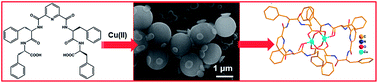
Peptides and proteins offer interesting starting points for triggering self-assembly processes owing to the chemical diversity of side-chains, ease of chemical modifications and the possibility of exploiting several non-covalent and metal-assisted interactions, to stabilize higher order ensembles. Consequently, a variety of nanoscale morphologies such as fibers, vesicles, nanotubes are observed for modified amino acids and short peptides and these biocompatible soft materials have been used for diverse biological, medical and material applications. Herein, we report metal-mediated modification of spherical soft assemblies, by introducing a coordinating linker for the Phe–Phe dipeptide, which results in the coalescence of soft structures. The possibility of copper ion coordination, with the metal-binding peptide conjugate, was confirmed by single crystal analysis. Based on these observations, a model depicting possible interactions leading to soft structure formation and metal-aided coalescence is also presented. The coalescence could be reversed in the case of Au-mediated soft structures with the help of thiol interference. Such an approach, exploiting interfacial metal ion interactions, is expected to provide an entry into novel metallopeptide materials.


Introduction

Self-assembling peptides offer a versatile platform towards formation of ordered nanoscale systems, with various applications in catalysis, delivery, and tissue engineering, to name a few.1–8 Control through different physicochemical properties of constituent amino acids, possibility of predictable design based on secondary structural signatures, and high compatibility interaction with other biological systems, are the hallmarks of peptide-based nanoscale materials. Consequently, it is quite advantageous to employ short self-assembling peptides, to obtain diverse hierarchical structures with desired functions. Recent advances along these lines concern interaction of metal ions with self-assembling peptides to afford bio-inspired metal–peptide frameworks (MPF),9,10 which may offer combined investigated properties of metal–organic frameworks (MOF) with biocompatibility.

Three different approaches could be envisaged for constructing MPFs: (i) introducing amino acids side chains capable of metal binding11 (ii) use of exogenous ligands supporting MOF architecture12 (iii) use of covalently linked chiral/achiral connectors. These approaches represent facile route for the design of novel, abiotic oligomers for supramolecular studies. As a specific example, Lehn, Huc and other groups have reported family of oligoamides based on achiral connectivity of 2,6-pyridinedicarboxylic acid, where the resultant oligomers not only self-organized into single helices, but also support formation of double helices under specific conditions.13,14 Notably, short oligomers with 2,6-pyridinedicarboxylic acid and related connectors afford planar ‘crescent-like’ conformation, while longer oligomers exhibit more pronounced propensity to form helical structures.15 In particular, pyridine diacid oligomers offer hydrogen bonding at the inner rim of helices resulting in ∼4.5 monomer per turn of the ensuing helix.

Diphenylalanine (FF) dipeptide motif exhibits spontaneous self-assembly to generate peptide nanotubes in solution.16 FF nanotubes offer interesting applications as scaffolds for the synthesis of metal nanowires, possess improved mechanical properties, semi-conductivity and serve as potential drug delivery agents.17–23 Side chain aromaticity in phenylalanine is implicated for self-assembly properties and key structural features in the solid state.24 In current study, given our continued interest in hierarchical self-assembled peptides systems and our ongoing efforts to prepare covalently linked conjugates of FF dipeptide,18,25,26 we decided to synthesize a symmetric FF conjugate using pyridinedicarboxylic acid as an achiral linker. The ability of this new peptide conjugate to self-assemble and its binding to various metals including copper, silver and gold leading to formation of metallopeptide soft structures is described.

Results and discussions

Molecular design of peptide conjugate, 4

We designed and synthesized 4, which is a hybrid of two diphenylalanine moieties conjugated using 2,6-pyridinedicarboxylic acid as a linker (Scheme 1). We envisioned the following advantages for the proposed conjugate: [i] possibility to engender a structure containing the ability of ‘crescent-like’ conformation in an aggregating dipeptide (Fig. 1a); [ii] introduction of metal ion co-ordination sites to further interrogate peptide self-assembly process.
image file: c4ra10532j-s1.tif
Scheme 1 Synthetic scheme of peptide conjugate 4: (i) 75% TFA-DCM, 4 h, N2 atm. (ii) HOBt, DCC, 0 °C, TEA, N2 atm. (iii) THF, 1 N NaOH, 6 h, N2 atm.

image file: c4ra10532j-f1.tif
Fig. 1 (a) Molecular structure of 4. (b) Predicted curvature of 4.

2,6-Pyridyl disubstituted linkers have been utilized to engender a crescent conformation or a curvature in many foldameric systems.15,27 In addition, pyridyl ring also offers additional advantage of metal ion interaction, which may further lead to useful coordination architectures and varied geometries. It can thus be surmised that such curved conformation (Fig. 1), upon solution phase self-assembly, may eventually culminate into spherical structures.

Crystal structure of 4 with Cu ions

Presence of pyridyl group as a linker gave us an impetus to focus our attention to metal ion coordination. As envisaged, we surmised that such interaction may offer an entry metal-mediated peptide soft matter, perhaps with tunable size and cavities. To start, we decided to assess metal ion interaction by growing appropriate crystal to ascertain the possible site of metal binding. 4 was interacted with Cu(II) ions and studied through single-crystal diffraction analysis (Fig. 2a and Table S1). Methanolic solution of 4 was treated with CuSO4·5H2O and diffraction quality crystals were grown by slow evaporation method. Crystal analysis revealed M2L2 coordination, via carboxylate groups, to afford a Cu(II)-supported paddle-wheel like structure.28–31 The dimeric units were further stabilized by hydrogen bonding between –N–H and –C[double bond, length as m-dash]O of adjacent dimeric units in the range of 2.11–2.12 Å (Fig. S1a) as well as through C–H⋯π interactions (2.47 Å)32–35 (Fig. S1b), to form a 3D lattice structure (Fig. 2b; Table S2). Notably, Cu(II) ions in this case did not interact with pyridyl nitrogen.
image file: c4ra10532j-f2.tif
Fig. 2 Crystal structure of Cu2L2 metallopeptide (L = 4): (a) dinuclear paddle wheel-type of structure (b) lattice structure showing peripheral interactions.

Formation of M2L2-type of discreet dinuclear copper cluster evinces interest not only due to interesting coordination geometry, but also due to the fact that coordination-driven self-assembled structures offer facile entry to the construction of novel metallosupramolecular ensembles such as two-dimensional polygons and three-dimensional spherical cages, prisms, and polyhedral.36–39 It is envisaged that these designed superstructures, which combine multiple weak interactions including π–π stacking, CH–π interactions, hydrogen bonds and metal-coordination, could extend application in domains ranging from optoelectronic materials to drug delivery to storage and catalysis. In case of 4, it could be imagined from crystal structure that metal–carboxylate interactions offer a beneficial cooperative effect, along with other stabilizing non-covalent interactions, to form interesting superstructures. Thus, we started with the determination of solution-phase morphology of 4, followed by investigating the effect of metal ions, in the solution phase, to generate metallopeptide ensembles.

Solution phase self-assembly of 4

The propensity of peptide conjugate 4 to self-assemble was studied. It was dissolved in 50% methanol–water at a concentration of 1 mM and unlike nanotube forming FF dipeptide,16 4 leads to instantaneous formation of spherical structures with a diameter of 350–650 nm, as confirmed by atomic force microscopy (AFM) (Fig. 3a and b) and dynamic light scattering (DLS) analysis (Fig. 3c). Spherical nature of these assemblies could be attributed to the crescent-shaped structural design of this conjugate. These spherical morphologies were further analysed using scanning electron microscopy (SEM) and transmission electron microscopy (TEM) (Fig. 3d and e).
image file: c4ra10532j-f3.tif
Fig. 3 Microscopy analysis of 4 (a and b) AFM micrographs. (c) DLS spectrum showing the spherical structure size distribution. (d) SEM image. (e) TEM image. (f) FIB-SEM image of the sphere after ion beam damage revealing the inner surface (1 mM, 50% aqueous methanol) on silicon wafer.

Stability of these soft structures was probed by subjecting them to high-energy ion beam manipulation (Fig. 3f). Consequently, a focused ion beam (FIB) system, using a gallium ion beam, was employed to site-selectively machine a spherical particle, at an operating voltage of 15.00 kV. This experiment revealed a solid inner core in the soft structure, suggesting formation of highly compact structures that are able to withstand ion beams without any measurable loss of morphological integrity.40

Stabilization of these structures could be attributed to known aromatic amino acid side-chain interactions during peptide aggregation process to form parallel-stacked or T-shaped geometries. Such instances lead to formation of hydrophobic microenvironments by excluding water. Moreover, it is also known that aromatic–aromatic interactions further help short peptides to efficiently access energetically favored conformations.41–44 It should be emphasized here that mixed solvent used in this case, due to solubility constraints, may also exert some effect in stabilizing FF dipeptide side-chain interactions. Notably, stacked arrangements are more stable in water, while amphiphilicity of methanol interferes with both stacked and T-shaped geometries, by attenuating aromatic–aromatic interactions.45

Interaction of 4 with copper ions

Cu(II) ions form stable four-, five- and six-coordinate complexes. These complexes show their potential use as anti-inflammatory,46 antimicrobial,47 antitumor agents,48–51 enzyme inhibitors, and chemical nucleases, generally bind through nitrogen of the ligands. Conjugate 4 offers pyridyl ring nitrogen as well as free carboxylate groups as probable metal binding sites. Samples were prepared by co-incubating 4 (1 mM, 0.5 mL, MeOH) with CuCl2·2H2O solution (1 mM, 0.5 mL, H2O), followed by microscopy evaluation. Minor morphological changes appeared within 2 h of mixing, which became prominent during prolonged incubation as confirmed by SEM and TEM microscopy (Fig. 4). These images clearly showed vesicle coalescence in presence of metal ions, leading to the formation of larger-sized vesicles in 24 h (Fig. 4b). The monitoring was further continued for 120 h through DLS measurements. The size of spherical structures increased up to ∼3.4 μm in 120 h upon copper interaction perhaps due to fusion of these vesicles. Notably, the presence of copper ions in vesicles was confirmed by energy-dispersive X-ray spectroscopy (EDX) measurements (Fig. 4d).
image file: c4ra10532j-f4.tif
Fig. 4 Microscopic images of 4 + Cu(II) complex. (a and b) SEM images on silicon wafer after 12 h and 24 h respectively. (c) TEM image (inset: magnified view of a metal-bound spherical structure). (d) EDX spectrum after 24 h.

One of the hallmarks of Cu(II) interaction was the coalescence of self-assembled structures. As Phe–Phe interactions are primarily guided by stacking interactions, phenyl rings prefer to be buried inside the spherical structures. Thus, it is likely that free carboxylic groups and pyridyl ring nitrogens in 4 would possibly be exposed at the surface of spherical structures for metal ion binding.52

Interaction of 4 with silver ions

Ag(I) ions generally interact through nitrogen centers in the ligands53 and many such complexes are known to exhibit biological activities.54,55 They are also known to trigger conformational changes in self-assembled peptides.56 As pyridyl nitrogen of 4 could be an interesting metal binding site for Ag(I), samples were prepared in presence of AgNO3 as described before. We were able to observe changes in morphology 12 h after the addition of metal ions (Fig. 5a). Interestingly, coalescence of spherical vesicles was observed again, where soft structures of 4 exhibited formation of fused structures, as evidenced by SEM and TEM micrographs (Fig. 5b and c). As this morphology change was slightly different compared to copper-mediated morphology, it is possible that Ag(I) coordination in this case is distinct to Cu(II) interactions.57 Unfortunately, our efforts to grow single crystals with Ag ions were not successful.
image file: c4ra10532j-f5.tif
Fig. 5 Microscopic images of 4 + Ag(I) complex. (a and b) SEM images on silicon wafer after 12 h and 24 h respectively. (c) TEM image (inset: magnified view of a Ag(I)-mediated soft spherical structures). (d) EDX spectrum after 24 h.

Interaction of 4 with Au(III) ions

Chloroauric acid (HAuCl4) protonates pyridyl ring with counter anion AuCl4 ion balancing overall charge.58–61 Latter possesses square-planar geometry, relatively diffuse charge distribution and a weak interaction with solvent (water) molecules, with nearest solvent molecules occupying two orthogonal quasi-elliptical surfaces.62 Dissolving 4 along with HAuCl4 also revealed morphological changes, where spherical structures were found to afford a network-like morphology within 12 h incubation (Fig. 6). Given the anionic nature of AuCl4, it is likely that carboxylate anions will not have any role in coordination. Thus, observed coalescence could be attributed to pyridyl nitrogen coordination of Au(III). Indeed, several groups have reported formation of Au(III) coordination complexes using pyridyl and phenanthronyl ligands.63 Vesicles linked in a network-like fashion were confirmed by various microscopy methods and EDX analysis data.
image file: c4ra10532j-f6.tif
Fig. 6 Microscopic images of 4 + Au(III) complex. (a and b) SEM images on Si wafer after 12 h and 24 h respectively. (c) TEM image, (inset: magnified view of a spherical structure). (d) EDX spectrum after 24 h.

Thus, different possibilities could be envisaged for metal–peptide interactions: in case of Cu(II) ions, free carboxyl group side-chains in 4 could interact as suggested by the solid state structure; but, this does not preclude solution interaction of pyridyl nitrogens with copper ions. In case of Ag(I) and Au(III) ions, interaction could possibly be supported by pyridyl nitrogen. Despite repeated attempts, we were unable to grow crystals with Ag(I) and Au(III) ions to arrive at precise sites of Ag or Au interaction with 4. Finally, role of surface in these experiments was also probed by doing this procedure on copper grids (Fig. S2). Similar results were obtained in all three cases irrespective of the surface used.

Thiol displacement studies

Au(III) ion binds very effectively with 4 to form network-like metal–peptide framework. As Au(III) has thiophilic binding preference over nitrogen or oxygen centers,64,65 we decided to treat 4 + Au(III) complex with 1-mercaptopropanoic acid (MPA) to achieve its displacement from the framework. Samples were prepared by mixing 4 (0.5 mL, 1 mM, MeOH) with HAuCl4 solution (0.5 mL, 1 mM, H2O), incubated for 12 h. This was followed by addition of MPA solution (0.5 mL, 10 mM, 50% methanol–water) and further incubation of 5 h (Fig. 7). Notably, network-like structure disappeared confirming crucial role of Au(III) ions in causing metalized vesicles to stick together.
image file: c4ra10532j-f7.tif
Fig. 7 (a) SEM image of 4. (b) SEM image of 4 + Au(III) complex after 12 h. (C) Magnified view of 4 + Au(III) complex. (d) SEM image of 4 + Au(III) + MPA after 5 h incubation.

Dynamic light scattering analysis

Effect of metal ions on vesicle coalescence was confirmed by DLS analysis using similar metal–peptide solutions used for microscopy studies.66 The samples were prepared by mixing 4 (1 mL, 10 μm, MeOH) with metal ion solutions (1 mL, 10 μm, H2O). It was observed that the peptide vesicles alone have a hydrodynamic diameter of ∼350–650 nm (Fig. 8a), which upon Cu(II) ions addition changes to ∼1.2 μm (Fig. 8b) after 24 h incubation and it further matured to an average diameter of ∼3.4 μm after 120 h (Fig. 8c). For Ag(I) ions, average size of metalated vesicles changed to ∼1 μm after 24 h (Fig. 8d) and matured to an average diameter of 2.2 μm after 120 h (Fig. 8e). Similar trend was also observed for Au(III) interactions (Fig. 8f and g). Such a change in size may be ascribed to metal-mediated vesicle fusion process.
image file: c4ra10532j-f8.tif
Fig. 8 DLS spectra: (a) size distribution of 4 (50% MeOH–H2O). (b, d and f) Size distributions after 24 h. (c, e and f) Size distributions after 120 h.

Peptide conjugate 4 afforded formation of spherical structures supported by H-bonding and π–π stacking interactions. Moreover, possible crescent-shape of monomers, engendered by pyridyl linker, further assisted self-organization of 4 to maximize non-covalent interactions.

Based on the occurrence of these forces, a model could be proposed to depict formation of spherical vesicles from crescent-shaped conjugate (Fig. 9). Observation of coalescence upon metal ion addition was attributed to metal-coordination, which could possibly bring these soft peptide-based structures in close proximity. As seen in Au experiment, exogenous addition of MPA could revert coalescence perhaps by complexing metal ions thus releasing spherical structures.


image file: c4ra10532j-f9.tif
Fig. 9 Proposed model of metal–peptide interactions.

Conclusions

Conjugate 4 was designed having a crescent-type of structure, synthesized and studied for its interaction with metal ions in the solid state and in solution. Crystal analysis of copper complex of 4 revealed M2L2 coordination, via carboxylate groups, to afford a Cu(II)-supported paddle-wheel like structure. Lattice structure offered insight into further interactions of these dinuclear units via noncovalent interactions. Solution phase self-assembly peptide conjugate revealed formation of spherical structures, which possessed metal ion binding properties leading to coalescence, as confirmed by microscopy. Cu(II), Ag(I) and Au(III) to form large sized vesicles: while Cu(II) ions bound free acid side chains of 4 as confirmed by solid state structure, Ag(I) and Au(III) ions could also invoke pyridyl nitrogen as additional binding sites. Reversal of coalescence was achieved in the case of Au(III) ions supported metal–peptide structures, when treated with 1-mercaptopropanoic acid. Thus, it can be stated that metal coordination plays an important role in connecting these spherical structures and exploit an interesting interfacial interaction to provide expeditious entry into novel metallopeptide materials.

Materials and methods

General procedures

N,N′-Dicyclohexylcarbodiimide (DCC), N-hydroxybenzotriazole (HOBt), di-tert-butyl dicarbonate (Boc), trifluoroacetic acid (TFA), and L-phenylalanine were purchased from SRL, Mumbai, India and used without further purification. Dichloromethane (DCM), N,N-dimethylformamide (DMF), methanol (MeOH) and triethylamine (TEA) were distilled according to standard procedures prior to use. 1H and 13C NMR spectra were recorded on JEOL-DELTA2 500 model operating at 500 and 125 MHz, respectively. HRMS (ESI+ and ESI) was recorded at IIT Kanpur, India, on WATERS, Q-Tof Premier Micromass HAB 213 mass spectrometer using capillary voltage 2.6–3.2 kV. IR spectra were recorded as KBr pellets on a Perkin-Elmer Model 1320 spectrophotometer operating from 400 to 4000 cm−1. For thin layer chromatography (TLC), Merck pre-coated TLC plates (MEARK 60 F254) were used, and compounds were visualized with a UV light at 254 nm. Chromatographic separations were performed on S. D. Fine-Chem 100–200 mesh silica gel.

Synthesis of peptide conjugate

4 was prepared by solution phase peptide synthesis.

N-(tert-Butyloxycarbonyl)-L-phenylalanine-L-phenylalanine methyl ester (1)

N-(Boc)-L-phenylalanine (5 g, 18.8 mmol), and HOBt (2.55 g, 18.8 mmol) were dissolved in dry DMF (25 mL) under nitrogen atmosphere and the reaction mixture was cooled to 0 °C in an ice bath. A solution of DCC in DCM (4.66 g, 22.6 mmol) was then added drop wise to the reaction mixture. The reaction mixture was stirred at 0 °C for 1 h, after which, L-phenylalanine methyl ester hydrochloride (4.05 g, 22.6 mmol) was added to it followed by triethylamine (13.1 mL, 94.2 mmol). The reaction mixture was monitored and stirred for 24 h at room temperature under nitrogen atmosphere. Reaction mixture was concentrated in vacuo, redissolved in ethyl acetate and filtered to remove DCU. The organic layer was then washed with 1 N HCl (3 × 30 mL), 10% NaHCO3 (3 × 30 mL) and brine (30 mL). The organic layer was dried over anhydrous sodium sulfate and concentrated in vacuo. The crude compound was purified through silica gel column chromatography by using hexane and ethyl acetate (80[thin space (1/6-em)]:[thin space (1/6-em)]20) solvent system to isolate pure 1 (6.17 g, 76.76% yield). m.p. 102–105 °C, Rf [30% ethyl acetate in hexane] = 0.5, 1H NMR (500 MHz, CDCl3, TMS, δ ppm): 1.38 (s, 9H); 2.99–3.08 (m, 4H); 3.65 (s, 3H); 4.32 (m, 1H); 4.77–4.93 (m, 1H); 6.96–6.97 (m, 2H); 7.17–7.29 (m, 10H, overlapped aromatic signal and CDCl3 peak); 13C NMR (125 MHz; CDCl3, δ ppm): 28.3, 38.0, 38.3, 52.3, 53.3, 55.7, 77.3, 80.4, 127.2, 128.6, 128.7, 129.3, 129.4, 170.8, 171.4; HRMS (M + H)+ for C24H30N2O5: 427.2233 (calcd), 427.2236 (anal.).

L-Phenylalanine-L-phenylalanine methyl ester (2)

1 (5 g, 9.5 mmol) was dissolved in 75% TFA-DCM and stirred for 1 h under nitrogen atmosphere. After completion of the reaction, the solvent was evaporated in vacuo and was subsequently washed with diethylether resulting in a white solid. The white solid then dissolved in methanol and passed through activated anion exchange resin and evaporated under reduced pressure to obtain pure 2 (3.27 g, 85.4% yield). Rf [50% ethyl acetate in hexane] = 0.5; 1H NMR (500 MHz, DMSO-d6, TMS, δ ppm): 2.89–3.09 (m, 4H), 3.57 (s, 3H); 4.02 (m, 1H); 4.51–4.55 (m, 1H); 7.18–7.30 (m, 10H), 8.16 (s, 3H); 13C NMR (125 MHz; DMSO-d6, δ ppm): 39.8, 40.0, 40.1, 40.3, 52.2, 54.1, 54.3, 128.4, 128.7, 129.5, 162.8, 172.1; HRMS (M + H)+ for C19H22N2O3: 327.1709 (calcd), 327.1700 (anal).

Pyridyl-bis-L-phenylalanine-L-phenylalanine methyl ester (3)

2,6-Pyridinedicarboxylic acid (0.25 g, 1.49 mmol) was dissolved in DMF and cooled to 0 °C in ice bath under nitrogen atmosphere. To this solution, HOBt (0.404 mg, 2.99 mmol) and a solution of DCC (0.617 g, 2.99 mmol) in DCM was added and stirred for 1 h under nitrogen atmosphere at 0 °C. Subsequently 2 (1.07 g, 3.29 mmol) was added into the reaction mixture followed by triethylamine (0.76 mL, 9.48 mmol). The reaction mixture was monitored and stirred for 24 h at room temperature under nitrogen atmosphere. Reaction mixture was concentrated in vacuo, redissolved in ethyl acetate and filtered to remove DCU. The organic layer was then washed with 1 N HCl (3 × 30 mL), 10% NaHCO3 (3 × 30 mL) and brine (30 mL). The organic layer was dried over anhydrous sodium sulfate and concentrated in vacuo. Crude compound was then purified through silica gel column chromatography by using methanol–DCM (4[thin space (1/6-em)]:[thin space (1/6-em)]96) resulting in pure 3. (0.45 g, 38.5% yield). m.p. = 142–144 °C, Rf [5% methanol in DCM] = 0.5; 1H NMR (500 MHz, DMSO-d6, TMS, δ ppm) = 2.46–3.18 (m, 8H), 3.54 (s, 6H), 4.48–4.53 (m, 2H), 4.70–4.77 (m, 2H), 7.08–7.29 (m, 20H) 8.07 (m, 2H), 8.59–8.60 (m, 1H), 8.92–8.93 (2s, 4H); 13C NMR (125 MHz; DMSO-d6, δ ppm): 37.1, 37.8, 39.5, 40.0, 40.4, 40.5, 52.3, 54.3, 54.7, 125.2, 126.8, 127.0, 128.7, 137.5, 139.9, 149.0, 163.5, 170.3, 172.1; HRMS (M + Cl) for C45H45ClN5O8: 818.2957 (calcd), 818.2957 (anal.).

Pyridyl-bis-L-phenylalanine-L-phenylalanine conjugate (4)

3 (0.1 g, 0.127 mmol) was dissolved in tetrahydrofuran to form a clear solution. To this 1 N NaOH (0.03 g, 0.765 mmol) was added and stirred for 6 h at room temperature. The solution obtained was passed over activated cation exchange resin. The filtrate was evaporated under reduced pressure to obtain pure 4. (0.07 g, 72.9% yield); m.p. 120–122 °C; 1H NMR (500 MHz, DMSO-d6, TMS, δ ppm): 2.46–3.34 (m, 8H), 4.46–4.73 (m, 2H), 4.74–4.83 (m, 2H), 7.06–7.34 (m, 20H), 8.12–8.13 (m, 1H), 8.41–8.46, (m, 2H), 8.85–8.95 (m, 4H), 9.23–9.31 (m, 2H); 13C NMR (125 MHz; DMSO-d6, δ ppm): 37.1, 37.4, 39.5, 40.0, 40.2, 40.3, 54.3, 55.5, 125.3, 126.7, 126.9, 128.6, 129.6, 139.0, 148.9, 157.1, 163.9, 171.8, 173.4; FTIR (KBr, cm−1): 1220, 1528, 1655 (C[double bond, length as m-dash]O, amide I, II and III), 1736 (C[double bond, length as m-dash]O, acid), 2931 (CH-aliph.), 3029 (CH-Ar), 3310 (NH, str); HRMS (M[thin space (1/6-em)][thin space (1/6-em)]H) for C43H41N5O8: 754.2877 (calcd), 754.2877 (anal).

Field emission scanning electron microscopy (FE-SEM)

10 μL aliquots of the samples (1 mM in 50% methanol–water) were deposited on a silicon wafer (100) and allowed to dry at room temperature. Subsequently the samples were dried in vacuo for 30 min prior to imaging. The samples were imaged with and without (for EDX) gold coating. SEM images were acquired on FEI Quanta 200 microscope, equipped with a tungsten filament gun, operating at WD 3 mm and 10 kV.

Atomic force microscopy (AFM)

10 μL aliquots of samples (1 mM in 50% methanol–water) was placed on a silicon wafer (100) at room temperature and allowed to dry by slow evaporation. Subsequently the samples were dried in vacuo for 30 min prior to imaging. AFM Samples were imaged with an atomic force microscope (Molecular Imaging, USA) operating under the Acoustic AC mode (AAC), with the aid of a cantilever (NSC 12(c) from MikroMasch). The force constant was 0.6 N m−1, while the resonant frequency was 150 kHz. The images were taken at room temperature, with the scan speed of 1.5–2.2 lines per s. The data acquisition was done using PicoView 1.4® software, while the data analysis was done using PicoView.

Focused ion beam-scanning electron microscopy (FIB-SEM)

10 μL aliquots of the samples (1 mM in 50% methanol–water) were deposited on a silicon wafer (100) and allowed to dry at room temperature. Subsequently the samples were dried in vacuo for 30 min prior to imaging. FIB-SEM images were acquired on FEI make Nova 600 Nanolab workstation equipped with a field emission Ga ion source, operating at 15 kV.

Transmission electron microscopy (TEM)

10 μL aliquots of the sample (1 mM in 50% methanol–water) were placed on a 400-mesh copper grid. After 1 min any excess fluids were removed. Due to the present of metal samples were imaged without any further staining. Samples were viewed using a JEOL 1200EX electron microscope operating at 80 kV.

Dynamic light scattering (DLS)

Particle size distribution of 4, 4 + Cu(II), 4 + Ag(I) and 4 + Au(III) complexes were measured using dynamic light scattering analyzer at a wavelength 657 nm, with Delsa Nano C Particle analyzer (Beckman Coulter). Samples were measured at 25 °C.

Acknowledgements

We thank Ion Beam Centre, Nuclear Physics unit, IIT Kanpur, for FIB-SEM and DST Thematic Unit of Excellence in Soft Nanofabrication, IIT Kanpur, for FE-SEM and DLS. We thank Z. Barkay for help with the SEM analysis and Y. Delarea for help with TEM experiments. We thank member of the Gazit group for helpful discussions. G.K. thanks IIT Kanpur and UGC, India for financial support. This work is supported by an Outstanding Investigator Award to S.V. from DAE-SRC, Department of Atomic Energy, India, J. C. Bose National Fellowship, DST, India, and by MHRD supported Centre for Excellence in Chemical Biology, IIT Kanpur.

Notes and references

  1. D. W. Pack, A. S. Hoffman, S. Pun and P. S. Stayton, Nat. Rev. Drug Discovery, 2005, 4, 581–593 CrossRef CAS PubMed.
  2. X. Guo and F. C. Szoka, Acc. Chem. Res., 2003, 36, 335–341 CrossRef CAS PubMed.
  3. O. S. Rabotyagova, P. Cebe and D. L. Kaplan, Biomacromolecules, 2011, 12, 269–289 CrossRef CAS PubMed.
  4. J. D. Brodin, X. I. Ambroggio, C. Tang, K. N. Parent, T. S. Baker and F. A. Tezcan, Nat. Chem., 2012, 4, 375–382 CrossRef CAS PubMed.
  5. M. C. Branco and J. P. Schneider, Acta Biomater., 2009, 5, 817–831 CrossRef CAS PubMed.
  6. R. Hirst, B. Escuder, J. F. Miravet and D. K. Smith, Angew. Chem., Int. Ed., 2008, 42, 8002–8018 CrossRef PubMed.
  7. E. F. Banwell, E. S. Abelardo, D. J. Adams, M. A. Birchall, A. Corrigan, A. M. Donald, M. Kirkland, L. C. Serpell, M. F. Butler and D. N. Wollfson, Nat. Mater., 2009, 7, 596–600 CrossRef PubMed.
  8. Z. S. Al-Ahmady, W. T. Al-Jamal, J. V. Bossche, T. T. Bui, A. F. Drake, A. J. Mason and K. Kostarelos, ACS Nano, 2012, 6, 9335–9346 CrossRef CAS PubMed.
  9. C. Mart-Gastaldo, J. E. Warren, K. C. Stylianou, N. L. O. Flack and M. J. Rosseinsky, Angew. Chem., Int. Ed., 2012, 51, 11044–11048 CrossRef PubMed.
  10. C. Martí-Gastaldo, D. Antypov, J. E. Warren, M. E. Briggs, P. A. Chater, P. V. Wiper, G. J. Miller, Y. Z. Khimyak, G. R. Darling and N. G. Berry, Nat. Chem., 2014, 6, 343–351 CrossRef PubMed.
  11. A. Mantion, L. Massüger, P. Rabu, C. Palivan, L. B. McCusker and A. Taubert, J. Am. Chem. Soc., 2008, 130, 2517–2526 CrossRef CAS PubMed.
  12. Y. Ikezoe, G. Washino, T. Uemura, S. Kitagawa and H. Matsui, Nat. Mater., 2012, 11, 1081–1085 CAS.
  13. V. Berl, I. Huc, R. G. Khoury, M. J. Krische and J. M. Lehn, Nature, 2000, 407, 720–723 CrossRef CAS PubMed.
  14. V. Berl, I. Huc, R. G. Khoury and J. M. Lehn, Chem.–Eur. J., 2001, 7, 2810–2820 CrossRef CAS.
  15. H. Jiang, J. M. Léger, C. Dolain, P. Guionneau and I. Huc, Tetrahedron, 2003, 59, 8365–8374 CrossRef CAS PubMed.
  16. M. Reches and E. Gazit, Science, 2003, 300, 625–627 CrossRef CAS PubMed.
  17. R. Ischakov, L. Alder-Abramovich, L. Buzhansky, T. Shekhter and E. Gazit, Bioorg. Med. Chem., 2013, 21, 3517–3522 CrossRef CAS PubMed.
  18. S. Ghosh, L. Alder-Abramovich, E. Gazit and S. Verma, Tetrahedron, 2013, 69, 2004–2009 CrossRef CAS PubMed.
  19. R. J. A. Hill, V. L. Sedman, S. Allen, P. M. Williams, M. Paoli, L. Alder-Abramovich, E. Gazit, L. Eaves and S. J. B. Tendler, Adv. Mater., 2007, 19, 4474–4479 CrossRef CAS.
  20. M. Reches and E. Gazit, Nat. Nanotechnol., 2006, 1, 195–200 CrossRef CAS PubMed.
  21. L. Alder-Abramovich, N. Kol, I. Yanai, D. Barlam, R. Z. Shneck, E. Gazit and I. Rousso, Angew. Chem., Int. Ed., 2010, 49, 9939–9942 CrossRef PubMed.
  22. L. Alder-Abramovich, D. Aronov, P. Beker, M. Yevnin, S. Stempler, L. Buzhansky, G. Rosenman and E. Gazit, Nat. Nanotechnol., 2009, 4, 849–854 CrossRef PubMed.
  23. C. A. Hauser and S. Zhang, Nature, 2010, 468, 516–517 CrossRef CAS PubMed.
  24. H. C. Görbitz, Chem.–Eur. J., 2001, 7, 5153–5159 CrossRef.
  25. S. Mondal, A. K. Barman and S. Verma, Chimia, 2012, 66, 930–935 CrossRef CAS PubMed.
  26. N. Gour, A. K. Braman and S. Verma, J. Pept. Sci., 2012, 18, 405–412 CrossRef CAS PubMed.
  27. D. W. Zhang, X. Zhao, J. L. Hou and T. Li, Chem. Rev., 2012, 112, 5271–5316 CrossRef CAS PubMed.
  28. A. Pichon, C. M. Fierro, M. Nieuwenhuyzen and S. L. James, CrystEngComm, 2007, 9, 449–451 RSC.
  29. J. S. Zhao, R. L. Zhang, S. Y. Yang and S. W. Ng, Acta Crystallogr., Sect. E: Struct. Rep. Online, 2004, 60, m264–m266 CAS.
  30. H. T. Xia, Y. F. Liu and S. A. Li, Acta Crystallogr., Sect. E: Struct. Rep. Online, 2006, 62, m2653–m2655 CAS.
  31. L. L. Kong, L. H. Huo, S. Gao and J. G. Zhao, Acta Crystallogr., Sect. E: Struct. Rep. Online, 2005, 61, m2289–m2290 CAS.
  32. A. Otero, A. Lara-Sánchez, J. Fernández-Baeza, C. Alonoso-Moreno, J. Tejeda, A. Castro-Osma, I. Márquez-Segovia, L. F. Sánchez-Barba, A. M. Rodríguez and M. V. Gómez, Chem.–Eur. J., 2010, 16, 8615–8619 CrossRef CAS PubMed.
  33. M. Nishio, CrystEngComm, 2004, 6, 130–158 RSC.
  34. J. Kumar, C. S. Purohit and S. Verma, Chem. Commun., 2008, 2526–2528 RSC.
  35. S. Bracco, A. Comotti, P. Valsesia, M. Beretta and P. Sozzani, CrystEngComm, 2010, 12, 2318–2321 RSC.
  36. T. R. Cook, Y. R. Zheng and P. Stang, Chem. Rev., 2013, 113, 734–777 CrossRef CAS PubMed.
  37. M. Spokoyny, D. Kim, A. Sumrein and C. A. Mirkin, Chem. Soc. Rev., 2009, 38, 1218–1227 RSC.
  38. G. H. Clever, W. Kawamura, S. Tashiro, M. Shiro and M. Shionoya, Angew. Chem., Int. Ed., 2012, 51, 2606–2609 CrossRef CAS PubMed.
  39. A. Carné-Sánchez, I. Imaz, M. Cano-Sarabia and D. A. Maspoch, Nat. Chem., 2013, 5, 203–211 CrossRef PubMed.
  40. K. B. Joshi and S. Verma, Angew. Chem., Int. Ed., 2008, 47, 2860–2863 CrossRef CAS PubMed.
  41. E. Gazit, FEBS J., 2005, 272, 5971–5978 CrossRef CAS PubMed.
  42. E. Gazit, FASEB J., 2002, 16, 77–83 CrossRef CAS PubMed.
  43. M. L. Waters, Curr. Opin. Chem. Biol., 2002, 6, 736–741 CrossRef CAS.
  44. E. Gloaguen, Y. Loquais, J. A. Thomas, D. W. Pratt and M. Mons, J. Phys. Chem. B, 2013, 117, 4945–4955 CrossRef CAS PubMed.
  45. R. Chelii, F. L. Gervasio, P. Procacci and V. Schettino, J. Am. Chem. Soc., 2002, 124, 6133–6143 CrossRef PubMed.
  46. T. Chaviara, P. C. Charistidis, A. Papageorgiou, E. Chrysogelou, J. Hadjipavlou-Litina and C. A. Bolos, J. Inorg. Biochem., 2005, 99, 2102–2109 CrossRef PubMed.
  47. K. Y. Djoko, B. M. Paterson, P. S. Donnelly and A. G. McEwan, Metallomics, 2014, 6, 854–863 RSC.
  48. N. H. Gokhale, S. S. Padhye, S. B. Psdhye, C. E. Anson and A. K. Powell, Inorg. Chim. Acta, 2001, 319, 90–94 CrossRef CAS.
  49. Z. Trávníček, M. Maloň, Z. Šindelář, K. Doležal, J. Rolčík, V. Kryštof, M. Strnad and J. Marek, J. Inorg. Biochem., 2001, 84, 23–32 CrossRef.
  50. A. Marín-Hernández, I. Gracia-Mora, L. Ruiz-Ramírez and R. Moreno-Sánchez, Biochem. Pharmacol., 2003, 65, 1979–1989 CrossRef.
  51. M. Barceló-Oliver, Á. Garcia-Raso, Á. Terrón, E. Molins, M. J. Prieto, V. Moreno, J. Martínez, V. Lladó, I. López and A. Gutiérrez, J. Inorg. Biochem., 2007, 101, 649–659 CrossRef PubMed.
  52. S. M. Butterfield, P. R. Patel and M. L. Waters, J. Am. Chem. Soc., 2002, 124, 9751–9755 CrossRef CAS PubMed.
  53. T. Hu and T. C. W. Mak, Eur. J. Inorg. Chem., 2013, 5476–5486 CrossRef CAS.
  54. N. Gerasimchuk, A. Gamian, G. Glover and B. Szponar, Inorg. Chem., 2010, 49, 9863–9874 CrossRef CAS PubMed.
  55. F. F. Fan and A. J. Bard, J. Phys. Chem. B, 2002, 106, 279–287 CrossRef CAS.
  56. S. N. Dublin and V. P. Conticello, J. Am. Chem. Soc., 2008, 130, 49–51 CrossRef CAS PubMed.
  57. P. Halder, E. Zangrando and T. P. Paine, Dalton Trans., 2009, 5386–5394 RSC.
  58. J. P. Spatz, S. Sheiko and M. Moller, Macromolecule, 1996, 29, 3220–3226 CrossRef CAS.
  59. J. P. Spatz, S. Mossmer, C. Hartmann and M. Moller, Langmuir, 2000, 16, 407–415 CrossRef CAS.
  60. X. Li, S. Zhao, S. Zhang, D. H. Kim and W. Knoll, Langmuir, 2007, 23, 6883–6888 CrossRef CAS PubMed.
  61. S. N. Sidorov, L. M. Bronstein, Y. A. Kabachii, P. M. Valetsky, P. L. Soo, D. Maysinger and A. Eisenberg, Langmuir, 2004, 20, 3543–3550 CrossRef CAS.
  62. Q. Ye, J. Zhou, T. Zhao, H. Zhao, W. Chu, Z. Sheng, X. Chen, A. Marcelli, Y. Luo and Z. Wu, J. Phys. Chem. B, 2012, 116, 7866–7873 CrossRef CAS PubMed.
  63. Z. D. Hudson, C. D. Sanghvi, M. A. Rhine, J. J. Ng, S. D. Bunge, K. I. Hardcastle, M. R. Saadein and C. E. MacBethc, Dalton Trans., 2009, 7473–7480 RSC.
  64. T. A. Rodina, A. V. Ivanov, A. V. Gerasimenko, O. V. Loseva, O. N. Antzutkin and V. I. Sergienko, Polyhedron, 2012, 40, 53–64 CrossRef CAS PubMed.
  65. J. M. Tunny, A. J. Blake, E. S. Davies, J. McMaster, C. Wilson and C. D. Garner, Polyhedron, 2006, 25, 591–598 CrossRef PubMed.
  66. H. Jin, W. Huang, Y. Zheng, Y. Zhou and D. Yan, Chem.–Eur. J., 2012, 18, 8641–8646 CrossRef CAS PubMed.

Footnote

Electronic supplementary information (ESI) available: Crystallographic data, details structures showing H-bonding and CH⋯π interactions in Cu242 complex. HR-SEM images of 4 + metal ions on copper grid and spectral characterisation. CCDC 1005702. For ESI and crystallographic data in CIF or other electronic format see DOI: 10.1039/c4ra10532j

This journal is © The Royal Society of Chemistry 2014