Graphene oxide incorporated collagen–fibrin biofilm as a wound dressing material

R. Deepachitra, V. Ramnath and T. P. Sastry*
Bio-Products Laboratory, Central Leather Research Institute, Adyar, Chennai 600020, India. E-mail: sastrytp@hotmail.com; Fax: +91 44 24912150; Tel: +91 44 24911386

Received 10th September 2014 , Accepted 5th November 2014

First published on 5th November 2014


Abstract

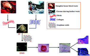
Functionalized graphene oxide offers many advantages as a biomaterial in various biomedical applications. In the present study, graphene oxide (GO) was incorporated in the collagen–fibrin composite film (CFGO) and used as wound dressing material in both in vitro and in vivo studies. A 3-(4,5-dimethylthiazol-2-yl)-2,5-diphenyltetrazolium bromide (MTT) assay using NIH 3T3 cells revealed that CFGO film is biocompatible. CFGO was also used as a wound dressing material on the experimental wounds of rats. Histopathological, biochemical and hematology studies have shown that the CFGO treated wounds healed faster than control and CF treated wounds. Based on the results, CFGO can be used as a wound dressing material in small and large animals.


Introduction

Graphene oxide (GO) is a single atomic plane of graphite containing abundant epoxide, hydroxyl and carboxyl functional groups on its basal plane and its edges provide many reactive sites for chemical functionalization and interaction.1,2 Interactions between graphene and biological molecules are quite interesting in cell imaging, drug delivery, biosensors, etc. Graphene nanosheets identically show some similar properties to carbon nanotubes (CNTs), but in the case of cytotoxicity studies, single-wall and multi-wall CNTs show more toxicity to human and animal cells compared to graphene derivatives. This is due to the fact that GO is a two-dimensional structure, whereas CNTs have only a single dimension.3 Due to concentration dependent toxicity, GO cannot be directly used in cell culture applications, however, GO can be functionalized with biomacromolecules for biomedical application. The potential of functionalized GO in cellular imaging, cancer therapy, drug delivery, etc. has been reported by several authors.4–6 These studies clearly indicate the bioactivities of surface modified GO. Graphene derivatives functionalized with peptides, proteins, aptamers, avidin–biotin and other small biomolecules are used as building blocks for biodevices.5 Multivalent functionalization and efficient loading of molecules on the substrate of GO suggests its biocompatibility in in vitro as well as in in vivo conditions.7–9 Currently, few applications of GO derivatives are available in previously studied reports. Recently, it was proven that GO-based nanomatrices can act as promising system for the production of insulin.10 A process for a multi-functional GO has been developed and was used as a fluorescence marker for in vitro and in vivo imaging.11 GO was used as an efficient vascular endothelial growth factor (VEGF) delivery system in animal models, thereby suggesting its therapeutic application for treating ischemic disease.12 Chitosan–polyvinyl alcohol–graphene composites were prepared by an electrospinning method and studied for their antibacterial properties and wound healing capacity and it was found that its antibacterial properties helped to heal wounds faster.13

Chrome containing leather waste (CCLW) is the most prominent solid waste in the tanning industry. CCLW is mainly used as a resource of collagen and chromium(III) complexes.14 Using alkaline protease enzyme under mild conditions, protein products (gelatin and collagen hydrolyte) were isolated from chrome shavings, which were potentially used in cosmetics, adhesive, printing, photography, micro encapsulation and as additives in the leather industry.15 Collagen is one of the structural proteins in the extracellular matrix of eukaryotes. It is the most abundant protein in vertebrates and constitutes about 30% of the total proteins present in skin, tendons, bones, etc. Collagen molecules secreted by cells form characteristic fibers, which are responsible for the functional integrity of tissues such as the bone cartilage, skin and tendon.16–18 Collagen has a vital role in biomedical applications as wound dressing material, vitreous implant, drug carriers, etc. Among various types, type I collagen is commonly found in mammalian tissues. It shows good biocompatibility, biodegradability, weak antigenicity and superior surface-active properties when employed as a scaffold in tissue engineering.19,20

Fibrinogen is converted to fibrin, which is a provisional matrix in the wound healing process; its structural composition binds the cells and proteins and promotes cell adhesion, migration and proliferation.21–23 It is a naturally occurring scaffold that is used in angiogenesis and tissue repair and available in the form of sponge, film, powder, sheets, etc. Fibrin glue helps in haemostasis and accelerates the wound healing. Fibrin sealant is used as improved wound healing material in diabetic patients. Bensaid et al.21 have reported that fibrin-based biomaterials are biocompatible, biodegradable and have high affinity to various biological surfaces. Sastry et al.24 have reported the preparation of physiologically clotted fibrin (PCF) from slaughterhouse wastes that could be used for biomedical purposes such as wound healing and osteoinduction.

Research on wound healing agents is one of the developing areas in modern biomedical sciences. Wound healing involves continuous cell–cell and cell–matrix interactions in response to an injury to restore the function and integrity of damaged tissues. There are many commercially available wound dressing materials in market based on gelatin, carboxymethylcellulose, collagen, pectin, etc.25,26 These materials are used individually as well as in various composite forms. The aim of the present work is to prepare a wound dressing material in the form of composite film containing collagen and fibrin impregnated with GO.

Results and discussion

Normally, GO cannot be used directly in animal models due to its toxicity, hence it was functionalized with collagen and fibrin (CFGO). The prepared films were characterized to establish the functionalization of GO. CFGO was used as a wound dressing material on the experimental wounds of rats and its efficacy was studied using biochemical, histological and general observations (Fig. 1 schematic illustration).
image file: c4ra10150b-f1.tif
Fig. 1 Schematic illustration of CFGO as wound dressing material.

The mechanical properties of a wound dressing material play a vital role when it is used in in vivo applications. The mechanical properties of composites are given in Tables 1–3. When compared to collagen, fibrin alone has lesser tensile strength, whereas the combination of both shows an improved mechanical strength. Among the different stoichiometric ratios used, the CF films, which exhibited better tensile strengths, were selected and were further used to prepare CFGO films with different stoichiometric ratios of GO (Table 3); 0.01% of GO has shown approximately two fold higher tensile strength. Hence, this composite film was used in further experiments. Ethylene glycol was used to improve the flexibility of the films.27,28

Table 1 Physiochemical properties of various stoichiometric ratios of collagen
Sample no. F[thin space (1/6-em)]:[thin space (1/6-em)]C (%composite) Elongation at break (%) Tensile strength (MPa)
1 1[thin space (1/6-em)]:[thin space (1/6-em)]0 0.78 ± 0.87 16.43 ± 4.67
2 1[thin space (1/6-em)]:[thin space (1/6-em)]0.2 3.23 ± 0.94 18.04 ± 5.27
3 1[thin space (1/6-em)]:[thin space (1/6-em)]0.4 7.18 ± 1.56 27.53 ± 4.37
4 1[thin space (1/6-em)]:[thin space (1/6-em)]0.6 7.77 ± 1.45 29.22 ± 3.54
5 1[thin space (1/6-em)]:[thin space (1/6-em)]0.8 3.59 ± 2.67 20.16 ± 5.16
6 1[thin space (1/6-em)]:[thin space (1/6-em)]1 3.24 ± 1.42 18.54 ± 3.24


Table 2 Physiochemical properties of various stoichiometric ratios of fibrin
Sample no. C[thin space (1/6-em)]:[thin space (1/6-em)]F (%composite) Elongation at break (%) Tensile strength (MPa)
1 1[thin space (1/6-em)]:[thin space (1/6-em)]0 1.78 ± 0.34 24.43 ± 3.67
2 1[thin space (1/6-em)]:[thin space (1/6-em)]0.2 3.23 ± 1.45 29.5 ± 3.56
3 1[thin space (1/6-em)]:[thin space (1/6-em)]0.4 4.57 ± 1.67 37.36 ± 4.23
4 1[thin space (1/6-em)]:[thin space (1/6-em)]0.6 6.62 ± 2.45 45.5 ± 4.67
5 1[thin space (1/6-em)]:[thin space (1/6-em)]0.8 10.8 ± 3.81 48.55 ± 5.87
6 1[thin space (1/6-em)]:[thin space (1/6-em)]1 8.41 ± 2.1 41.41 ± 3.52


Table 3 Physiochemical properties of various stoichiometric ratios of graphene oxide composites
Sample no. C[thin space (1/6-em)]:[thin space (1/6-em)]F[thin space (1/6-em)]:[thin space (1/6-em)]GO (%composite) Elongation at break (%) Tensile strength (MPa)
1 1[thin space (1/6-em)]:[thin space (1/6-em)]0.8[thin space (1/6-em)]:[thin space (1/6-em)]0.001 6.4 ± 0.67 40.43 ± 3.56
2 1[thin space (1/6-em)]:[thin space (1/6-em)]0.8[thin space (1/6-em)]:[thin space (1/6-em)]0.003 14.8 ± 1.34 47.36 ± 4.45
3 1[thin space (1/6-em)]:[thin space (1/6-em)]0.8[thin space (1/6-em)]:[thin space (1/6-em)]0.005 11.8 ± 2.35 57.5 ± 5.34
4 1[thin space (1/6-em)]:[thin space (1/6-em)]0.8[thin space (1/6-em)]:[thin space (1/6-em)]0.01 9.62 ± 1.25 78.55 ± 5.74
5 1[thin space (1/6-em)]:[thin space (1/6-em)]0.8[thin space (1/6-em)]:[thin space (1/6-em)]0.02 9.43 ± 2.45 71.41 ± 3.67


Infra-red spectra (Fig. 2a) of C, F and CF composites show the amide I peaks at 1637, 1644 and 1634 cm−1, amide II peaks at 1549, 1539 and 1541 cm−1, and amide III peaks at 1241, 1238 and 1243 cm−1, respectively. The peaks at 1453, 1451 and 1453 cm−1 correspond to the –CH2 moiety of C, F and CF, respectively.29 The IR spectrum of GO shows the presence of ν (C–O) of –COOH groups at 1734 and 1626 cm−1, carboxyl group at 1401 and alkoxy group at 1058 cm−1.10 The CFGO composite shows the absence of a peak at 1734 cm−1, which represents the reduction of the carboxyl moiety on the surface of GO with the addition of C and F. CFGO presents amide I, II and III peaks at 1634, 1541 and 1242 cm−1 respectively. XRD spectra (Fig. 2b) of GO shows a peak at 11.2°, which represents the presence of more oxygen functional groups on the basal plane of the graphene sheet. This sharp narrow peak intensity is lesser with the addition of C and F in CFGO; in addition the presence of a peak at 20.6° indicates the presence of protein addition, in CF this peak is present at 20.4°. This shows that the addition of GO does not alter the structure of CF.


image file: c4ra10150b-f2.tif
Fig. 2 (a) FTIR spectra of collagen, fibrin, CF, GO and CFGO. (b) XRD spectra of GO, CF and CFGO. (c) Raman spectra of GO and CFGO.

The Raman spectrum of GO shows the presence of a D band at 1366 cm−1, which represents the vibrations of sp3 carbon atoms, and the band at 1601 cm−1 represents the sp2 hybridized carbon atom of GO (Fig. 2c).30 CFGO represents amide II and III peaks at 1543 and 1242 cm−1, however, the amide I peak is absent; in addition, lesser intensity in the peaks of D band at 1347 and G band at 1598 cm−1 were observed (Fig. 2c) when compared to those of GO, this may be due to the interaction between the GO and CF.

Scanning electron microscopy (SEM) images of GO, CF and CFGO are shown in (Fig. 3a). GO shows agglomeration of ultrathin sheets; the surface of CF was smooth and porous in nature. The presence of GO was evident in CFGO and it had retained its porosity even after the addition of GO. AFM images (Fig. 3b) of GO, CF and CFGO also indicate the smooth surface of the films and have similar morphology compared to those of SEM. The porous nature of the films helps in absorbing wound fluids and keeps the wound surface dry, and it also helps with the oxygen supply to the wound.29 Water absorption capacity (Fig. 3c) is an important property to keep the wound surface dry and to enhance wound healing. The water absorption capacity of CF and CFGO show that CFGO has high water absorption values when compared to CF, and this may be attributed to the presence of more hydrophilic groups like –OH, –NH2, etc., on its surface. GO already contains functional groups like –COOH, –C[double bond, length as m-dash]O, –OH, and –C–O–C– on its surface. Functionalization with collagen and fibrin has further increased the hydrophilic groups of the protein networks.31 The degradation rate of CFGO (Fig. 3d) is higher when compared to CF; this may be due to its more hydrophilic nature.


image file: c4ra10150b-f3.tif
Fig. 3 (a) SEM images of GO, CF and CFGO. (b) AFM images of GO, CF and CFGO. (c) Water absorption studies of CF and CFGO composite films. (d) Degradation rates of CF and CFGO composite films using PBS buffer.

MTT assay was carried out using normal fibroblast (NIH 3T3) cells. Cells treated with GO, CF and CFGO were incubated for 1, 4 and 7 days. The results (Fig. 4a) revealed that CF exhibited 100% cell viability till 7 days, and showed higher cell proliferation rate compared to control. GO treated cells showed significant (p ≤ 0.05) decrease in cell viability, whereas addition of GO with CF improved the cell viability percentage. There was no significant difference between the control and CFGO treated groups throughout the study. According to previous studies, GO enhances more cell adherence and proliferation than pristine graphene due to the abundant oxide groups on its surface. At low concentrations, GO induces stronger metabolic cell activity, and this is reversed at high concentrations. GO produces cytotoxicity in concentration and time dependent manners and can enter into the cytoplasm (lysosome, mitochondrion, and endoplasm) and nucleus, which decreases cell adhesion and induces cell floating and apoptosis.32 But in the case of CFGO, reduced toxicity was observed because of the functionalization of GO by CF. Fig. 4b shows the inverted microscopic images of the cell viability on day 1 of all treatments and control.


image file: c4ra10150b-f4.tif
Fig. 4 (a) MTT assay representation of GO, CF and CFGO. (b) The microscopic images show the cell viability of those treatments on 1st day. Significant difference (p ≤ 0.05) occurred between GO and the control. (c) Hemolytic assay representation of GO, CF, CFGO and Triton X-100. Significant difference (p ≤ 0.05) occurred between Triton X-100 and GO. (d) ROS generation of GO, CF, CFGO and LPS treated cells and (e) fluorescence microscopic images of treated cells. Significant difference (p ≤ 0.05) occurred between control and other treatments.

Hemolysis assay revealed the effect of GO, CF and CFGO against blood components (Fig. 4c). GO revealed significant hemolytic properties compared to the control, which could be due the presence of abundant negatively charged carboxyl groups that are highly concentrated at the edges of GO; there may be strong interactions (electrostatic and hydrophobic interactions) between the amphiphilic GO and the lipid bilayer of the blood cell membrane.5 The chemical structure of GO endows it with surfactant-like performance, which has adsorption ability at the interfaces and could lower the surface or interfacial tension.33 Thus, small sized GO may act as a molecular surfactant, and large sized GO can behave like a molecule–colloid dualistic surfactant. Thus, when GO is exposed to blood cells, it may lead to severe hemolysis.34,35 However, the effect was less when compared with the positive control (Triton X 100). Interestingly, hemolytic properties were considerably reduced in CFGO when compared to GO, which could be attributed to the functionalization of GO with CF. CFGO might have difficulty in interacting with the lipid bilayers,34 thereby suppressing the hemolysis.

Reactive oxygen site (ROS) is a common toxicity mechanism of carbon-based and other nanoscale materials; ROS initiates the dysfunction of mitochondria and induces the cell cytotoxicity. According to clinical studies, materials with small size and large surface area enhance the ROS.36–38 In the present study, GO has induced ROS in the cells, and results are shown in Fig. 4d. GO induces cytotoxicity by causing physical damage to the cell membrane, which results from direct interactions between the cell membrane and the GO nanosheets. Electrostatic interactions occur between negatively charged oxygen functional groups in GO and positively charged phosphatidylcholine lipid of the cell membranes.39 Significant reduction (p ≤ 0.05) in ROS levels was observed in CF and CFGO treated cells compared to those subjected to GO treatment. Fluorescence microscopic images of treated cells are shown in Fig. 4e. 2′,7′-Dichlorofluorescindiacetate (DCFH) reacts with ROS to form DCF, the fluorescent product. More number of ROS induced fluorescence cells appeared at the positive control lipopolysaccaride (LPS), however, CF and CFGO did not show considerable ROS induced cells.

Surface of the wounds were photographed periodically from a constant distance for both control and experimental animals (Fig. 5a). A faster rate of healing was observed in the experimental wounds compared to those of control. The results are in agreement with those of planimetric observations. On the 4th day, the wound contraction of control animals was 28%, whereas 33% and 42% closure were observed in CF and CFGO treated wounds, respectively. However, by the 12th day, 82% of the wound was closed in the CF experimental groups and 97% of the wound was closed in the CFGO experimental groups, whereas 72% closure was observed in control. These results show the efficacy of CF and CFGO as wound dressing materials. The presence of GO in CF films accelerated wound healing on the 12th day. According Lu et al. 2012, graphene accelerated wound healing in experimental animals due to its antibacterial activity.13 According to our results, the presence of GO exhibited better wound healing properties compared to CF and the control. The following mechanism might have enhanced the wound healing process: the presence of collagen and fibrin in CFGO would have caused moderate ROS generation in wound healing process.40 Graphene impede the prokaryotic cells and not the eukaryotic cells; therefore, this antibacterial efficiency enhances faster wound healing. This may occur due to the electron transfer from graphene into the nucleus of prokaryotic cells, which are devoid of nuclear membrane.13 Furthermore, due to the better water absorption properties of CFGO, the surface of the wound is dry and this might have avoided the bacterial infection on wound surface.29,41,42 The higher rate of 3T3 fibroblast cell proliferation in CFGO treated cells in in vitro is an indication of the faster wound healing mechanism in in vivo.


image file: c4ra10150b-f5.tif
Fig. 5 (a) Photographic evaluation of wound healing. Faster healing was observed on 12th day in the CFGO treated rats. (b) Histological section of H&E staining on control, CF and CFGO treated wounds up to 12 days post wound. (c) Histological section of Masson trichrome staining on control, CF and CFGO treated wounds up to 12 days post wound.

Histological studies of treated and control groups were performed by using H&E and Masson trichrome staining (Fig. 5b and c). On the 4th day, CFGO and CF treated groups have shown acute and chronic inflammatory cells, such as neutrophils and lymphocytes, with blood vessels and extravasated RBC's. Inflammatory cells promote the migration and proliferation of endothelial cells, which leads to neovascularisation. This in turn aids in re-epithelialization of the wounded tissue.43 On the 8th day, when compared to control, the treated group showed moderate inflammatory cells. The fibrous nature of connective tissues with less inflammatory cells and red blood vessels were clearly monitored in CFGO treated groups, which facilitated the faster wound healing. On the 12th day, CFGO treated groups showed epithelium and connective tissue formation; the density of collagen fibers is high in CFGO when compared to those of CF and control. On the 12th day, CF treated groups showed moderate epithelialization. The collagen deposition and faster epithelial formation in CFGO could have significantly accelerated the better wound healing compared to other treatments. Masson's trichrome staining helped to clearly visualize the changes in collagen fibers. On the 8th day, the formation of new collagen fibers was more prominent in the CFGO treated groups compared to CF and control. Proliferation of fibroblasts, which are the major producer of collagen, was also observed. Masson's trichrome staining clearly shows the blue colour of collagen fibers. Collagen remodeling and degradation occurs simultaneously to provide the tensile strength and reduce scar formation.44 Other than collagen, MT staining also helps histopathologists to differentiate other anatomical structures and organelles in the healed skin such as scab, fine and coarse collagen fibers, hair follicle and adipose tissue.45 Faster wound healing and deposition of collagen did not contribute to any scar formation in the wounded area. This indicates that CFGO might be used as a wound dressing material, which affects a scarless healing.

The biochemical parameters i.e., collagen (Fig. 6a), hexosamine (Fig. 6b) and uronic acid (Fig. 6c) were analyzed in the granulation tissues of experimental rats on different days after wound creation. The concentration of collagen in CFGO treated wounds increased till 8th day and later decreased on 12th day. This noticeable increase might be due to the increased synthesis of collagen and could be correlated to the effective healing of wounds. Because collagen is the predominant extracellular protein in the granulation tissue of a healing wound, rapid increase in the synthesis of this protein indicates faster wound healing. Hexosamine, a matrix molecule, acts as a ground substratum for the synthesis of new extracellular matrix. The glycosaminoglycans are known to stabilize the collagen fibres by enhancing electrostatic and ionic interactions and possibly they control their ultimate alignment. Their ability to bind and alter protein–protein interactions has identified them as important determinants of cellular responsiveness in development, homeostasis, and disease.46 In our study, a prominent increase in the levels of hexosamine in the treated groups was observed compared to that of the control group. Similar trend was observed in the contents of uronic acid. Increase in the levels of collagen, uronic acid, and hexosamine in the treated groups gave an indication of the faster rate of wound healing compared to controls.26


image file: c4ra10150b-f6.tif
Fig. 6 (a) Collagen content, (b) hexosamine values and (c) uronic acid values of granulation tissue in control and experimental wounds. Collagen content of CFGO treated rats increased up to the 8th day and later decreased. Hexosamine and uronic acid values show the decreasing trend in both control and treated rats. Significant difference (p ≤ 0.05) occurred between control and CFGO treated tissues.

Tensile strength values of healed wounds of treated groups were higher when compared to those of control group (Table 4). Increased tensile strength indicates the maturation of collagen by formation of inter- and intra-molecular crosslinking. In the case of control wounds, the decreased tensile strength may be due to the delay in the maturation of collagen. CFGO might have enhanced the epithelization process and hastened the deposition of collagen, which resulted in the better tensile strength in the experimental wounds. Normally, tensile strength is directly related to the amount of collagen synthesized at the wound site.47 The hematological parameters were studied on 12th day on both the treated and control rats (Table 5). There was a significant (p ≤ 0.05) increase in the values of hemoglobin, platelets, and lymphocytes in the CFGO treated groups compared to those of control. The results revealed that the films caused no adverse effects under in vivo conditions, which was in agreement with the in vitro biocompatibility studies.48

Table 4 Tensile strengths of healed skins of rats of control and experimental groups
Group Elongation at break (%) Tensile strength (MPa)
Control 5.67 ± 3.67 0.35 ± 0.89
CF 9.90 ± 5.78 0.94 ± 0.67
CFGO 42.50 ± 7.89 1.63 ± 0.98


Table 5 Comparison of hematological values between treated and control groups of rats after 20 days post wounding
  Control CF CFGO
WBC (109 μL−1) 8.85 ± 2.3 5.2 ± 0.4 3.6 ± 1.3
RBC (1012 μL−1) 4.09 ± 2.6 8.65 ± 0.6 10.09 ± 2.7
Hb (g dL−1) 11.6 ± 1.3 13.78 ± 1.2 15.4 ± 1.4
HCT (%) 27.3 ± 2.4 50.7 ± 1.2 40.8 ± 2.1
MCV (fL) 66 ± 1.7 53 ± 0.6 58 ± 2.1
MCH (pg) 28 ± 2.3 17.8 ± 0.3 15.8 ± 0.6
MCHC (%) 42 ± 2.4 33 ± 0.5 30 ± 0.9
Platelet (103 μL−1) 665 ± 1.4 553 ± 0.7 498 ± 3.6
Lym (%) 43.7 ± 1.6 60.5 ± 0.8 78.65 ± 1.2


Conclusion

Collagen–fibrin biocomposite films incorporated with GO were prepared. The presence of GO increased the mechanical strength of collagen/fibrin composite films. In vitro studies revealed the biocompatible nature of CFGO. Faster wound healing was observed in CFGO treated rats compared to those with CF and control. The present study indicates that functionalized GO enhances wound healing and CFGO may be tried on more clinical wounds of smaller and larger animals before its application on humans.

Experimental section

CCLW was obtained from a local leather tanning industry; crude fibrin was collected from nearby municipal slaughterhouse and other chemicals used in this study were analytical reagents purchased form Sigma Aldrich Co., India.

CFGO bio film synthesis

Graphene oxide was prepared according to a modified Hummers method.49 The obtained GO was further dialyzed for 24 h using water and then it was dried at 60 °C. The powdered GO was further sonicated to get a well dispersed solution with different ratios (0.001%, 0.003%, 0.005%, 0.01% and 0.02%).

Collagen was isolated from chrome containing leather waste, as described earlier.47 The collagen was further purified by dialyzing against 0.1 M acetic acid and distilled water for 24 h. The sample was freeze-dried and used as such. Physiologically clotted crude fibrin was separated from fresh blood by churning, as described earlier.23 Fibrin was further purified by a wet precipitation method. Then, 1 g of the final content was dissolved in 10 ml of 1 N NaOH solution; the pH was adjusted to 5 to get white precipitation of purified fibrin. This was dialyzed against water for 24 h to get purified fibrin. The sample was freeze-dried and used as such.

Collagen–fibrin (CF) films were prepared by mixing the different stoichiometric ratios of fibrin and collagen, as described in Tables 1 and 2. From Table 2, the CF composite, which exhibited better tensile strength, was selected and further mixed with different stoichiometric ratios of GO to prepare the CFGO films described in Table 3. From Tables 2 and 3, the better composites, which exhibited better tensile strength, were selected and used for further experiments. Ethylene glycol was added as a plasticizing agent. This mixture was poured into polythene trays (measurement 12 cm × 7.5 cm) and dried at room temperature (30 °C) to get CFGO in sheet form.

Characterization

Tensile strength properties were measured using three dumb-bell shaped specimens of 4 mm wide and 10 mm length of prepared films. Mechanical properties, such as tensile strength (MPa) and percentage of elongation at break (%), were measured using a universal testing machine (INSTRON model 1405) at an extension rate of 5 mm min−1.

Fourier transform infrared (FTIR) measurements were carried out to determine the formation and changes in the functional groups on the prepared composite films. The spectra were measured at a resolution of 4 cm−1 in the frequency range of 4000–500 cm−1 using Nicolet 360 FTIR spectrometer. The X-ray powder diffraction (XRD) patterns were done with a Seifert JSO Debye flex 2002. X-ray Diffractometer (30 mA, 40 kV) using Cu Kα radiation (k = 0.154056 nm). The FT-Raman spectrum was carried out using Bruker Rfs 27 FT-Raman spectrometer, with a scanning range of 50–4000 cm−1.

The surface morphology of the samples was visualized by scanning electron microscope (SEM Model LEICA stereo scan 440). The samples were coated with gold ions using an ion coater (Fisons sputter coater) with the following parameters: 1 Torr pressure, 20 mA current, and 70 s coating time, using a 15 kV as accelerating voltage. Atomic force microscopy (AFM) was carried out with an Agilent Pico LE Scanning Probe Microscope model. The Agilent instrument is a tip scan instrument, equipped with small (10 ml) and large AFM scanners (150 ml). This instrument is equipped with an environmental chamber, capable of heating up to 200 °C.

The water absorption capacities of CF and CFGO biocomposites were determined according to the method followed by Sastry and Rao.50 To measure the degradation rate, the weight of the scaffold was measured as a function of degradation time. Three specimens of all scaffolds were equally weighed (W0) and then immersed in a phosphate buffer solution (PBS) (pH 7.4) for 24 h. The temperature was maintained at 37 °C. After predetermined periods of soaking time, each specimen was taken out and kept for 24 h. These were then weighed (WT). The percentage of weight loss was given by the following equation.

Weight loss (%) = [(W0WT)/W0] × 100

In vitro studies

NIH-3T3 cells were grown in a 96-well plate. The cell viability was measured after 1st day, 4th day and 7th day using 3-(4,5-dimethylthiazol-2-yl)-2,5-diphenyltetrazolium bromide (MTT) and 10% dimethyl sulfoxide (DMSO). The assay was performed in triplicates and the untreated wells were maintained as control.51

The haemolytic assay was carried out to evaluate the safety of films for in vivo applications.52 Blood was collected from healthy rats [Ethical Approval: IAEC no. 11/2014 (a)] in a heparin sodium-containing tube and centrifuged at 10[thin space (1/6-em)]000 × g for 10 min. The pellet containing RBCs was diluted in 20 mM HEPES buffered saline (pH 7.4) to 5% v/v solution. The RBC suspension was treated with CF and CFGO and incubated at 37 °C for 60 min. RBCs treated with 1% Triton X-100 served as positive control.53 After incubation, all the samples were centrifuged and the supernatants were used for measuring optical density at 540 nm.

The oxidant-sensitive dye 2′,7′-dichlorofluorescein-diacetate (DCFH-DA) was used for ROS detection. Lipopolysaccharides (LPS) treated macrophage cells were treated with CF and CFGO films for 12 and 24 h. The effect of films on ROS was studied with the help of DCFH-DA, which enters cells passively and is deacetylated by esterase to non-fluorescent DCFH. DCFH reacts with ROS to form DCF, the fluorescent product. Fluorescence was read at wavelengths of 485 nm for excitation and 530 nm for emission with a fluorescence plate reader (TECAN). Untreated wells served as control and LPS (alone) treated wells served as positive control.54

In vivo studies

In vivo experiments were preformed according to the Institutional Animal Ethical Committee approval and guidelines: IAEC no. 11/2014 (a). 30 male albino Wistar rats (150–200 g weight) were chosen and they were divided into three groups (n = 3). Throughout the experiment, rats were maintained in an air-conditioned room at 25 ± 1 °C with a proper lighting schedule of 12 h light and 12 h dark cycle. The animals received commercial rat diet and water ad libitum.

Each animal was given a dose of sodium pentobarbital (40 mg per kg body weight) intraperitoneally. The dorsal surface of the rat below the cervical region was epilated under aseptic conditions. An open excision wound of 2 cm × 2 cm was created on the shaved dorsal side of rats using a sterile surgical blade. The control group wounds were dressed with sterile cotton gauze dipped with gentamicin, and wound dressing films of CF and CFGO were applied separately on experimental wounds. The dressings were periodically changed at an interval of 4 days with the respective materials. Three rats were sacrificed periodically on the 4th, 8th and 12th day of post wound creation and the granulation tissues formed were removed and stored at −70 °C until analysis. The progress of wound healing in the three groups was evaluated visually, planimetrically, histologically and biomechanically by periodical monitoring of the wound surface.

Hair was clipped around the scar for proper visualization and the individual contour of the wounds of both control and experimental animals was measured periodically, using a transparent graph sheet and the rate of healing was calculated and expressed as percentage contraction.

In the present study, collagen, hexosamine and uronic acid levels were estimated in the granulation tissue of control and experimental wounds on 4th, 8th and 12th day. The granulation tissue was collected after sacrificing the animals on the respective days. Collagen and hexosamine were determined in defatted dried granulation tissues by the methods of Woessner,55 Elson56 and Morgan.57 Extraction of uronic acid from the tissue was carried out according to the method of Schiller58 and estimated by the method of Bitter and Muir.59

The animals were sacrificed periodically on 4th, 8th, and 12th day post wound creation and the tissue from the wound site of the individual animal was removed. These samples were then separately fixed in 10% formalin, dehydrated through graded alcohol series, cleared in xylene, and embedded in paraffin wax (m.p. 56 °C). Serial sections of 5 μm of thickness were cut and stained with hematoxylin, eosin and Masson's trichrome. The sections were examined under a microscope and photomicrographs were taken.

Blood was collected from rats when they were sacrificed at the 12th day, during the wound healing process. Hematological parameters, such as hemoglobin, total WBC count, differential leucocyte count, erythrocyte sedimentation rate, total RBC count, platelets, packed cell volume, mean corpuscular volume, mean corpuscular hemoglobin, mean corpuscular haemoglobin concentration and colour index ratio, were carried out using Sigma Diagnostic kits.48

On the 30th day after creating the wound, one animal from each group was anesthetized. Healed tissue along with normal skin at the two ends was excised for measuring tensile strength (MPa) and percentage of elongation at break (%) using a universal testing machine (Instron model 4501). The break load was measured and the tensile strength was calculated using the following equation:

Tensile strength = break load/strip cross sectional area

Statistical analysis

The results were expressed as mean ± standard deviation (SD) of three individual experiments (n = 3). The statistical analysis was performed using a t-test. The significant difference level is p ≤ 0.05.

Acknowledgements

R. Deepachitra gratefully acknowledges the financial support provided by the Department of Science and Technology (DST).

References

  1. S. Park and R. S. Rodney, Nat. Nanotechnol., 2009, 4, 217–224 CrossRef CAS.
  2. O. C. Compton and S. T. Nguyen, Small, 2010, 6, 711–723 CrossRef CAS.
  3. A. K. Geim and K. S. Novoselov, Nat. Mater., 2007, 6, 183–191 CrossRef CAS PubMed.
  4. C. Chung, Y. K. Kim, D. Shin, S. R. Ryoo, B. H. Hong and D. H. Min, Acc. Chem. Res., 2013, 46, 2211–2224 CrossRef CAS PubMed.
  5. S. Goenka, V. sant and S. Sant, J. Controlled Release, 2014, 173, 75–88 CrossRef CAS PubMed.
  6. Y. Yang, A. M. Asiri, Z. Tang, D. Du and Y. Lin, Mater. Today, 2013, 16, 365–373 CrossRef CAS PubMed.
  7. K. Yang, J. Wan, S. Zhang, Y. Zhang, S. T. Lee and Z. Liu, ACS Nano, 2011, 5, 516–522 CrossRef CAS PubMed.
  8. W. Hu, C. Peng, L. V. Min, X. Li, Y. Zhang, N. Chen, C. Fen and Q. Huang, ACS Nano, 2011, 5, 3693–3700 CrossRef CAS PubMed.
  9. O. N. Ruiz, K. A. Fernando, B. Wang, N. A. Brown, P. G. Luo, N. D. McNamara and M. Vangsness, ACS Nano, 2011, 5, 8100–8107 CrossRef CAS PubMed.
  10. K. Turcheniuk, M. Khanal, A. Motorina, P. Subramanian, A. Barras, V. Zaitsev, V. Kuncser, A. Lec, A. Martoriati, K. Cailliau, J. F. Bodart, R. Boukherroub and S. Szunerits, RSC Adv., 2014, 4, 865–875 RSC.
  11. G. Gollavelli and Y. C. Ling, Biomaterials, 2012, 33, 2532–2545 CrossRef CAS.
  12. Z. Sun, P. Huang, G. Tong, J. Lin, A. Jin, P. Rong, L. Zhu, L. Nei, G. Niu, F. Cao and X. Chen, Nanoscale, 2013, 5, 6857–6866 RSC.
  13. B. Lu, T. Li, H. Zhao, X. Li, C. Gao, S. Zhang and E. Xie, Nanoscale, 2012, 4, 2978–2982 RSC.
  14. S. Swarnalatha, A. Ganesh Kumar, S. Tandaiah and G. Sekaran, J. Chem. Technol. Biotechnol., 2009, 84, 751–760 CrossRef CAS.
  15. L. F. Cabeza, M. M. Taylor, G. L. DiMaio, E. M. Brown, W. N. Marmer, R. Carrio, P. J. Celma and J. Cot, Waste Management, 1998, 18, 211–218 CrossRef CAS.
  16. G. Bilgen, G. Oktay, Z. Tokgoz, G. Guner and S. Yalcin, Turk. J. Vet. Anim. Sci., 1999, 23, 483–487 Search PubMed.
  17. T. Nagai, M. Izumi and M. Ishii, Int. J. Food Sci. Technol., 2004, 39, 239–244 CrossRef CAS PubMed.
  18. M. Sadowska, I. Kolodziejska and C. Niecikowska, Food Chem., 2003, 81, 257–262 CrossRef CAS.
  19. M. J. Fonseca, M. A. Alsina and F. Reig, Biochim. Biophys. Acta, 1996, 1279, 259–265 Search PubMed.
  20. M. Maeda, S. Tani, A. Sano and K. Fujioka, J. Controlled Release, 1999, 62, 313–324 CrossRef CAS.
  21. W. Bensaid, J. T. Triffitt, C. Blanchat, K. Oudina, L. Sedel and H. Petite, Biomaterials, 2003, 24, 2497–2502 CrossRef CAS.
  22. Q. Ye, G. Zund, P. Benedikt, S. Jockenhoevel, S. P. Hoerstrup, S. Sakyama, J. A. Hubbell and M. Turina, Eur. J. Cardio Thorac. Surg., 2000, 17, 587–591 CrossRef CAS.
  23. S. L. Rowe, S. Lee and J. P. Stegemann, Acta Biomater., 2007, 3, 59–67 CrossRef CAS PubMed.
  24. S. E. Noorjahan and T. P. Sastry, J. Biomed. Mater. Res., Part B, 2004, 71, 305–312 CrossRef CAS PubMed.
  25. J. S. Boateng, K. H. Matthews, H. N. Stevens and G. M. Eccleston, J. Pharm. Sci., 2008, 97, 2892–2923 CrossRef CAS PubMed.
  26. S. E. Noorjahan and T. P. Sastry, J. Polym. Sci., Part A: Polym. Chem., 2004, 42, 2241–2252 CrossRef CAS.
  27. J. Sathian, T. P. Sastry, L. Suguna, Y. Lakshminarayana and G. Radhakrishnan, J. Biomed. Mater. Res., Part A, 2003, 65, 435–440 CrossRef CAS PubMed.
  28. T. P. Sastry, C. Rose, S. Gomathinayagam and G. Radhakrishnan, J. Appl. Polym. Sci., 1998, 68, 1109–1115 CrossRef CAS.
  29. N. Natarajan, V. Shashirekha, S. E. Noorjahan, M. Rameshkumar, C. Rose and T. P. Sastry, J. Macromol. Sci., Part A: Pure Appl. Chem., 2005, 42, 945–953 CrossRef PubMed.
  30. R. Y. N. Gengler, A. Veligura, A. Enotiadis, E. K. Diamanti, D. Gournis, C. Jozsa, B. J. Van Wees and P. Rudolf, Small, 2009, 6, 35–39 CrossRef PubMed.
  31. J. M. Pachence, R. A. Berg and F. H. Silver, Med. Device Diagn. Ind., 1987, 9, 49–55 CAS.
  32. K. Wang, J. Ruan, H. Song, J. Zhang, Y. Wo, S. Guo and D. Cui, Nanoscale Res. Lett., 2011, 6, 1–8 Search PubMed.
  33. J. Kim, L. J. Cote, F. Kim, W. Yuan, K. R. Shull and J. Huang, J. Am. Chem. Soc., 2010, 132, 8180–8186 CrossRef CAS.
  34. D. Lichtenberg, E. Opatowski and M. M. Kozlov, Biochim. Biophys. Acta, 2000, 1508, 1–19 CrossRef CAS.
  35. N. Muruthy, I. Chang, P. Stayton and A. Hoffman, Macromol. Symp., 2001, 172, 49–55 CrossRef.
  36. K. Yang, J. Wan, S. Zhang, B. Tian, Y. Zhang and Z. Liu, Biomaterials, 2012, 33, 2206–2214 CrossRef CAS PubMed.
  37. A. Thubagere and B. M. Reinhard, ACS Nano, 2010, 4, 3611–3622 CrossRef CAS PubMed.
  38. J. R. Gurr, A. S. S. Wang, C. H. Chen and K. Y. Jan, Toxicology, 2005, 213, 66–73 CrossRef CAS PubMed.
  39. C. Peng, W. Hu, Y. Zhou, C. Fan and Q. Huang, Small, 2010, 6, 1686–1692 CrossRef CAS PubMed.
  40. N. Bryan, H. Ahswin, N. Smart, Y. Bayon, S. Wohler and J. A. Hunt, Eur. Cells Mater., 2012, 24, 249–265 CAS.
  41. J. S. Boateng, K. H. Mathews, H. N. E. Stevens and G. M. Eccleston, J. Pharm. Sci., 2008, 97, 2892–2923 CrossRef CAS PubMed.
  42. C. C. Wang, C. H. Su and C. C. Chen, J. Biomed. Mater. Res., 2008, 84, 1006–1017 CrossRef PubMed.
  43. J. Adam, M. D. Singer, A. F. Richard and M. D. Clark, N. Engl. J. Med., 1999, 341, 738–746 CrossRef PubMed.
  44. S. Enoch and D. J. Leaper, Surgery, 2008, 26, 31–37 Search PubMed.
  45. M. L. Noorlander, P. Meli, A. Jonker and C. J. F. Van Noordeen, J. Histochem. Cytochem., 2002, 50, 1469–1474 CrossRef CAS PubMed.
  46. J. M. Trownbridge and R. L. Gallo, Glycobiology, 2002, 12, 117–125 CrossRef PubMed.
  47. V. Ramnath, S. Sekar, S. Sankar, C. Sankaranarayanan and T. P. Sastry, J. Mater. Sci.: Mater. Med., 2012, 23, 3083–3095 CrossRef CAS PubMed.
  48. S. A. Bidgoli, M. Mahdavi, S. M. Rezayat, M. Korani, A. Amani and P. Ziarati, Acta Med. Iran., 2013, 51, 203–208 Search PubMed.
  49. W. S. Hummers and R. E. Offeman, J. Am. Chem. Soc., 1958, 80, 1339 CrossRef CAS.
  50. T. P. Sastry and K. P. Rao, J. Bioact. Compat. Polym., 1990, 5, 430–438 CrossRef CAS PubMed.
  51. T. Mosmann, J. Immunol. Methods, 1983, 65, 55–63 CrossRef CAS.
  52. P. Venkatesan, N. Puvvada, R. Dash, B. N. Prashanth Kumar, D. Sarkar, B. Azab, A. Pathak, S. C. Kundu, P. B. Fisher and M. Mandal, Biomaterials, 2011, 32, 3794–3806 CrossRef CAS PubMed.
  53. P. Khullar, V. Singh, A. Mahal, P. N. Dave, S. Thakur, G. Kaur, J. Singh, S. S. Kamboj and M. S. Bakshi, J. Phys. Chem., 2012, 116, 8834–8843 CAS.
  54. T. Wang, L. Qin, B. Liu, Y. Liu, B. Wilson, T. E. Eling, R. Langenbach, S. Taniur and J. S. Hong, J. Neurochem., 2004, 88, 939–947 CrossRef CAS.
  55. J. F. Woessner, Biochem. J., 1962, 83, 304–314 CAS.
  56. L. A. Elson and W. T. J. Morgan, Biochem. J., 1933, 27, 1824–1828 CAS.
  57. P. W. Morgan, A. G. Binnington, C. W. Miller, D. A. Smith, A. Valliant and J. F. Prescott, Vet. Surg., 1994, 23, 494–502 CrossRef CAS.
  58. S. Schiller, A. G. Slover and A. Dorfman, J. Biol. Chem., 1961, 236, 983–987 CAS.
  59. T. Bitter and H. M. Muir, Anal. Biochem., 1962, 4, 330–334 CrossRef CAS.

This journal is © The Royal Society of Chemistry 2014
Click here to see how this site uses Cookies. View our privacy policy here.