Solvothermal synthesis, crystal structure and photoluminescence properties of four Cd(II) coordination polymers with different topological structures

Zhi-Hao Yanab, Xiao-Wei Zhangab, Haiduo Panga, Yiheng Zhanga, Daofeng Sun*b and Lei Wang*a
aKey Laboratory of Eco-chemical Engineering, Ministry of Education, College of Chemistry and Molecular Engineering, Qingdao University of Science and Technology, Qingdao 266042, P. R. China. E-mail: inorchemwl@126.com
bCollege of Science, China University of Petroleum (East China), Qingdao, Shandong 266580, P. R. China. E-mail: dfsun@upc.edu.cn

Received 9th September 2014 , Accepted 15th October 2014

First published on 16th October 2014


Abstract

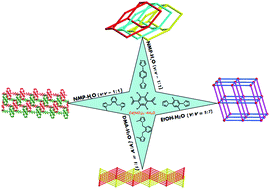
Solvothermal reactions of tetrabromoterephthalic (H2tbtpa) acid and different bis-imidazoles with cadmium nitrate provided four new Cd(II) coordination polymers (CPs), namely, {[Cd(bip)(tbtpa)]·H2O}n (1), {[Cd2(bibp)(tbtpa)2(H2O)4]·H2O}n (2), {[Cd(1,2-mbix)(tbtpa)]·H2O}n (3) [Cd(1,2-mbix)(tbtpa)]n (4), (bip = 1,4-di(1H-imidazol-1-yl)benzene, bibp = 1,1′-(2,5-dimethyl-1,4-phenylene)bis(1H-imidazole), 1,2-mbix = 1,2-bis((2-methyl-1H-imidazol-1-yl)methyl)benzene, H2tbtpa = tetrabromoterephthalic acid). All of the compounds have been structurally characterized by single-crystal X-ray diffraction analyses and further characterized by elemental analyses, IR spectroscopy, powder X-ray diffraction (PXRD), and thermogravimetric analyses (TGA). Single crystal X-ray diffraction analysis reveals that compound 1 exhibits a 3D diamond-type (dia, 66) framework of 3-fold interpenetration. Compound 2 displays a noninterpenetrated 3D network with the classical pcu topology. Compound 3 exhibits a polyrotaxane-like 2D + 2D → 2D layer with 44-sql topology and the 2D sheets further formed a 3D framework by π⋯π interaction. Compound 4 is a 2D 44-sql network and the adjacent 2D networks are packed parallel in a ⋯ABAB⋯fashion. Moreover, the thermal stability and photoluminescence properties of the compounds were investigated.

1. Introduction

Recently, mixed ligand assembly strategies incorporating pyridine or imidazole-based ligands and polycarboxylates have been verified as an effective approach for the construction of coordination networks.1,2 Among these, entangled networks have attracted particular attention for crystal engineers not only due to their intrinsic aesthetic appeal and potential as functional materials in gas storage, magnetism, and optics, et al.,3,4 but also for their intricate molecular architectures and topologies.5 There have been some comprehensive reviews by Robson, Batten, and Ciani's groups about fascinating entangled structures and their classification.6 However, the controlled assembly of coordination polymers with tailored structures and properties is still a significant challenge fraught with difficulties, closely related to the unpredictable impact on the composition of resulting compounds imposed by the modulation of the chemical environment, such as the temperature, pH value, solvents and counter anions.7 Nevertheless, more and more novel structures obtained through changing the synthetic conditions, at the same time, so many parameters also create obstacles to the exploration of the suitable reaction conditions and prediction of resulting structures.

Significantly, although polycarboxylates are widely utilized in the construction of coordination polymers,8,9 CPs built by H2tbtpa and N-donor ligands, especially bis(imidazole) have been investigated relatively rare. In addition, a careful selection of N-donor ligands with different conformations as secondary auxiliary ligands is a key step for the rational design of structures with specific physical and chemical properties. With the inspiration from the aforementioned points, we used such a ligand, tetrabromoterephthalic acid (H2tbtpa) to construct coordinative frameworks. Moreover, in order to explore the influence of N-donor ligands on the tuning of entangled networks and achieve different topological structures, a series of bis(imidazole) ligands (bip = 1,4-di(1H-imidazol-1-yl)benzene, bibp = 1,1′-(2,5-dimethyl-1,4-phenylene)bis(1H-imidazole), 1,2-mbix = 1,2-bis((2-methyl-1H-imidazol-1-yl)methyl)benzene) with different conformations were introduced into the system (Scheme 1). The aromatic dicarboxylate ligand incorporating bis(imidazole) ligands with Cd(II) ions under solvothermal conditions constructs four 2D and 3D coordination polymers, namely, {[Cd(bip)(tbtpa)]·H2O}n (1), {[Cd2(bibp)(tbtpa)2(H2O)4]·H2O}n (2), {[Cd(1,2-mbix)(tbtpa)]·H2O}n (3), [Cd(1,2-mbix)(tbtpa)]n (4). All complexes are characterized by single crystal X-ray diffraction, infrared spectroscopy, thermogravimetry, elemental analysis, and powder X-ray diffraction measurements.


image file: c4ra10039e-s1.tif
Scheme 1 Structures of H2tbtpa and bis(imidazole) ligands used in this work.

2. Experimental

2.1. Materials and physical measurements

The IR absorption spectra of these complexes were recorded in the range 40–4000 cm−1 by means of a Nicolet (Impact 410) spectrometer with KBr pellets. Element analyses (C, H, N) were carried out on a Perkin-Elmer model 240C analyzer. PXRD measurements were performed on a Bruker D8 Advance X-ray diffractometer using Mo Kα radiation (λ = 0.71073 nm), in which the X-ray tube was operated at 40 kV and 30 mA. Luminescence spectra were recorded with a SHIMAZU VF-320 X-ray fluorescence spectrophotometer at room temperature (25 °C). Thermogravimetric analysis (TGA) was performed under a N2 atmosphere (20 mL min−1) with a heating rate of 10 K min−1 by using a Perkin-Elmer thermogravimetric analyzer.

2.2. X-ray crystallography

Single crystals of the compounds 1–4 with appropriate dimensions were chosen under an optical microscope and quickly coated with high vacuum grease (Dow Corning Corporation) before being mounted on a glass fiber for data collection. Data of compound 1, 3 and 4 were collected on a Super nova CCD diffractometer with graphite-monochromated Cu Kα radiation source (λ = 1.54184 Å), and data for compound 2 was collected with a Bruker Apex II CCD diffractometer with graphite-monochromated Mo Kα radiation source (λ = 0.71073 Å). There was no evidence of crystal decay during data collection. A preliminary orientation matrix and unit cell parameters were determined from 3 runs of 12 frames each, each frame corresponds to a 0.5° scan in 5 s, followed by spot integration and least-squares refinement. For 1–4, data were measured using ω scans of 0.5° per frame for 10 s until a complete hemisphere had been collected. Cell parameters were retrieved using SMART software and refined with SAINT on all observed reflections.10 Data reduction was performed with the SAINT software and corrected for Lorentz and polarization effects. Absorption corrections were applied with the program SADABS.10 In all cases, the highest possible space group was chosen. All structures were solved by direct methods using SHELXS-97 (ref. 11) and refined on F2 by full-matrix least-squares procedures with SHELXL-97.12 Atoms were located from iterative examination of difference F-maps following least squares refinements of the earlier models.

Hydrogen atoms were placed in calculated positions and included as riding atoms with isotropic displacement parameters 1.2–1.5 times Ueq of the attached C atoms. All structures were examined using the Addsym subroutine of PLATON13 to assure that no additional symmetry could be applied to the models. Pertinent crystallographic data collection and refinement parameters are collated in Table 1. Selected bond lengths and angles are collated in Table 2.

Table 1 Crystal data for 1–4
Compound 1 2 3 4
Empirical formula C20H10Br4CdN4O4 C15H11Br4CdN2O6 C24H18Br4CdN4O4 C24H18Br4CdN4O4
Formula weight 802.36 747.30 858.46 858.46
Temperature/K 291(2) 293(2) 293(2) 293(2)
Crystal system Monoclinic Triclinic Triclinic Monoclinic
Space group C2/c P[1 with combining macron] P[1 with combining macron] C2/c
a 12.8805(12) 9.3940(6) 10.4982(4) 32.3224(7)
b 9.6455(8) 9.4050(7) 11.9704(4) 13.1789(3)
c 18.5453(13) 13.5440(8) 12.5963(5) 16.2944(3)
α 90.00 69.748(6) 66.271(4) 90.00
β 94.942(7) 88.094(5) 80.316(4) 119.738(2)
γ 90.00 63.280(6) 75.709(3) 90.00
Volume/Å3 2295.5(3) 992.01(11) 1400.05(11) 6026.9(2)
Z 4 2 2 8
ρcalc/mg mm−3 2.322 2.502 2.036 1.892
μ/mm−1 16.103 9.192 6.525 6.063
F(000) 1512.0 702.0 820.0 3280.0
2Θ range for data collection 9.58 to 134.1° 4.9 to 50° 3.54 to 50.19° 5.8 to 50°
Reflections collected 5706 7124 14[thin space (1/6-em)]481 15[thin space (1/6-em)]446
Independent reflections 2017 [R(int) = 0.0258] 3491 [R(int) = 0.0278] 4986 [R(int) = 0.0424] 5303 [R(int) = 0.0275]
Data/restraints/parameters 2017/0/150 3491/0/256 4986/0/336 5303/0/336
Goodness-of-fit on F2 1.103 1.087 1.101 1.037
Final R indexes [I ≥ 2σ(I)] R1 = 0.0300, wR2 = 0.0750 R1 = 0.0325, wR2 = 0.0673 R1 = 0.0533, wR2 = 0.1515 R1 = 0.0309, wR2 = 0.0746
Final R indexes [all data] R1 = 0.0404, wR2 = 0.0821 R1 = 0.0441, wR2 = 0.0720 R1 = 0.0609, wR2 = 0.1566 R1 = 0.0405, wR2 = 0.0782
Largest diff. peak/hole/e Å−3 0.59/−1.19 0.54/−0.74 2.24/−1.23 0.88/−0.89


Table 2 Selected bond lengths (Å) and angles (°) for 1–4
Compound 1
Cd1–O1i 2.513 (4) Cd1–O1i 2.513 (4) Cd1–O2i 2.279 (3) Cd1–O2 2.279 (3)
Cd1–N1 2.263 (3) Cd1–N1i 2.263 (3)        
O1–Cd1–O1i 103.15 (17) O2i–Cd1–O1 103.94 (12) O2–Cd1–O1 54.04 (11) O2–Cd1–O1i 103.94 (12)
O2i–Cd1–O1i 54.04 (11) O2i–Cd1–O2 147.65 (18) N1–Cd1–O1i 153.00 (12) N1i–Cd1–O1i 87.16 (13)
N1i–Cd1–O1 153.00 (12) N1–Cd1–O1 87.16 (13) N1i–Cd1–O2i 102.29 (12) N1i–Cd1–O2 99.46 (12)
N1–Cd1–O2i 99.46 (12) N1–Cd1–O2 102.29 (12) N1i–Cd1–N1 94.8 (2)    
Symmetry codes: (i) −x + 1, y, −z + 1/2; (ii) −x + 3/2, −y − 1/2, −z + 1; (iii) −x + 1, −y + 1, −z + 1.
[thin space (1/6-em)]
Compound 2
Cd1–O1 2.252 (3) Cd1–O3i 2.482 (3) Cd1–O3 2.307 (3) Cd1–O5 2.531 (3)
Cd1–O6 2.346 (3) Cd1–N1 2.238 (4)        
O1–Cd1–O3 91.25 (12) O1–Cd1–O3i 106.01 (11) O1–Cd1–O5 87.02 (12) O1–Cd1–O6 82.14 (12)
O3–Cd1–O3i 73.81 (11) O3i–Cd1–O5 164.18 (11) O3–Cd1–O5 115.64 (11) O3–Cd1–O6 161.39 (12)
O6–Cd1–O3i 91.23 (11) O6–Cd1–O5 81.53 (12) N1–Cd1–O1 168.18 (13) N1–Cd1–O3i 85.68 (12)
N1–Cd1–O3 93.75 (13) N1–Cd1–O5 81.16 (13) N1–Cd1–O6 96.14 (13)    
Symmetry codes: (i) −x + 1, −y + 1, −z + 1; (ii) −x + 1, −y, −z + 2; (iii) −x, −y + 2, −z + 1; (iv) −x + 1, −y + 1, −z.
[thin space (1/6-em)]
Compound 3
Cd1–O1 2.518 (6) Cd1–O2 2.261 (6) Cd1–O3 2.221 (6) Cd1–O4 2.803 (6)
Cd1–N1 2.270 (6) Cd1–N3iii 2.240 (6)        
O2–Cd1–O1 54.4 (2) O2–Cd1–N1 114.2 (2) O1–Cd1–O4 130.37 (3) O2–Cd1–O4 90.60 (1)
O3–Cd1–O4 50.61 (2) O4–Cd1–N1 135.96 (2) O4–Cd1–N3iii 80.72 (2) O3–Cd1–N3iii 106.8 (2)
O3–Cd1–O1 157.0 (2) O3–Cd1–O2 104.9 (2) O3–Cd1–N1 86.9 (2)    
Symmetry codes: (i) x, y − 1, z + 1; (ii) −x, −y, −z + 1; (iii) −x, −y + 1, −z + 2; (iv) x, y + 1, z − 1.
[thin space (1/6-em)]
Compound 4
Cd1–O1 2.217 (3) Cd1–O2 2.878 (4) Cd1–O3 2.224 (3) Cd1–N1 2.222 (3)
Cd1–N3iii 2.229 (3)            
O1–Cd1–O3 100.48 (11) O1–Cd1–N1 110.33 (11) O1–Cd1–N3iii 121.79 (11) O3–Cd1–N3iii 90.83 (11)
O1–Cd1–O2 49.58 (11) O3–Cd1–O2 139.46 (11) N1–Cd1–O2 97.53 (11) N3iii–Cd1–O2 86.26 (11)
N1–Cd1–O3 120.87 (11) N1–Cd1–N3iii 111.60 (11)        
Symmetry codes: (i) −x − 1/2, −y + 3/2, −z; (ii) −x − 1, −y + 1, −z; (iii) −x − 1/2, −y + 1/2, −z.


2.3. Synthesis of compounds 1–4

2.3.1. Synthesis of {[Cd(bip)(tbtpa)]·H2O}n (1). A mixture of Cd(NO3)2·4H2O (11 mg, 0.036 mmol), bip (3.1 mg, 0.015 mmol), H2tbtpa (7.0 mg, 0.015 mmol), KOH (0.1 M, 0.05 mL) and 1.0 mL NMP–H2O (v/v = 1[thin space (1/6-em)]:[thin space (1/6-em)]1) were sealed in a glass tube, and heated to 120 °C in 8 hours, kept 120 °C for 72 hours then slowly cooled to 30 °C in 13 hours. The colorless crystals were collected, washed with EtOH and dried in the air (yield: 80%). Elemental analysis calcd (%) for 1 (C20H10Br4N4O4Cd): C 29.94, H 1.26, N 6.98. Found: C 28.70, H 1.61, N 7.11. Selected IR peaks (cm−1): 3484 (s), 3126 (s), 2946 (s), 1650 (s), 1400 (m), 1306 (m), 1012 (w), 744 (m), 649 (w).
2.3.2. Synthesis of {[Cd2(bibp)(tbtpa)2(H2O)4]·H2O}n (2). A mixture of Cd(NO3)2·4H2O (16 mg, 0.050 mmol), bibp (5 mg, 0.021 mmol), H2tbtpa (20 mg, 0.050 mmol), KOH (0.1 M, 0.05 mL) and 1.2 mL EtOH–H2O (v/v = 5[thin space (1/6-em)]:[thin space (1/6-em)]7) were sealed in a glass tube, and heated to 120 °C in 10 hours, kept 120 °C for 83 hours then slowly cooled to 30 °C in 13 hours. The pale-yellow crystals were collected, washed with EtOH and dried in the air (yield: 80%). Elemental analysis calcd (%) for 2 (C15H11Br4N2O6Cd): C 24.11, H 1.48, N 3.75. Found: C 23.51, H. 1.29, N 3.95. Selected IR peaks (cm−1): 3372 (s), 1598 (s), 1532 (s), 1462 (s), 1400 (s), 1379 (s), 1344 (s), 1164 (m), 925 (m), 828 (m), 758 (m), 738 (m), 717 (m), 685 (m).
2.3.3. Synthesis of {[Cd(1,2-mbix)(tbtpa)]·H2O}n (3). A mixture of Cd(NO3)2·4H2O (11 mg, 0.036 mmol), 1,2-mbix (3 mg, 0.011 mmol), H2tbtpa (7.0 mg, 0.015 mmol), KOH (0.1 M, 0.05 mL) and 1.2 mL DMA–H2O (v/v = 1[thin space (1/6-em)]:[thin space (1/6-em)]1) were sealed in a glass tube, and heated to 120 °C in 8 hours, kept 120 °C for 72 hours then slowly cooled to 30 °C in 13 hours. The colorless crystals were collected, washed with EtOH and dried in the air (yield: 80%). Elemental analysis calcd (%) for 3 (C24H18Br4N4O4Cd): C 33.58, H 2.11, N 6.53. Found: C 34.76, H. 2.50, N 6.84. Selected IR (KBr): v(cm−1) = 3433 (s), 1613 (s), 1504 (m), 1404 (m), 1331 (m), 1240 (w), 1113 (w), 1072 (w), 1013 (w), 649 (w), 562 (w).
2.3.4. Synthesis of [Cd(1,2-mbix)(tbtpa)]n (4). The synthetic procedure was similar to that of 3, except that the solvent system was changed to 1.2 mL NMP–H2O (v/v = 1[thin space (1/6-em)]:[thin space (1/6-em)]1). The colorless crystals were collected, washed with EtOH and dried in the air (yield: 80%). Elemental analysis calcd (%) for 4 (C24H18Br4N4O4Cd): C 33.58, H 2.11, N 6.53. Found: C 34.76, H. 2.50, N 5.54. Selected IR (KBr): v(cm−1): 3441 (s), 1615 (s), 1585 (s), 1506 (m), 1382 (s), 1334 (w), 1243 (w), 1073 (w), 1012 (w), 819 (w), 562 (w).

3. Result and discussion

3.1. Synthesis and general characterization

The synthesis for the target four compounds were performed in seal glass tube (1.2 mL) by utilizing the solvothermal method. As will be described later in this paper, compounds 1–4 possess different polydimensional frameworks. Such structural diversities may be attributed to the different auxiliary N-donor ligand, which exhibited different coordination modes in these structures.

Besides, metal ions with different coordination environments, geometries of the auxiliary N-donor ligands, and the solvent may also play important roles in the formation of 1–4. For example, the same H2tbtpa and 1,2-mbix ligands were employed in the formation of 3 and 4; their different entangled structures may be due to the different solvent. For 1 and 2, the introduction of two different auxiliary N-donor ligands (bip, and bibp) was likely the main reason for the formation of the different Cd(II) topological arrays.

All the compounds 1–4 are stable in the solid state upon extended exposure to air. They have poor solubility in common organic solvents and only are slightly soluble in very high polarity solvents, such as DMF, DEF and DMSO. Powder X-ray diffraction (PXRD) has been used to check the phase purity of the bulk samples in the solid state. For compounds 1–4, the measured PXRD patterns closely match the simulated patterns generated from the results of single-crystal diffraction data (Fig. S1, ESI), indicative of pure products. In their IR spectra (Fig. S2, ESI), the absorption bands resulting from the skeletal vibrations of the aromatic ring were observed in the 1400–1600 cm−1 region. The as(COO), s(COO) bands appear at 1616, 1390 cm−1; 1606, 1345 cm−1; 1622, 1360 cm−1, 1613, 1381 cm−1, for 1–4. Broad peaks at around 3440 cm−1 for the spectra of 1–4 can be attributed to the O–H stretch of lattice or coordinated water molecules. These results are also confirmed by single-crystal structure analysis.

3.2. Structure descriptions

3.2.1.{[Cd(bip)(tbtpa)]·H2O}n (1). Single-crystal X-ray diffraction analysis reveals that 1 is a three-dimensional 3-fold interpenetrating diamondoid network and crystallizes in the monoclinic group C2/c. As shown in Fig. 1a, the asymmetric unit of 1 consists of one crystallographically independent Cd(II) ion, one half bip and one half tbtpa. Each Cd(II) center is coordinated by two nitrogen atoms from two different bip ligands and four carboxylate oxygen atoms from two tbtpa ligands to give a slightly distorted CdN2O4 octahedral geometry (Cd1–O1 = 2.513(4) Å, Cd1–O2 = 2.280(3) Å, Cd1–N1 = 2.262(4) Å). The bond angles for Cd1 are in the range of 54.04(12)°–153.00(13)°, with an average value of 109.31(10), which slightly deviates from an angle of 90 in a perfect octahedron.
image file: c4ra10039e-f1.tif
Fig. 1 (a) The coordination environment of Cd(II) ion in 1 with the thermal ellipsoids at the 50% probability level. Hydrogen atoms and water molecules were omitted for clarity. (Symmetry codes: (i): −x + 1, y, −z + 1/2; (ii): −x + 1, −y + 1, −z + 1; (iii): −x + 3/2, −y − 1/2, −z + 1). (b) Perspective (left) and simplified (right) view of the single super adamantane. (c) Three interpenetrated adamantane cages.

A better insight into the nature of this 3D framework can be provided by a topology analysis. By considering Cd(II) ions as the 4-connected nodes and all ligands as the 2-connected spacers (Fig. 1b), the 3D structure of 1 can be clarified as a classical 4-connected diamond (dia) lattice. As can be seen, the large channel in a single framework has an approximate pore size (corresponding to the longest intracage Cd⋯Cd distances) of 12.88 Å × 28.94 Å × 38.20 Å. This large super adamantane gives the single diamond network a huge chamber, which is filled via mutual interpenetration of three independent equivalent networks, generating a 3-fold interpenetrated 3D architecture (Fig. 1c). It is to be noted that many diamond-related nets displaying various interpenetration modes ranging from 2- to 11-fold including the 3-fold interpenetrating CPs in the presence of the mixed organic ligands have been reported in previous works.14

Moreover, the Br⋯Otbtpa (3.218(4) Å, ∠C–Br⋯O = 151.2°) and Br⋯Br (3.2867(9) Å, ∠C–Br⋯Br = 146.8°) halogen bonds also exist in the 3D framework.

3.2.2. {[Cd2(bibp)(tbtpa)2(H2O)4]·H2O}n (2). Compared to compound 1, the bibp ligand was used as the substitute of bip in 2 (Fig. 2a), which could be generated the larger steric hindrance by the resulting structure is a noninterpenetrating 3D network with the classic pcu topology. Recently, Yaghi's group and Zhou's group have successful approaches for control of interpenetration in MOFs or coordination polymers.15 Single-crystal X-ray diffraction reveals that 2 crystallizes in triclinic system with P[1 with combining macron] space group. The asymmetric unit consists of one crystallographically independent Cd(II) ion, two half tbtpa and one bibp ligands besides two H2O molecules in an octahedron geometry. As shown in Fig. 2a, the Cd(II) [Cd–O = 2.253(3)–2.531(3) Å] and one nitrogen atoms from one bibp ligands [Cd–N = 2.239(4) Å]. Two crystallographically equivalent Cd(II) cations are bridged by two carboxylate groups adopting a bidentate coordination mode to generate a [Cd2(CO2)2] secondary building unit (SBU) with a Cd⋯Cd distance of 3.831(10) Å (Fig. 2e).
image file: c4ra10039e-f2.tif
Fig. 2 (a) The coordination environment of Cd(II) ion in 2 with the thermal ellipsoids at the 50% probability level. Hydrogen atoms and water molecules were omitted for clarity. (Symmetry codes: (i): −x + 1, −y + 1, −z + 1; (ii): −x + 1,−y,−z + 2; (iii): −x, 2 − y, −z + 1; (iv): −x + 1, −y + 1, −z). (b) Ball-and-stick presentation of the 2D (4,4) layer. (c) The linking of the 6-connected Cd(II) center in 2. (d) Schematic representation of the 6-connected single 3D network with pcu topology. (e) Perspective view of the SBU.

The SBU is bridged by tbtpa ligands to form a 2D (4,4) flat network with square grids. Because of the tortile SBU, the tbtpa forms a rectangle with a size of 9.86 × 13.55 Å2 (Fig. 2b). Such 2D sheets are further connected together in the third dimension by axially coordinating bibp ligands to give a 3D open framework that possesses largely distorted cube-like cavities approximately 13.55 × 9.86 × 13.54 Å3 in size (Fig. 2c and d). To simplify the intricate structure of compound 2, each dinuclear units can be treated as a 6-connected node, thus, compound 2 represents a {412·63} pcu topology (Fig. 4d). However compounds 2 have non-interpenetrating frameworks may be because of the steric hindrance from methyl groups of bibp ligands. The framework is reinforced by Owater–H⋯Otbtpa hydrogen bonds (2.99(2)–3.13(2) Å) and Br⋯Br halogen bonds.

3.2.3. {[Cd(1,2-mbix)(tbtpa)]·H2O}n (3). Structural analysis indicates that compound 3 crystallizes in the triclinic space group P[1 with combining macron]. The asymmetric unit of 3 is composed of one crystallographically independent Cd(II) atom, one 1,2-mbix ligand and two halves of tbtpa ligands. As shown in Fig. 3a, each Cd(II) center is coordinated by two nitrogen atoms from different 1,2-mbix ligands and four carboxylate oxygen atoms from different tbtpa ligands in a distorted octahedral geometry. Cd–O bond lengths fall in the range of 2.221(6)–2.518(6) Å, and Cd–N bond lengths are 2.270(6) Å (Cd1–N1) and 2.240(6) Å (Cd1–N4), which are in accordance with the previous report.16 Further extension of the [CdN2O4] octahedral through the bis-chelating tbtpa ligands and bis-bridging 1,2-mbix ligands results in the formation of a 4-connected 2D corrugated layer structure with 44-sql topology (Fig. 3b). The wave shape and large rhomboid voids with dimensions of 11.27 × 13.43 Å2 of each layer allow the formation of catenation between adjacent layers in a parallel manner. The most striking feature of compound 3 is that two identical 2D single 44-sql sheets are interlocked with each other in a 2D → 2D parallel fashion thus directly leading to the formation of a 2D polyrotaxane-like structure containing rotaxane-like motifs (Fig. 3c). The interpenetrated layers are held together by multiple hydrogen bonds between the lattice water molecules and carboxyl groups.
image file: c4ra10039e-f3.tif
Fig. 3 (a) The coordination environment of the Cd(II) ion. (Symmetry codes: (i): −x, −y + 1, −z + 2; (ii): −x, −y, −z + 1; (iii): x, −y + 1, z + 1). (b) Ball and stick view of the 2D undulated 44-sql sheet. (c) Stereochemical relationship between a Cd-tbtpa-Cd rod and a [Cd2(1,2-mbix)2] loop. (d) Schematic drawing of the 2D + 2D → 2D polycatenated network. (e) The packing of the wavy sheets in the crystal. (f) The π⋯π interaction between adjacent layers.

The C–H⋯O hydrogen bonds (3.171(6)–3.406(6) Å) between adjacent layers further consolidate the 3D supramolecular framework. Particularly, one imidazole ring and one benzene of the 1,2-mbix ligands and another 1,2-mbix ligands with one imidazole ring and one benzene between the adjacent 2D layers are nearly parallel with a normal offset face-to-face π⋯π interaction as depicted in Fig. 3f, and two imidazole rings are separated by a centroid-to-centroid distance of 3.641(1) Å and distance of 3.837(2) Å for two benzene rings. Such π–π interactions link the 2D layers to a 3D framework (Fig. 3e).

3.2.4. [Cd(1,2-mbix)(tbtpa)]n (4). Single crystal X-ray structural study shows that crystallizes in the monoclinic crystal system with C2/c space group and exhibits a 2D wavy (4,4) net of rectangular grids with lattice water molecules. As illustrated in Fig. 4a, the structure 4 consists of one crystallographically independent Cd(II) cation, one 1,2-mbix ligand and two halves of tbtpa ligands. The Cd1 is located in a distorted trigonal bipyramidal geometry. and coordinated by two N atoms from two 1,2-mbix ligands and three O atoms from two tbtpa ligands (Cd1–O1 = 2.224(3) Å, Cd1–O2 = 2.878(4), Cd1–O3 = 2.234(3) Å, Cd1–N1 = 2.225(3), Cd1–N3iii = 2.239(3) Å). The bond angles for Cd are in the range of 49.58(11)–139.46(11), with an average value of 102.87(10), which slightly deviates from an angle of 90.00 in a perfect trigonal bipyramidal. And the distortion of the trigonal bipyramidal can be indicated by the calculated value of the τ5 parameter17 to describe the geometry of a five-coordinate metal system, which is 0.29 for Cd. As shown in Fig. 4c, a pair of symmetry-related 1,2-mbix ligands adopt bridging mode joining adjacent Cd(II) cations to form a [Cd2(1,2-mbix)2] loop, which is a 24-membered ring. Then, such loop is further linked by tbtpa ligands to form a 2D network (Fig. 4b).
image file: c4ra10039e-f4.tif
Fig. 4 (a) The coordination environment of Cd(II) ion in 4 with the thermal ellipsoids at the 50% probability level. (Symmetry codes: (i): −x + 1, −y + 1, −z + 1; (ii): −x + 1/2, −y + 1/2, −z + 1/2; (iii): −x + 1, −y, −z + 1). (b) Presentation of a 2D 44-sql net. (c) Perspective view of {Cd2(1,2-mbix)2} as its SBU. (d) 2D + 2D → 2D of the packing sheets in the crystal with a –ABAB– fashion. (e) Schematic representation of the layer structure of 4.

If the SBUs [Cd2(1,2-mbix)2] (Fig. 4c) are considered as 4-connected nodes, and all crystallographic independent ligands are considered as linkers; thus, the 2D network can be simplified to a (4,4) net with the window dimensions of 17.45 Å × 13.17 Å.

Furthermore, the adjacent 2D networks are packed parallel in a ⋯ABAB⋯ fashion, as depicted in Fig. 4d. However, compared to compounds 3, no interpenetration was observed in 4, which may be caused by the relatively smaller window dimensions in 4, precluding the insertion of a rod of adjacent windows.

3.3. Influence of organic ligands and solvent system on the structures of the compounds

With several factors in design and syntheses of the coordination polymers (CPs) taken into consideration, judicious choice of organic ligands is a key strategy. Herein, based on tetrabromoterephthalic, three N-donor ligands (bip, bibp, and 1,2-mbix) were selected with different length and configuration. In compounds 1–4, although the N-donor ligands adopts similar μ2 bridging coordination modes, their spatial orientations are quite different, which result in completely different framework structures. For compounds 1 and 2, our aim is to investigate the effects of the substituent group of the imidazole containing ligands on the self-assembly of coordination polymers in the different solvent system. The synthetic strategy was used to construct four Cd(II) coordination polymers. When the substituent group imidazole containing ligands were changed, we obtained compound 1 as a 3-fold interpenetrating 6-connected dia 3D network and compound 2 as a noninterpenetrating {412·63} pcu network. The different steric hindrance and substituent electronic effects play important roles in determining the resultant structures.

Furthermore, the dimensionality and topology of networks are also influenced by the solvent systems to some extent. The above analysis of compounds 3 and 4 directly indicated that the solvents play a key role in influencing the coordination environments of the Cd(II) ions and the linking modes of H2tbtpa ligand, and further influencing the final structures of the compounds. For compounds 3 and 4, both of them were obtained by using the same ligands but different solvent system. In compound 3, we obtain a 2D + 2D → 2D framework in mixed solvents of DMA–H2O (v/v = 1[thin space (1/6-em)]:[thin space (1/6-em)]1), however, in compound 4 the NMP–H2O (v/v = 1[thin space (1/6-em)]:[thin space (1/6-em)]1) was used which generated a 2D 44-sql network. The results indicate that the solvent systems have a remarkable effect on the formation of CP structures. In this paper, we provide an approach toward tuning structural topologies of coordination polymers through modifying the substituent group in the organic ligand and in different solvent systems.

3.4. The thermal stability of 1–4

The thermogravimetric (TG) measurements were performed in N2 atmosphere on polycrystalline samples of compounds 1–4 in order to characterize the compounds more fully in terms of thermal stability, and the TG curves are shown in Fig. 5. In four compounds, all CPs have two identifiable weight loss steps. For compound 1, the first weight loss in the temperature range of 100 to 321 °C is attributed to the loss of lattice water molecules (obsd 1.7%, calcd 2.19%). the solvent-free network does not decompose until 340 °C, then the collapse of the network of 1 occurs. For compound 2, the weight loss corresponding to the release of lattice water molecules is observed from 20 to 266 °C (obsd 1.13%, calcd 1.19%), and then the framework begins to collapse, accompanying the release of bibp, tbtpa ligands and coordinated water molecules. Upon further heating, the coordinated ligands were lost until the residue was CdO. For compound 3, the loss of uncoordinated water molecules (obsd 1.89%, calcd 2.05%) is observed before 295 °C. The abrupt weight loss corresponding to the release of organic ligands. The TGA curve of 4 shows that the framework is stable to 230 °C, and then the framework begins to collapse, accompanying the release of organic ligands. The remaining residue corresponds to the formation of CdO.
image file: c4ra10039e-f5.tif
Fig. 5 TGA curves for CPs 1–4.

3.5. Photoluminescence properties

Recently, CPs based on d10 metal ions have attracted intensive attention because of their fluorescence properties and potential application in chemical optical sensors,18 such as in chemical sensors, photochemistry, electroluminescent display, and so on.19 The solid-state excitation and emission spectra of compounds 1–4 were measured at ambient temperature (Fig. 6). The free N-donor ligands emit strong fluorescence centered at 410 nm (λex = 367 nm) for bip, 421 nm (λex = 362 nm) for bibp, and 338 nm (λex = 290 nm) for 1.2-mbix respectively,20 and the main emission peak of the H2tbtpa ligand is at 415 nm (λex = 275 nm), which the fluorescence observed for these organic ligands arises from π* → n and π* → π transitions of intraligand.21
image file: c4ra10039e-f6.tif
Fig. 6 Photoluminescence of CPs 1–4 at room temperature in solid state.

On metallization of these ligands with Cd(II) atom, the main emission peaks occur at 455 nm (λex = 350 nm) for 1, 418 nm (λex = 364 nm) for 2, 439 nm (λex = 310 nm) for 3, and 438 nm (λex = 313 nm) for 4. The emissions of 1–4 are mainly based on the luminescence of tbtpa anions. Compared to the free H2tbtpa ligand, their peaks are red-shifted by 40 nm, 3 nm, 24 nm, and 23 nm respectively. These emissions are neither metal-to-ligand charge transfer (MLCT) nor ligand-to-metal charge transfer (LMCT) in nature,22 since Cd(II) ions are difficult to oxidize or reduce due to their d10 configuration.23 The emission discrepancy of these compounds is probably due to the differences of organic ligands and coordination environments of central metal ions, which have a close relationship to the photoluminescence behavior.24

4. Conclusions

In summary, we have successfully synthesized and characterized four new compounds by the self-assembly of the rigid H2tbtpa ligand, different bis(imidazole) ligands and Cd(II) salts under solvothermal conditions. X-ray analysis revealed that these compounds exhibit different intriguing architectures in which various halogen bonding are found as well. Compound 1 shows a 3-fold interpenetrating 3D networks with dia topology, compound 2 exhibits a 3D noninterpenetrating network structure with the classical pcu topology and compound 3 is a 2D network composed of loop of [Cd2(1,2-mbix)2] with 44-sql topology, and with the π⋯π interaction the 2D sheets were further formed a 3D framework. Compound 4 is 2D 44-sql network and the adjacent 2D networks are packed parallel in a ⋯ABAB⋯ fashion. The results indicate that the introduction of polycarboxylate ligands with different geometry, length and flexibility as well as the solvent system show significant effects on the formation of the resulting structures.

Acknowledgements

This work was supported by the National Natural Science Foundation of China (no. 51372125, 21176128 and 21203106), and the Scientific and Technical Development Project of Qingdao (no. 13-1-4-184-jch).

References

  1. (a) X. B. Zhao, B. Xiao, A. J. Fletcher, K. M. Thomas, D. Bradshaw and M. J. Rosseinsky, Science, 2004, 306, 1012 CrossRef CAS PubMed; (b) L. J. Murray, M. Dincă and J. R. Long, Chem. Soc. Rev., 2009, 38, 1294 RSC; (c) J. R. Li, R. J. Kuppler and H. C. Zhou, Chem. Soc. Rev., 2009, 38, 1477 RSC; (d) Z. J. Lin, D. S. Wragg, J. E. Warren and R. E. Morris, J. Am. Chem. Soc., 2007, 129, 10334 CrossRef CAS PubMed; (e) Y. C. Liao, F. L. Liao, W. K. Chang and S. L. Wang, J. Am. Chem. Soc., 2004, 126, 1320 CrossRef CAS PubMed; (f) M. H. Mir, L. L. Koh, G. K. Tan and J. J. Vittal, Angew. Chem., Int. Ed., 2010, 49, 390 CrossRef CAS PubMed; (g) Y. F. Han, W. G. Jia, W. B. Yu and G. X. Jin, Chem. Soc. Rev., 2009, 38, 3419 RSC.
  2. (a) S. Yoon and S. J. Lippard, J. Am. Chem. Soc., 2005, 127, 8386 CrossRef CAS PubMed; (b) D. Bradshaw, T. J. Prior, E. J. Cussen, J. B. Claridge and M. J. Rosseinsky, J. Am. Chem. Soc., 2004, 126, 6106 CrossRef CAS PubMed; (c) S.-M. Fang, M. Hu, Q. Zhang, M. Du and C.-S. Liu, Dalton Trans., 2011, 40, 4527 RSC; (d) Q. X. Jia, H. Tian, J. Y. Zhang and E. Q. Gao, Chem.–Eur. J., 2011, 17, 1040 CrossRef CAS PubMed.
  3. (a) N. W. Ockwig, O. Delgado-Friederichs, M. O'Keeffe and O. M. Yaghi, Acc. Chem. Res., 2005, 38, 176 CrossRef CAS PubMed; (b) R. Kitaura, K. Seki, G. Akiyama and S. Kitagawa, Angew. Chem., Int. Ed., 2003, 42, 428 CrossRef CAS PubMed; (c) L. Carlucci, G. Ciani and D. M. Proserpio, Coord. Chem. Rev., 2003, 246, 247 CrossRef CAS; (d) S. R. Batten, CrystEngComm, 2001, 3, 67 RSC; (e) B. Moulton and M. J. Zaworotko, Chem. Rev., 2001, 101, 1629 CrossRef CAS PubMed; (f) O. R. Evans and W. B. Lin, Acc. Chem. Res., 2002, 35, 511 CrossRef CAS PubMed; (g) Z. J. Lin, J. Lv, M. C. Hong and R. Cao, Chem. Soc. Rev., 2014, 43, 5867 RSC.
  4. (a) J. P. Zhang, Y. B. Zhang, J. B. Lin and X. M. Chen, Chem. Rev., 2012, 112, 1001 CrossRef CAS PubMed; (b) Q. R. Fang, G. S. Zhu, M. Xue, J. Y. Sun, Y. Wei, S. L. Qiu and R. R. Xu, Angew. Chem., Int. Ed., 2005, 44, 3845 CrossRef CAS PubMed; (c) J. R. Li, J. Sculley and H. C. Zhou, Chem. Rev., 2012, 112, 869 CrossRef CAS PubMed; (d) X. J. Gu, Z. H. Lu, H. L. Jiang, T. Akita and Q. Xu, J. Am. Chem. Soc., 2011, 133, 11822 CrossRef CAS PubMed; (e) G. C. Xu, W. Zhang, X. M. Ma, Y. H. Chen, L. Zhang, H. L. Cai, Z. M. Wang, R. G. Xiong and S. Gao, J. Am. Chem. Soc., 2011, 133, 14948 CrossRef CAS PubMed; (f) J. P. Zhang and X. M. Chen, J. Am. Chem. Soc., 2008, 130, 6010 CrossRef CAS PubMed; (g) Z. M. Hao, X. Z. Song, M. Zhu, X. Meng, S. N. Zhao, S. Q. Su, W. T. Yang, S. Y. Song and H. J. Zhang, J. Mater. Chem. A, 2013, 1, 11043 RSC.
  5. (a) K. M. Park, D. Whang, E. Lee, J. Heo and K. Kim, Chem.–Eur. J., 2002, 8, 498 CrossRef CAS PubMed; (b) V. Niel, A. L. Thompson, M. C. Munoz, A. Galet, A. E. Goeta and J. A. Real, Angew. Chem., Int. Ed., 2003, 42, 3760 CrossRef CAS PubMed; (c) J. A. Aitken and M. G. Kanatzidis, J. Am. Chem. Soc., 2004, 126, 11780 CrossRef CAS PubMed; (d) L. Q. Ma and W. B. Lin, Angew. Chem., Int. Ed., 2009, 48, 3637 CrossRef CAS PubMed; (e) S. Wang, T. T. Zhao, G. H. Li, L. Wojtas, Q. S. Huo, M. Eddaoudi and Y. L. Liu, J. Am. Chem. Soc., 2010, 132, 18038 CrossRef CAS PubMed; (f) R. L. LaDuca, Coord. Chem. Rev., 2009, 253, 1759 CrossRef CAS; (g) X. F. Kuang, X. Y. Wu, R. M. Yu, J. P. Donahue, J. S. Huang and C. Z. Lu, Nat. Chem., 2010, 2, 461 CrossRef CAS PubMed; (h) F. Wang, X. M. Jing, B. Zheng, G. H. Li, G. Zeng and Y. L. Liu, Cryst. Growth Des., 2013, 13, 3522 CrossRef CAS; (i) B. Zheng, J. H. Luo, F. Wang, Y. Peng, G. H. Li, Q. S. Huo and Y. L. Liu, Cryst. Growth Des., 2013, 13, 1033 CrossRef CAS; (j) H. He, D. Yuan, H. Ma, D. Sun, G. Zhang and H.-C. Zhou, Inorg. Chem., 2010, 49, 7605 CrossRef CAS PubMed; (k) X. Zhao, H. He, T. Hu, F. Dai and D. Sun, Inorg. Chem., 2009, 48, 8057 CrossRef CAS PubMed.
  6. (a) V. A. Blatov, L. Carlucci, G. Ciani and D. M. Proserpio, CrystEngComm, 2004, 6, 378 RSC; (b) I. A. Baburin, V. A. Blatov, L. Carlucci, G. Ciani and D. M. Proserpio, CrystEngComm, 2008, 10, 1822 RSC; (c) S. R. Batten and R. Robson, Angew. Chem., Int. Ed., 1998, 37, 1460 CrossRef; (d) S. R. Batten, CrystEngComm, 2001, 3, 67 RSC; (e) L. Carlucci, G. Ciani and D. M. Proserpio, Coord. Chem. Rev., 2003, 246, 247 CrossRef CAS.
  7. (a) S. Q. Su, Y. B. Zhang, M. Zhu, X. Z. Song, S. Wang, S. N. Zhao, S. Y. Song, X. G. Yang and H. J. Zhang, Chem. Commun., 2012, 48, 11118 RSC; (b) S. Q. Su, S. Wang, X. Z. Song, S. Y. Song, C. Qin, M. Zhu, Z. M. Hao, S. N. Zhao and H. J. Zhang, Dalton Trans., 2012, 41, 4772 RSC; (c) X. L. Wang, C. Qin, E. B. Wang and Z. M. Su, Chem.–Eur. J., 2006, 12, 2680 CrossRef CAS PubMed; (d) H. Wu, J. Yang, Z. M. Su, S. R. Batten and J. F. Ma, J. Am. Chem. Soc., 2011, 133, 11406 CrossRef CAS PubMed; (e) H. M. Zhang, J. Yang, Y. C. He and J. F. Ma, Chem.–Asian J., 2013, 8, 2787 CAS; (f) S. Dang, X. Min, W. T. Yang, F. Y. Yi, H. P. You and Z. M. Sun, Chem.–Eur. J., 2013, 19, 17172 CrossRef CAS PubMed.
  8. (a) S. Y. Yang, L. S. Long, Y. B. Jiang, R. B. Huang and L. S. Zheng, Chem. Mater., 2002, 14, 3229 CrossRef CAS; (b) X. Guo, G. Zhu, Z. Li, F. Sun, Z. Yang and S. Qiu, Chem. Commun., 2006, 3172 RSC; (c) F. Guo, F. Wang, X. L. Zhang and J. Zhang, Inorg. Chem., 2012, 51, 9677 CrossRef CAS PubMed.
  9. (a) R. Q. Zou, R. Q. Zhong, M. Du, T. Kiyobayashi and Q. Xu, Chem. Commun., 2007, 2467 RSC; (b) L. Carlucci, G. Ciani and D. M. Proserpio, Networks, Topologies, and Entanglements, in Making Crystals by Design-Methods, Techniques and Applications, ed. D. Braga and F. Grepioni, Wiley-VCH, Weinheim, 2007, ch. 1.3 Search PubMed; (c) R. Natarajan, G. Savitha, P. Dominiak, K. Wozniak and J. N. Moorthy, Angew. Chem., Int. Ed., 2005, 44, 2115 CrossRef CAS PubMed; (d) L. Carlucci, G. Ciani and D. M. Proserpio, Coord. Chem. Rev., 2003, 246, 247 CrossRef CAS; (e) S. R. Batten and R. Robson, Angew. Chem., Int. Ed., 1998, 37, 1460 CrossRef; (f) S. R. Batten, CrystEngComm, 2001, 3, 67 RSC; (g) F. Guo, B. Y. Zhu, G. L. Xu, M. M. Zhang, X. L. Zhang and J. Zhang, J. Solid State Chem., 2013, 199, 42 CrossRef CAS.
  10. Bruker SMART, SAINT and SADABS, Bruker AXS Inc., Madison, Wisconsin, USA, 1998 Search PubMed.
  11. G. M. Sheldrick, SHELXS-97, Program for X-ray Crystal Structure Determination, University of Gottingen, Germany, 1997 Search PubMed.
  12. G. M. Sheldrick, SHELXL-97, Program for X-ray Crystal Structure Refinement, University of Gottingen, Germany, 1997 Search PubMed.
  13. A. L. Spek, Implemented as the PLATON Procedure, A Multipurpose Crystallographic Tool, Utrecht University, Utrecht, The Netherlands, 1998 Search PubMed.
  14. (a) L. Song, S.-W. Du, J.-D. Lin, H. Zhou and T. Li, Cryst. Growth Des., 2007, 7, 2268 CrossRef CAS; (b) J. H. Park, W. R. Lee, Y. Kim, H. J. Lee, D. W. Ryu, W. J. Phang and C. S. Hong, Cryst. Growth Des., 2014, 14, 699 CrossRef CAS; (c) T.-F. Liu, J. Lu, Z. G. Guo, D. M. Proserpio and R. Cao, Cryst. Growth Des., 2010, 10, 1489 CrossRef CAS; (d) Q. X. Yang, L. F. Huang, M. D. Zhang, Y. Z. Li, H. G. Zheng and Qi. Y. Lu, Cryst. Growth Des., 2013, 13, 440 CrossRef CAS; (e) C. M. Nagaraja, B. Ugalea and A. Chanthapally, CrystEngComm, 2014, 16, 4805 RSC; (f) X.-F. Wang, Y.-B. Zhang and Y.-Y. Lin, CrystEngComm, 2013, 15, 3470 RSC.
  15. (a) H. L. Jiang, T. A. Makal and H. C. Zhou, Coord. Chem. Rev., 2013, 257, 2232 CrossRef CAS; (b) H. Furukawa, K. E. Cordova, M. O'Keeffe and O. M. Yaghi, Science, 2013, 341, 123044 CrossRef PubMed; (c) F. Guo, B. Y. Zhu, M. L. Liu, X. L. Zhang and J. P. Zhao, CrystEngComm, 2013, 15, 6191 RSC.
  16. (a) F. Zeng, J. Ni, Q. Wang, Y. Ding, S. W. Ng, W. Zhu and Y. Xie, Cryst. Growth Des., 2010, 10, 1611 CrossRef CAS; (b) C. A. Bauer, T. V. Timofeeva, T. B. Settersten, B. D. Patterson, V. H. Liu, B. A. Simmons and M. D. Allendorf, J. Am. Chem. Soc., 2007, 129, 7136 CrossRef CAS PubMed.
  17. A. W. Addison, T. N. Rao, J. Reedijk, J. V. Rijn and G. C. Verschoor, Dalton Trans., 1984, 1349 RSC.
  18. (a) L. Qin, M. Zhang, Q. Yang, Y. Li and H. Zheng, Cryst. Growth Des., 2013, 13, 5045 CrossRef CAS; (b) Y. Gong, Z. Hao, J. L. Sun, H.-F. Shi, P.-G. Jiang and J.-H. Lin, Dalton Trans., 2013, 42, 13241 RSC; (c) X.-G. Guo, W.-B. Yang, X.-Y. Wu, Q.-K. Zhang, L. Lin, R. Yu and C.-Z. Lu, CrystEngComm, 2013, 15, 3654 RSC.
  19. Q. Wu, M. Esteghamatian, N.-X. Hu, Z. Popovic, G. Enright, Y. Tao, M. D'Iorio and S. Wang, Chem. Mater., 2000, 12, 79 CrossRef CAS.
  20. (a) J. Xu, Z. S. Bai, M. S. Chen, Z. Su, S. S. Chen and W. Y. Sun, CrystEngComm, 2009, 11, 2728 RSC; (b) F. Guo, B. Y. Zhu, G. L. Xu, M. M. Zhang, X. L. Zhang and J. Zhang, J. Solid State Chem., 2013, 199, 42 CrossRef CAS; (c) B.-F. Huang, T. Sun, Z. Sharifzadeh, M.-Y. Lv, H.-P. Xiao, X.-H. Li and A. Morsali, Inorg. Chim. Acta, 2013, 405, 83 CrossRef CAS.
  21. (a) X. Shi, G. Zhu, Q. Fang, G. Wu, G. Tian, R. Wang, D. Zhang, M. Xue and S. Qiu, Eur. J. Inorg. Chem., 2004, 185 CrossRef; (b) X. M. Zhang, M. L. Tong, M. L. Gong and X. M. Chen, Eur. J. Inorg. Chem., 2003, 138 CrossRef; (c) G. Tian, G. S. Zhu, Q. R. Fang, X. D. Guo, M. Xue, J. Y. Sun and S. L. Qiu, J. Mol. Struct., 2006, 787, 45 CrossRef CAS.
  22. (a) H. Y. Liu, H. Wu, J. F. Ma, Y. Y. Liu, B. Liu and V. Yang, Cryst. Growth Des., 2010, 10, 4795 CrossRef CAS; (b) W. Q. Kan, Y. Y. Liu, J. Yang, Y. Y. Liu and J. F. Ma, CrystEngComm, 2011, 13, 4256 RSC; (c) H. Y. Bai, J. Yang, B. Liu, J. FMa, W. Q. Kan and Y. Y. Liu, CrystEngComm, 2011, 13, 5877 RSC.
  23. (a) L.-L. Wen, D.-B. Dang, C.-Y. Duan, Y.-Z. Li, Z.-F. Tian and Q.-J. Meng, Inorg. Chem., 2005, 44, 7161–7170 CrossRef CAS PubMed; (b) T.-L. Hu, R.-Q. Zou, J.-R. Li and X.-H. Bu, Dalton Trans., 2008, 1302–1311 RSC.
  24. (a) J. Yang, Q. Yue, G.-D. Li, J.-J. Cao, G.-H. Li and J.-S. Chen, Inorg. Chem., 2006, 45, 2857 CrossRef CAS PubMed; (b) X.-H. Jin, C.-X. Ren, J.-K. Sun, X.-J. Zhou, L.-X. Cai and J. Zhang, Chem. Commun., 2012, 48, 10422 RSC.

Footnote

Electronic supplementary information (ESI) available: IR, and PXRD patterns. CCDC 995996–995999. For ESI and crystallographic data in CIF or other electronic format see DOI: 10.1039/c4ra10039e

This journal is © The Royal Society of Chemistry 2014