Prabhat Gautam,
Ramesh Maragani,
Shaikh M. Mobin and
Rajneesh Misra*
Department of Chemistry, Indian Institute of Technology, Indore 452 017, India. E-mail: rajneeshmisra@iiti.ac.in; Fax: +91 7312361482; Tel: +91 7312438710
First published on 14th October 2014
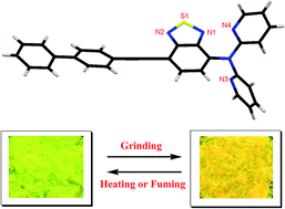
We report the design and synthesis of push–pull benzothiadiazoles (BTDs) of type D1–π–A–π–D2 and D1–π–A–D2. These BTDs show strong charge transfer interaction. BTD 3 shows reversible mechanochromism with color contrast between yellow (crystalline state) and orange (amorphous state). Photophysical and computational studies reveal that the planar orientation of the pyridyl and BTD unit in 2 results in no change in solid state emission whereas non-planar orientation of the dipyridylamine and BTD unit in 3 results in efficient mechanochromism.
2,1,3-Benzothiadiazole (BTD) is a strong acceptor owing to its high electron affinity.5 Our group is involved in the design and synthesis of symmetrical and unsymmetrical donor-substituted BTDs.6 In this contribution we wish to report the synthesis, photophysical and reversible mechanochromic response of dipyridylamine substituted unsymmetrical BTD. The design of the dipyridylamine substituted unsymmetrical BTD is based on the following considerations: (1) possibility of potential intermolecular hydrogen bonding interactions via the N-atoms of dipyridylamine unit. (2) The orientation of the two pyridyl rings of the dipyridylamine unit can provide twisted arrangement to endorse non-parallel packing in solid state. (3) The incorporation of BTD acceptor results in strongly luminescent systems.7
The synthetic route to the push–pull BTDs 2 and 3 are shown in Scheme 1. The Pd-catalyzed Sonogashira cross-coupling reaction of dibromo-BTD with 4-ethynylbiphenyl resulted BTD 1 in 52% yield. In order to study the effect of orientation of pyridyl rings on the mechanochromic behavior, we designed BTD 2 and 3 with acetylene linked pyridyl unit and N-linked dipyridylamine unit respectively. The Pd-catalyzed Sonogashira cross-coupling reaction of BTD 1 with 3-ethynylpyridine resulted BTD 2 in 78% yield. The Ullmann coupling reaction of BTD 1 with dipyridylamine in the presence of CuSO4 and K2CO3 in dichlorobenzene at 180 °C for 48 h resulted BTD 3 in 65% yield. The BTDs 2 and 3 were well characterized by 1H, 13C NMR and HRMS techniques.
![]() | ||
Scheme 1 Synthesis of BTDs 2 and 3. Reaction conditions: (i) PdCl2(PPh3)2, CuI, THF–TEA (1![]() ![]() |
The thermal stability of the push–pull BTDs 2 and 3 were studied using thermogravimetric analysis (TGA) at a heating rate of 10 °C min−1, under nitrogen atmosphere (Fig. S10, ESI†). The decomposition temperatures (5% weight loss) for BTD 2 and 3 were 430 °C and 321 °C respectively. The pyridyl-substituted BTD 2 exhibits higher thermal stability compared to dipyridylamine-substituted BTD 3.
The electronic absorption and emission spectra of BTDs 2 and 3 were recorded in dichloromethane at room temperature (Fig. 1) and the data are compiled in Table S1.† The BTDs 2 and 3 exhibit strong absorption between 300–320 nm, corresponding to π → π* transition and a charge transfer (CT) band between 399–450 nm.8 The low energy transition in BTDs 2 and 3 exhibits red-shift, on increasing the solvent polarity indicating the CT character (Fig. S19; ESI†).9 There is a large red shift (∼65 nm) in the low energy transition of BTD 3 compared to BTD 2, reflecting strong electronic communication between dipyridylamine and BTD moiety. The BTDs 2 and 3 emit green and orange fluorescence at the wavelength of ∼513 nm and ∼578 nm respectively. The emission maxima exhibit red-shift with the increase in solvent polarity.
In order to explore the electronic structure of the BTDs 2 and 3 density functional theory (DFT) calculations were performed at the B3LYP/6-31G** level. In BTD 2 and 3 the HOMO is localized on the electron donating biphenyl, pyridyl, dipyridylamine and the hydrocarbon portion of the BTD unit and LUMO on the electron withdrawing BTD unit reflecting strong donor–acceptor interaction. The incorporation of dipyridylamine in BTD 3 results in lowering of the HOMO–LUMO gap, leading to red shift of the absorption spectrum (Fig. 2).
DFT optimized structure of BTD 2 and 3 exhibits planar and non-planar alignment of the pyridyl rings with respect to BTD core respectively. This indicates that the incorporation of dipyridylamine results in twisted arrangement to endorse non-parallel packing in solid state (Fig. 3).
The mechanochromic properties of unsymmetrical BTDs 2 and 3 were studied by the absorption and emission studies. The crystalline samples of BTDs 2 and 3 absorb at 443 and 481 nm respectively (Fig. S15 and S16; ESI†). Upon grinding, the sample of BTD 2 shows no change in the absorption behavior whereas BTD 3 exhibits red-shifted absorption at 498 nm (Fig. S17; see ESI† for details). The solid samples of BTD 2 show greenish-yellow emission at 538 nm whereas BTD 3 exhibits yellow emission at 556 nm (Fig. S18† and 4). The solid sample of BTD 3 upon grinding using a spatula or a pestle exhibits drastic change in the emission behavior and the emission peak at 556 nm was red shifted to 581 nm (Fig. 4). The solid sample of BTD 2 exhibits no change in the emission upon grinding. The mechanochromic effect of BTD 3 can be reverted to its original color either by annealing or fuming with dichloromethane vapor (Fig. 4). The grinded sample of BTD 3 upon annealing at 150 °C for 35 min or fuming with dichloromethane vapor for 4 min restored the original yellow emission (Fig. 4 and S19 and S20†).
![]() | ||
Fig. 4 Solid state emission spectra and fluorescence colour change induced upon grinding the solid sample of BTD 3. |
The single crystal of BTD 3 was obtained via slow diffusion of ethanol into the dichloromethane solution at room temperature. BTD 3 crystallizes in the monoclinic space group P21/c and exhibits twisted structural arrangement (Fig. 5). The dihedral angle between the planes containing the BTD core and the pyridyl rings of dipyridylamine unit was found to be 69.90° and 82.82°. The important bond lengths and bond angles are listed in the Table S3 (see ESI† for details). The packing diagram of BTD 3 exhibits H-bonding interaction N4⋯H4 (2.59 Å) between the dipyridylamine nitrogen (N4) and the BTD hydrogen H4. The H-bonding interaction between the dipyridylamine and BTD results in twisted arrangement in the crystalline state.
In order to gain insight into the mechanism of mechanochromism in BTD 3 powder X-ray diffraction (PXRD) analysis was performed (Fig. 6). The BTD 3 exhibit intense and sharp diffraction peaks before grinding reflecting the crystalline character. The BTD 3 sample show very weak diffraction peaks upon grinding suggesting the transition to amorphous state.10 The ground sample of BTD 3 exhibits sharp diffraction peaks when subjected to heating or fuming indicating the transformation to the crystalline phase. This study clearly concludes that the mechanochromism in BTD 3 is associated with the morphology change from the crystalline state to the amorphous state and vice versa.
Footnote |
† Electronic supplementary information (ESI) available: Experimental procedures, NMR spectra, UV-vis spectra, the mechanochromic effect, computational data and crystal structural data for BTD 2 and 3. CCDC 1020099. For ESI and crystallographic data in CIF or other electronic format see DOI: 10.1039/c4ra09921d |
This journal is © The Royal Society of Chemistry 2014 |