Dicationic imidazolium-based ionic liquids: a new strategy for non-toxic and antimicrobial materials

Izabelle M. Gindria, Danyal A. Siddiquia, Pooja Bhardwajb, Lucas C. Rodrigueza, Kelli L. Palmerb, Clarissa P. Frizzoc, Marcos A. P. Martinsc and Danieli C. Rodrigues*a
aDepartment of Bioengineering, University of Texas at Dallas, Richardson, TX 75080, USA. E-mail: danieli@utdallas.edu
bDepartment of Molecular and Cell Biology, University of Texas at Dallas, Richardson, TX 75080, USA
cDepartment of Chemistry (NUQUIMHE), Universidade Federal de Santa Maria, Santa Maria, 97105-900, Brazil

Received 5th September 2014 , Accepted 12th November 2014

First published on 12th November 2014


Abstract

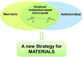
New dicationic imidazolium-based ionic liquids (ILs) were synthesized, characterized and tested in regards to cytotoxicity and antimicrobial activity. Insertion of a new cationic head and use of organic anions increased the biocompatibility of the ILs developed. IC50 (concentration necessary to inhibit 50% of enzymatic activity) values obtained were considerably higher than those described for monocationic ILs, which indicates an improvement in cytocompatibility. Antimicrobial activity against bacterial species of clinical relevance in wounds and the oral environment was tested. The results showed that ILs were effective in inhibiting bacterial growth even below the minimum inhibitory concentration (MIC). It was observed that structural features that confer higher hydrophobicity to ILs decreased both the IC50 and MIC simultaneously. However, it was possible to establish an equilibrium between those two effects, which gives the safe range of concentrations that ILs can be employed. The results demonstrated that the dicationic-imidazolium-based ILs synthesized may constitute a potent strategy for applications requiring non-toxic materials exhibiting antimicrobial activity.


1. Introduction

Ionic liquids (ILs) are a class of low temperature molten salts, comprised of an amphiphilic cationic moiety and a weakly coordinated anion.1 Even though they were described almost a century ago, ILs have recently attracted interest in an assorted array of applications, ranging from synthetic processes in chemistry,2–4 to a number of biological processes,5 and utilization as active pharmaceutical ingredients (API).6 The most attractive property of ILs is the flexibility or ‘tunability’ in the design of physical, chemical and biological properties by changing the structure of the cation and anion.7 Such possibilities have driven phenomenal interest in ILs synthesis. Commonly studied ILs are comprised of bulky, N-containing organic cations (e.g., imidazole and pyridine) in combination with anions, ranging from simple inorganic ions (e.g., halides) to more complex organic species (e.g., sugars and amino acids). Imidazolium-based ILs are among the most studied classes of ILs and recently, dicationic imidazolium-based ILs have emerged as a new option for applications, for instance, uses as solvents,8 surfactant,9,10 lubricant,11,12 and for nanoparticle coating.13 Although ILs have been proposed as new “green strategy”, problems associated with cytotoxicity and environmental contamination have been reported.12,14,15

Cytotoxicity is the property of a compound that can result in a toxic effect against human cells, and this effect has been broadly reported for monocationic ILs. For a homologous series, cationic alkyl chain length is the main factor associated with toxic effects.16–19 Increase in alkyl chain length is related to an increase in hydrophobicity and consequently cell damage.16 Anionic moieties have been discussed to also play an important role in toxicity, however to a lesser extent in comparison to cations. Following the same trend observed for cations, more hydrophobic anions tend to exert a higher toxic effect than those considered biocompatible and highly hydrophilic, such as chloride.20,21 Considering these findings, we hypothesize that introducing a new cationic moiety in the imidazolium cation can reduce the toxicity of ILs due to an increase in polarity of the IL structure. Recently, Steudte et al.,22 investigated the toxicity of pyridinium and imidazolium-based dicationic ILs. Dicationic imidazolium-based ILs were found to have considerably lower toxicity in comparison to analogous monocationic ILs, which supports our hypothesis. Furthermore, organic moieties such as amino acids and ascorbic acid are also considered as a strategy to design non-toxic ILs.23

Antimicrobial and antibiofilm activity of monocationic imidazolium-based ILs have been investigated.1,15,24–28 The introduction of longer alkyl chains on the imidazolium cation generally results in potent activity, which consequently lowers the minimal inhibitory concentration (MIC) against microorganisms.1,27 Łuczak et al. investigated the role of cation and anions on the IL antimicrobial activity.27 They observed that the higher hydrophobicity of both moieties played a key role in increasing antimicrobial effectiveness, but the anion influence was relatively smaller as observed in the cytotoxic experiment. The antimicrobial activity and cytotoxicity of ILs are directly related, as observed for conventional surfactants and cationic antimicrobial peptides.29 The antimicrobial mechanism of action of these compounds is the targeting of cell membranes, which can compromise both microbial and human cells.29 For example, cationic antimicrobial peptides are known to exert a more specific toxic effect against gram-negative bacterial strains. However, cell necrosis is also observed in treatments with these compounds, due to their intrinsic cytotoxicity.29 Generally, in a homologous series of ILs, more hydrophobic structures result both in lower MIC and IC50 (dose to inhibit 50% of enzymatic activity) values. This can be considered a nonspecific toxic effect triggered by these compounds, in which cell toxicity may be associated to a side effect of antimicrobial activity.30,31 Therefore, a current drawback in antimicrobial applications of ILs is that effectiveness against bacteria comes with the cost of toxicity to host cells, which restricts the biological applications of such compounds.

In this study, we have developed a series of non-toxic ILs with antimicrobial activity against clinically relevant bacteria for in vivo applications. ILs were designed with structural features such as dicationic moiety and organic anions, which were observed to have a reduced toxic effect. Two imidazolium-based cations with different alkyl chain length connecting imidazolium heads were investigated (n = 8 and n = 10) in order to study the differences in hydrophobicity provided by the cationic moiety. A monocationic IL, with analogous structure to the dicationic IL, was also evaluated to compare the structural effect on cytotoxicity. Anions amino acid- and ascorbate-based were selected, as well as bromide.

2. Results and discussion

IL compounds synthesized are liquid at a temperature of 25 °C, with the exception of IL-7. Structures were rationalized in terms of finding a balance between hydrophobicity and hydrophilicity. The structures of the designed compounds are illustrated in Fig. 1, as well as the monocationic IL. The additional imidazolium head on the cationic moiety was proposed in an attempt to reduce toxicity, which has been previously reported for imidazolium-based monocationic ILs.14,17,32 Antimicrobial activity against different groups of gram-positive and gram-negative clinically relevant bacterial strains in wounds and oral environment was also accessed for dicationic based ILs and correlated with IC50 values.
image file: c4ra09906k-f1.tif
Fig. 1 Structure of investigated ILs.

2.1. Cytotoxicity of dicationic imidazolium-based ILs

Cytotoxicity screening of all ILs synthesized (Fig. 1) was based on a 24 h toxicity assay using MC3T3-E1 pre-osteoblast cells. IC50 values were calculated using a dose–response model, which was obtained from sigmoidal fitting of response curves of percent inhibition versus logarithmic concentration of IL using Origin Software. Calculated IC50 results are shown in Table 1 while the graphs are demonstrated in Fig. 2(a) and (b) for ILs 1–6, IL-13, and ILs 7–12, respectively.
Table 1 IC50 values of dicationic imidazolium-based ILs
IL IC50 (mM)
IL-1 24.6 ± 3.5
IL-2 3.6 ± 0.6
IL-3 8.3 ± 3.0
IL-4 12.5 ± 0.2
IL-5 25.7 ± 8.7
IL-6 24.2 ± 10.3
IL-7 12.3 ± 0.1
IL-8 3.1 ± 1.2
IL-9 8.5 ± 1.5
IL-10 12.3 ± 0.5
IL-11 12.9 ± 1.1
IL-12 13.9 ± 2.7
IL-13 1.51 ± 0.2


image file: c4ra09906k-f2.tif
Fig. 2 Dose–response curves of ILs with (a) 8 and (b) 10 carbons in the cation alkyl chain with different anionic moieties as illustrated in Fig. 1.

From non-linear fitting, r2 values obtained were above 0.95. The relationship between chemical structure and toxicity was investigated. ILs from IL-1 to IL-7 and IL-13 had the alkyl chain of imidazolium dication with 8 carbons while ILs from IL-8 to IL-12 had the alkyl chain with 10 carbons. The comparison between IC50 values obtained for IL-1 and IL-13, revealed that dicationic IL had expressive higher values than the analogous monocationic. This result supports our hypothesis that dicationic IL toxicity was reduced, in comparison to monocationic IL, due to an additional cationic head. This result implies that there is a possibility of using higher concentrations of ILs without triggering toxic effects against bone cells. Cation hydrophobic effect has been previously discussed in the literature for monocationic ILs.16,17,32 McLaughlin et al.16 observed that the cytotoxicity of ILs was governed by alkyl chain length. It was found that the higher toxicity exerted by more hydrophobic ILs (with longer alkyl chains) is associated with an increase in membrane permeability and change in the physical properties of the lipid bilayer.16 We assume that, in dicationic IL, the alkyl chain is “trapped” between the two cationic imidazolium heads losing the ability to interact with cell membranes, which reduces its toxic effect. However, the effect of hydrophobicity of the two different dicationic moieties on the toxicity of ILs was also observed. The compounds with cationic alkyl chain with 10 carbons (ILs 7–12) showed an increase in toxicity compared with ILs with 8 carbons (ILs 1–6). The anion also played an important effect, ILs with more hydrophobic anions such as phenylalanine-based (IL-3 and IL-9) had lower IC50 values than those prepared with more hydrophilic anions such as bromide (IL-1 and IL-7) and histidine-based (IL-5 and IL-11). The anion influence on IL cytotoxicity has been studied both theoretically and experimentally by Stolte et al.20 The authors observed an increased toxic effect of more hydrophobic anions and this feature was related to stronger interactions with cell membranes and hydrophobic protein domains, which may potentially disrupt essential physiological functions.20 Moreover, results obtained in this present work corroborate the trend observed for cholinum-based ILs synthesized with amino acids as the anionic moiety,23 in which higher toxicity was correlated with hydrophobic features of amino acids structures.

The lowest IC50 values were found for ILs synthesized with ascorbate as anion (IL-2 and IL-8). This was in fact an unexpected behavior given the hydrophilic nature of this anion. To better understand this result, cell cultures exposed to ascorbate-based ILs were further investigated by optical microscopy. Microscopy revealed the formation of IL crystals in concentrations at and above 10 mM. An interesting finding was the affinity between those crystals with pre-osteoblast cells. In Fig. 3(a) and (b), it is possible to verify that the crystals (red arrows) tend to stay on cells surfaces (black arrows). We speculate that these IL crystals could be triggering an additional toxic effect to the cells, reducing the IC50. The affinity between ascorbate/ascorbic acid and osteoblasts-like cells has been previously reported in the literature.33 Furthermore, the transport of polar anionic compounds across biological membranes was investigated by Vincent et al.34 They observed that this process may be facilitated when anions are paired with lipophilic ammonium cations, which work as a phase transfer. Therefore, we hypothesize that cationic moieties could be acting as a phase transfer, increasing the affinity of ascorbate anionic moiety with cells.


image file: c4ra09906k-f3.tif
Fig. 3 Adsorption of ascorbic acid based IL crystals (red arrows) on the surface of cells (black arrows) with magnification of (a) 40× and (b) 20×.

2.2. Antimicrobial evaluation

The minimum inhibitory concentration (MIC) of all ILs under study was evaluated. MICs were determined for two groups of bacterial strains. The first group (group 1) was comprised of Enterococcus faecalis, Pseudomonas aeruginosa and Staphylococcus epidermidis (gram-positive), which are opportunistic pathogens associated with infections on biomedical devices and responsible for up to 60% of all prosthetic infections since 1980.35–38 These microorganisms have also been associated with oral diseases such as peri-implantitis.39–41 MIC values were determined and are given in Table 2. ILs were more effective towards S. epidermidis while a lower antimicrobial effect was observed for E. faecalis. Another interesting finding was the influence of structural features on antimicrobial activity. The two gram-positive organisms were more sensitive to differences in IL hydrophobicity than the gram-negative organisms, as can be observed in Table 2. While MIC varied for E. faecalis and S. epidermidis, the results of P. aeruginosa were similar regardless the IL used. This trend is in accordance with previous findings reported in the literature and can be explained by the differences in cell envelope composition of gram-positive and gram-negative microorganisms.1 The higher activity against gram-positive strains were observed for more hydrophobic ILs, composed by cationic moiety with n = 10 (IL-9–IL-12), as can be observed in Table 2.
Table 2 MIC (mM) for dicationic imidazolium-based ILs
Ionic liquid MIC (mM)
E. faecalis P. aeruginosa S. epidermidis
IL-1 79 79 79
IL-2 156 20 10
IL-3 79 20 10
IL-4 79 20 20
IL-5 79 20 10
IL-6 79 20 10
IL-7 39 39 2
IL-8 79 20 20
IL-9 5 20 5
IL-10 10 20 2
IL-11 20 20 5
IL-12 20 20 2


The second group (group 2) tested was comprised of gram-positive oral streptococcal species (Streptococcus mutans, Streptococcus salivarius, Streptococcus sanguinis, Streptococcus gordonii, and Streptococcus uberis). MIC results are summarized in Table 3. Streptococci are classified as cariogenic bacteria and produce acid metabolites, which decreases pH and leads to tooth surface demineralization.42 Lowered pH is also associated with surface damage of dental implants in which active dissolution of metal ions in vivo can be triggered, ultimately leading to implant failure.43 These bacterial strains are additionally associated with oral diseases such as root canal and peri-implantitis.42 In general, higher antimicrobial activity of ILs with 10 methylene groups in the cationic alkyl chain length (ILs 7–12) was observed. As mentioned above, gram-positive strains are more sensitive to a difference in hydrophobicity of ILs, which can explain this trend. IL-1 and IL-2 were observed to be less effective against those microorganisms, which can be related to a higher hydrophilicity of the anionic moiety of these compounds. Unlike the results observed with cells, the toxic effect of ILs with ascorbic acid as anionic moiety (IL-2 and IL-8) was not observed, which supports our hypothesis of increase in affinity between those ILs with osteoblast-like cells.

Table 3 MIC (mM) for dicationic imidazolium-based ILs
Ionic liquid MIC (mM)
S. sanguinis S. salivarius S. mutans S. gordonii S. uberis
IL-1 79 79 39 39 20
IL-2 79 39 39 39 39
IL-3 20 20 10 79 20
IL-4 39 20 10 10 10
IL-5 20 20 20 10 20
IL-6 20 20 10 20 20
IL-7 10 5 2 5 2
IL-8 20 20 10 20 10
IL-9 10 5 5 5 2
IL-10 10 5 5 5 5
IL-11 10 5 5 5 2
IL-12 10 5 5 5 5


In order to evaluate the correlation between MIC and IC50, these results were plotted in Fig. 4(a) and (b) for both groups of bacteria investigated. Interestingly, a conflict between cytotoxicity and antimicrobial activity does not occur for some ILs, mainly when considering oral bacteria. When the red line (corresponding to IC50 values) is above MIC bars, the IL can be considered a strong candidate for biological applications. This means that in contact with both bacteria and host cells, the IL is able to limit bacterial growth but not host cell proliferation.


image file: c4ra09906k-f4.tif
Fig. 4 Comparison between MIC and IC50 results of ILs for (a) group 1 and (b) group 2.

To further investigate the antimicrobial effects of ILs, bacteria growth rate (GR) experiments were performed to investigate IL inhibitory effect in sub-MIC concentrations. This was important because the MIC measurements gave only an endpoint (24 h) view of bacterial growth effects. Bacterial growth was monitored over a 24 h period using a 96-well spectrophotometer. Due to technical limitations in generating a microaerophilic atmosphere, only bacteria from testing group 1 were evaluated. We chose a gram-positive and gram-negative strains that ILs were effective against (P. aeruginosa and S. epidermidis, respectively). As expected, no growth was observed for cultures with MIC IL concentrations. P. aeruginosa showed a decreased growth rate at sub-MIC concentrations of IL-1–IL-6 and IL-7–IL-12 as shown in Fig. 5(a) and (b), respectively. Similar results were observed for S. epidermidis, in which bacterial proliferation was decreased in the presence of IL-7–IL-12 (Fig. 5(d)). However for IL-1–IL-6, in sub-MIC concentration, it was observed bacterial growth similar to positive control, indicating loss of ILs antimicrobial activity under those conditions. These observations indicate that even at sub-MIC concentrations for some ILs, inhibition of bacterial growth occurs. This further points towards a potential use of these compounds as antibacterial materials.


image file: c4ra09906k-f5.tif
Fig. 5 Growth rate experiments for P. aeruginosa (a) IL-1–IL-6, (b) IL-7–IL-12 and S. epidermidis (c) IL-1–IL-6, (d) IL-7–IL-12.

High efficiency of ILs against clinically relevant bacteria and low toxicity of tested ILs emerges as a powerful strategy for applications in the biomedical field. Although studies involving cytotoxicity and antimicrobial activity of ILs have been widely described, there are only a few reports exploring such features in dicationic imidazolium-based IL.22,25 Also, this is the first study involving antimicrobial activity of ILs against oral bacteria. Hence, ILs designed in this work, which demonstrated high biocompatibility with bone cells and antimicrobial activity have potential application in this field. Investigation of such strains provided a better idea about how these materials may work to protect the oral environment.

3. Experimental

3.1. Materials

The chemicals used were received as follow: 1,8-dibromooctane (Alfa Aesar, Ward Hill, MA, USA); L-phenylalanine and L-leucine (MP Biomedicals, Santa Ana, CA, USA); L-ascorbic acid (Sigma Aldrich, St. Louis, MO, USA); 1-methylimidazole, 1,10-dibromodecane, L-histidine, L-methionine, AMBERLITE IRN-78 OH and ethyl ether (Acros Organics, NJ, USA); acetonitrile and ethanol (Fisher Science, Waltham, MA, USA). All chemical products were of high-grade purity and were used without additional purification.

3.2. Synthesis and characterization

The ILs 1 and 7 were synthesized according to procedures described in the literature.13,44 Fifty mmol of 1-methylimidazole and acetonitrile (50 mL) were added to a flask connected to a reflux condenser under inert atmosphere and stirred for 2 minutes. Then, 25 mmol of dibromide alkyl were slowly added for synthesis of ILs 1 and 7 (Fig. 1). The reaction mixture was maintained at 70 °C for 72 h. Finally, the solvent was evaporated under reduced pressure, washed with diethyl ether, and the mixture was dried under vacuum (4 mbar, 50 °C, 48 h) to obtain a product with high purity. To synthesize the monocationic IL (IL-13), the same procedure was used with an equimolar (10 mM) ratio for 1-methylimidazole and 1-bromooctane, according to the literature.7

ILs 2–6 and 8–12 (Fig. 1) were synthesized according to the procedure proposed by Fukumoto et al. performed with slight modifications.45 1,8-Bis(3-methylimidazolium-1-yl)octane hydroxide and 1,10-bis(3-methylimidazolium-1-yl)decane hydroxide were prepared from 1,8-bis(3-methylimidazolium-1-yl)octane bromide and 1,10-bis(3-methylimidazolium-1-yl)decane bromide ethanolic solutions, respectively using anion exchange resin. ILs 2–6 and 8–12 (Fig. 1) were prepared by adding dropwise 1,8-bis(3-methylimidazolium-1-yl)octane hydroxide or 1,10-bis(3-methylimidazolium-1-yl)decane dihydroxide ethanolic to a slight excess equimolar ascorbic-acid or amino-acid ethanolic solution. The mixture was then stirred at 25 °C for 12 h. Then solvent was evaporated at 70 °C under vacuum. Nine mL of acetonitrile and 1 mL of methanol were added to the reaction mixture under vigorous stirring. The mixture was then filtered to remove excess amino acid or ascorbic acid. The filtrate was subsequently evaporated to remove solvents and the product was dried in vacuum for 48 h at 70 °C. The structures of the resulting ILs were confirmed by 1H and 13C NMR spectroscopy (500 MHz Bruker spectrometer, Billerica, MA) and mass spectrometry (Shimadzu, Kyoto, KYT). The NMR experiments were performed at 25[thin space (1/6-em)]°C, and for the analysis of each compound 20 mg of IL was dissolved in 0.5 mL of DMSO-d6. The thermal characterization was performed using differential scanning calorimetry at a heating rate of 10 °C/min (DSC, PerkinElmer, Waltham, MA).

1,8-Bis(3-methylimidazolium-1-yl)octane dibromide (IL-1). C16H28Br2N4, MW: 436.23 g mol−1; from 8.2 g (50 mmol) of 1H-methylimidazole, and 13.6 g (100 mmol) of 1,8-dibromooctane, 21.1 g of IL-1 was obtained (yield: 97%); Tg: −37.91 °C; 1H NMR (500 MHz, DMSO-d6): δ 9.42 (s, 2H), 7.91 (s, 2H), 7.81 (s, 2H), 4.21 (t, 4H), 3.89 (s, 6H), 1.78 (qui, 4H), 1.25 (m, 8H). 13C NMR (125 MHz, DMSO-d6): δ 136.5 (2C), 123.5 (2C), 122.2 (2C), 48.7 (2C), 35.7 (2C), 29.3 (2C), 28.1 (2C), 25.3 (2C). MS m/z molecular ion: 276.081 (cation), 79.332 (anion).
1,8-Bis(3-methylimidazolium-1-yl)octane diascorbate (IL-2). C28H44N4O12, MW: 628.676 g mol−1; from 4.3 g (10 mmol) of IL-1, and 3.5 g (20 mmol) of L-ascorbic acid, 4.4 g of IL-2 was obtained (yield: 71%); Tg: −26.65 °C; 1H NMR (500 MHz, DMSO-d6): δ 9.21 (s, 2H), 7.77 (s, 2H), 7.70 (s, 2H), 4.16 (t, 4H), 4.07 (d, 2H, ascorbate), 3.86 (s, 6H), 3.45 (m, 6H, ascorbate), 1.77 (qui, 4H), 1.27 (m, 8H). 13C NMR (125 MHz, DMSO-d6): δ 172.99 (2C, ascorbate), 136.43 (2C), 123.45 (2C), 122.08 (2C), 113.01 (2C, ascorbate), 79.19 (2C, ascorbate), 71.90 (2C, ascorbate), 63.81 (2C, ascorbate), 48.52 (2C), 35.58 (2C), 29.21 (2C), 28.01 (2C), 25.26 (2C). MS m/z molecular ion: 276.081 (cation), 175.059 (anion).
1,8-Bis(3-methylimidazolium-1-yl)octane diphenylalanine (IL-3). C36H52N6O4, MW: 632.850 g mol−1; from 4.3 g (10 mmol) of IL-1, and 3.3 g (20 mmol) of L-phenylalanine, 5.0 g of IL-3 was obtained (yield: 82%); Tg: −25.18 °C; 1H NMR (500 MHz, DMSO-d6): δ 9.67 (s, 2H), 7.78 (s, 2H), 7.75 (s, 2H), 7.21 (m, 8H, phenylalanine), 7.13 (t, 2H, phenylalanine), 4.16 (t, 4H), 3.86 (s, 6H), 3.07 (d, 2H, phenylalanine), 3.01 (d, 2H, phenylalanine), 2.46 (t, 2H, phenylalanine), 1.76 (qui, 4H), 1.25 (m, 8H). 13C NMR (125 MHz, DMSO-d6): δ 176.48 (2C, phenylalanine), 140.91 (2C, phenylalanine), 136.97 (2C), 128.95 (2C, phenylalanine), 127.51 (4C, phenylalanine), 125.05 (2C, phenylalanine), 123.24 (2C), 121.90 (2C), 57.71 (2C, phenylalanine), 48.32 (2C), 42.11 (2C, phenylalanine), 35.31 (2C), 29.09 (2C), 27.82 (2C), 25.08 (2C). MS m/z molecular ion: 276.081 (cation), 164.210 (anion).
1,8-Bis(3-methylimidazolium-1-yl)octane dileucine (IL-4). C28H52N6O4, MW: 536.762 g mol−1; from 4.3 g (10 mmol) of IL-1, and 2.6 g (20 mmol) of L-leucine, 4.2 g of IL-4 was obtained (yield: 78%); Tg: −40.07 °C; 1H NMR (500 MHz, DMSO-d6): δ 9.73 (s, 2H), 7.82 (s, 2H), 7.74 (s, 2H), 4.18 (t, 4H), 3.87 (s, 6H), 2.79 (t, 2H, leucine), 1.77 (qui, 4H), 1.70 (t, 2H, leucine), 1.41 (t, 2H, leucine), 1.27 (m, 8H), 1.06 (m, 2H, leucine), 0.85 (d, 6H, leucine), 0.81 (d, 6H, leucine). 13C NMR (125 MHz, DMSO-d6): δ 177.61 (2C, leucine), 137.26 (2C), 123.44 (2C), 122.12 (2C), 54.46 (2C, leucine), 48.51 (2C), 45.61 (2C, leucine), 35.53 (2C), 29.28 (2C), 28.04 (2C), 25.29 (2C), 24.58 (2C, leucine), 23.65 (3C, leucine), 21.83 (3C, leucine). MS m/z molecular ion: 276.081 (cation), 130.367 (anion).
1,8-Bis(3-methylimidazolium-1-yl)octane dihistidine (IL-5). C28H44N10O4, MW: 584.726 g mol−1; from 4.3 g (10 mmol) of IL-1, and 3.1 g (20 mmol) of L-histidine, 4.7 g of IL-5 was obtained (yield: 81%); Tg: −21.11 °C; 1H NMR (500 MHz, DMSO-d6): δ 9.54 (s, 2H), 7.80 (s, 2H), 7.73 (s, 2H), 7.42 (s, 2H, histidine), 6.64 (s, 2H, histidine), 4.16 (t, 4H), 3.87 (s, 6H), 3.06 (d, 2H, histidine), 2.88 (d, 2H, histidine), 2.45 (d, 2H, histidine), 1.77 (qui, 4H), 1.23 (m, 12H). 13C NMR (500 MHz, DMSO-d6): δ 176.36 (2C, histidine), 136.83 (2), 133.80 (2C, histidine), 123.30 (2C), 121.97 (2C), 56.14 (2C, histidine), 48.40 (2C), 35.41 (2C), 33.51 (2C, histidine), 29.09 (2C), 27.86 (2C), 25.12 (2C). MS m/z molecular ion: 276.081 (cation), 154.287 (anion).
1,8-Bis(3-methylimidazolium-1-yl)octane dimethionine (IL-6). C26H48N10O4, MW: 600.882 g mol−1; from 4.6 g (10 mmol) of IL-1, and 3.0 g (20 mmol) of L-methionine, 4.3 g of IL-6 was obtained (yield: 75%); Tg: −47.41 °C; 1H NMR (500 MHz, DMSO-d6): δ 9.66 (s, 2H), 7.82 (s, 2H), 7.75 (s, 2H), 4.16 (t, 4H), 3.88 (s, 6H), 2.92 (d, 2H, methionine), 2.48 (m, 4H, methionine), 2.00 (s, 6H, methionine), 1.78 (m, 4H), 1.78 (t, 2H, methionine), 1.51 (t, 2H), 1.27 (m, 12H). 13C NMR (125 MHz, DMSO-d6): δ 176.56 (2C, methionine), 137.19 (2C), 123.34 (2C), 122.05 (2C), 55.29 (2C, methionine), 48.39 (2C), 35.86 (2C, methionine), 35.42 (2C), 30.78 (2C, methionine), 29.18 (2C), 2.92 (2C), 25.18 (2C), 14.54 (2C, methionine). MS m/z molecular ion: 276.081 (cation), 148.271 (anion).
1,10-Bis(3-methylimidazolium-1-yl)decane dibromide (IL-7). C18H32Br2N4, MW:464.28 g mol−1; from 8.2 g (50 mmol) of 1H-methylimidazole, and 15.0 g (100 mmol) of 1,10-dibromodecane, 22.3 g of IL-7 was obtained (yield: 96%); MP: 130.77 °C or Tg: −21.21 °C; 1H NMR (500 MHz, DMSO-d6): δ 9.26 (s, 2H), 7.83 (s, 2H), 7.75 (s, 2H), 4.18 (t, 4H), 3.88 (s, 6H), 1.78 (m, 4H), 1.25 (m, 12H). 13C NMR (125 MHz, DMSO-d6): δ 136.3 (2CH), 123.4 (2CH), 122.1 (2CH), 48.7 (2CH2), 35.6 (2CH3), 29.2 (2CH2), 28.5 (2CH2), 28.1 (2CH2), 25.3 (2CH2). MS m/z molecular ion: 304.262 (cation), 79.350 (anion).
1,10-Bis(3-methylimidazolium-1-yl)decane diascorbate (IL-8). C28H44N4O12, MW: 654.714 g mol−1; from 4.6 g (10 mmol) of IL-7, and 3.5 g (20 mmol) of L-ascorbic acid, 4.9 g of IL-8 was obtained (yield: 75%); Tg: −60.13 °C; 1H NMR (500 MHz, DMSO-d6): δ 9.14 (s, 2H), 7.76 (s, 2H), 7.69 (s, 2H), 4.22 (d, 2H, ascorbate), 4.14 (t, 4H), 3.86 (s, 6H), 3.43 (m, 6H, ascorbate), 1.77 (qui, 4H), 1.25 (m, 12H). 13C NMR (125 MHz, DMSO-d6): δ 172.45 (2C, ascorbate), 136.47 (2C), 123.55 (2C), 122.18 (2C), 114.48 (2C, ascorbate), 77.70 (2C, ascorbate), 70.76 (2C, ascorbate), 63.23 (2C, ascorbate), 48.72 (2C), 35.58 (2C), 29.35 (2C), 28.70 (2C), 28.32 (2C), 25.46 (2C). MS m/z molecular ion: 304.262 (cation), 175.279 (anion).
1,10-Bis(3-methylimidazolium-1-yl)decane diphenylalanine (IL-9). C36H52N6O4, MW: 632.850 g mol−1; from 4.6 g (10 mmol) of IL-7, and 3.3 g (20 mmol) of L-phenylalanine, 4.7 g of IL-9 was obtained (yield: 74%); Tg: −48.25 °C; 1H NMR (500 MHz, DMSO-d6): δ 9.49 (s, 2H), 7.78 (s, 2H), 7.72 (s, 2H), 7.19 (m, 8H, phenylalanine), 7.12 (t, 2H, phenylalanine), 4.15 (t, 4H), 3.85 (s, 6H), 3.02 (d, 4H, phenylalanine), 2.41 (t, 2H, phenylalanine), 1.76 (qui, 4H), 1.23 (m, 12H). 13C NMR (125 MHz, DMSO-d6): δ 176.32 (2C, phenylalanine), 141.40 (2C, phenylalanine), 137.07 (2C), 129.12 (2C, phenylalanine), 127.68 (4C, phenylalanine), 125.16 (2C, phenylalanine), 123.44 (2C), 122.12 (2C), 57.98 (2C, phenylalanine), 48.57 (2C), 42.58 (2C, phenylalanine), 35.55 (2C), 29.34 (2C), 28.63 (2C), 28.26 (2C), 25.41 (2C). MS m/z molecular ion: 304.262 (cation), 164.184 (anion).
1,10-Bis(3-methylimidazolium-1-yl)decane dileucine (IL-10). C30H56N6O4, MW: 564.82 g mol−1; from 4.6 g (10 mmol) of IL-7, and 2.6 g (20 mmol) of L-leucine, 4.3 g of IL-10 was obtained (yield: 78%); Tg: −33.92 °C; 1H NMR (500 MHz, DMSO-d6): δ 9.87 (s, 2H), 7.84 (s, 2H), 7.77 (s, 2H), 4.18 (t, 4H), 3.88 (s, 6H), 2.78 (t, 2H, leucine), 1.77 (qui, 4H), 1.70 (t, 2H, leucine), 1.41 (t, 2H, leucine), 1.24 (m, 12H), 1.05 (m, 2H, leucine), 0.83 (d, 6H, leucine), 0.80 (d, 6H, leucine). 13C NMR (125 MHz, DMSO-d6): δ 179.72 (2C, leucine), 137.32 (2C), 123.25 (2C), 121.96 (2C), 54.39 (2C, leucine), 48.34 (2C), 45.85 (2C, leucine), 35.30 (2C), 29.23 (2C), 28.47 (2C), 28.10 (2C), 25.25 (2C), 24.41 (2C, leucine), 23.48 (3C, leucine), 21.63 (3C, leucine). MS m/z molecular ion: 304.262 (cation), 130.367 (anion).
1,10-Bis(3-methylimidazolium-1-yl)decane dihistidine (IL-11). C30H48N10O4, MW: 612.780 g mol−1; from 4.6 g (10 mmol) of IL-7, and 3.1 g (20 mmol) of L-histidine, 4.4 g of IL-11 was obtained (yield: 72%); Tg: −39.33 °C; 1H NMR (500 MHz, DMSO-d6): δ 9.55 (s, 2H), 7.81 (s, 2H), 7.73 (s, 2H), 7.42 (s, 2H, histidine), 6.64 (s, 2H, histidine), 4.16 (t, 4H), 3.87 (s, 6H), 3.06 (d, 2H, histidine), 2.88 (d, 2H, histidine), 2.46 (d, 2H, histidine), 1.77 (qui, 4H), 1.23 (m, 12H). 13C NMR (125 MHz, DMSO-d6): δ 177.08 (2C, histidine), 137.02 (2), 134.08 (2C, histidine), 123.46 (2C), 122.14 (2C), 56.53 (2C, histidine), 48.58 (2C), 35.55 (2C), 33.15 (2C, histidine), 29.34 (2C), 28.60 (2C), 28.24 (2C), 25.39 (2C). MS m/z molecular ion: 304.262 (cation), 154.287 (anion).
1,10-Bis(3-methylimidazolium-1-yl)decane dimethionine (IL-12). C28H52N6O4S2, MW: 600.882 g mol−1; from 4.6 g (10 mmol) of IL-7, and 3.0 g (20 mmol) of L-methionine, 4.8 g of IL-12 was obtained (yield: 80%); Tg: −56.42 °C; 1H NMR (500 MHz, DMSO-d6): δ 9.91 (s, 2H), 7.87 (s, 2H), 7.80 (s, 2H), 4.20 (t, 4H), 3.90 (s, 6H), 2.87 (d, 2H, methionine), 2.48 (m, 4H, methionine), 2.00 (s, 6H, methionine), 1.79 (m, 4H), 1.79 (t, 2H, methionine), 1.47 (t, 2H), 1.24 (m, 12H). 13C NMR (125 MHz, DMSO-d6): δ 176.21 (2C, methionine), 137.05 (2C), 123.36 (2C), 122.04 (2C), 55.18 (2C, methionine), 48.49 (2C), 35.57 (2C, methionine), 35.46 (2C), 30.72 (2C, methionine), 29.27 (2C), 28.56 (2C), 28.19 (2C), 25.34 (2C), 14.52 (2C, methionine). MS m/z molecular ion: 304.262 (cation), 148.236 (anion).
1-Octyl-3-methylimidazolium bromide (IL-13). C12H23BrN2, MW: 275.23 g mol−1; 1H NMR (500 MHz, DMSO-d6): δ 9.25 (s, 1H), 7.83 (s, 1H), 7.76 (s, 1H), 4.19 (t, 2H), 3.88 (s, 3H), 1.79 (m, 2H), 1.26 (m, 10H). 0.87 (t, 3H). 13C NMR (125 MHz, DMSO-d6): δ 136.46 (1C), 123.54 (1C), 122.22 (1C), 48.71 (1C), 35.74 (1C), 31.13 (1C), 29.37 (1C), 28.45 (1C), 28.30 (1C), 25.46 (1C), 22.02 (1C), 13.91 (1C).

3.3. Cytotoxicity evaluation

Cytotoxicity was evaluated in vitro using osteoblast cell culture (mouse pre-osteoblast cell line MC3T3-E1). Cells were cultured according to standard procedures (culture in alpha minimum essential media supplemented with 10% fetal bovine serum) and incubated at 37 °C in a humidified atmosphere. Osteoblasts were seeded at a density of 10[thin space (1/6-em)]000 cells per well in 96-well microtiter plates. After 24 hours of incubation, medium was removed and replaced with fresh medium containing IL dilutions at the concentration range of 10−8 M to 10−1 M. After 24 hours, the wells were washed with PBS, then 100 μL of media and 10 μL of 3-[4,5-dimethylthiazol-2-yl]-2,5-diphenyltetrazolium bromide (MTT) reagent were added to each microtiter plate well and cells returned to incubation for 4 h. After this period, 100 μL of detergent solution was added to each well and the plate was incubated overnight. Absorption was measured at 570 nm with a Spectrophotometer (Biotek, Winooski, VT). Percentage cell viability was calculated relative to untreated control wells at each time point after subtraction of the blank value.18 Microscopy was performed using an Olympus IX83 Microscope (Olympus, Tokyo, Tokyo, JA).

3.4. Antimicrobial activity

Enterococcus faecalis V583 (gram-positive), Staphylococcus epidermidis (gram-positive), Pseudomonas aeruginosa PA14 (gram-negative) and gram-positive human oral strains Streptococcus mutans UA159, Streptococcus salivarius 13419, Streptococcus sanguinis 10556, Streptococcus gordonii DL1.1 and Streptococcus uberis 13419 were used to evaluate the antimicrobial activity of synthesized ILs. Two-fold serial dilutions of each IL were made in Brain Heart Infusion (BHI) broth (100 μL) in a 96-well microtiter plate over the range of 350–0.6 mM. Overnight cultures of each bacterial strain in BHI were diluted to an optical density at 600 nm (OD600nm) of 0.01. Five μL of diluted culture was used to inoculate the wells of the 96-well IL testing plate, and the plate was incubated for 24 hours at 37 °C. Oral streptococcal strains were incubated in a microaerophilic environment (BD GasPak EZ Campy Container System) per manufacturer's recommendations for 24 hours at 37 °C. Positive (inoculated BHI with no IL) and negative (uninoculated BHI broth) growth controls were included in each assay. Four replicates were performed for each IL sample and twelve replicates were used for positive and negative controls. The lowest concentration of IL for which no bacterial turbidity (growth) was visible was recorded as the MIC.

3.5. Bacterial growth rate

Growth rates were determined by the broth microdilution method in a 96-well microtiter plate with BHI broth and ILs. Staphylococcus epidermidis (gram-positive) and Pseudomonas aeruginosa PA14 (gram-negative) were exposed to MIC and sub-MIC IL concentrations. Three replicates were performed for each IL concentration and twelve replicates were used for positive and negative controls. Culture conditions and bacterial inocula were identical to those used for MIC experiments. OD600nm readings were taken for 24 hours using an automated plate reader (Biotek, Winooski, VT, USA). Results were averaged and plotted against time using Origin Software (OriginLab Corporation, USA). IL-treated samples were compared with positive controls to evaluate bacterial growth inhibition.

4. Conclusion

A series of new non-toxic and antimicrobial dicationic imidazolium-based ILs was developed. New compounds were synthesized and characterized through 1H NMR, 13C NMR, mass spectrometry and thermal analysis. Toxicity was investigated and IC50 was determined for all ILs. In general, association of cations and anions with hydrophobic characteristics triggered higher toxicity toward osteoblast-like cells. ILs with ascorbate as anionic moiety were the only exception due to the crystallization of these compounds in cell medium. Interaction between these ILs and osteoblast cells will be further investigated in future studies. Antimicrobial activity was also examined and oral streptococci were sensitive to ILs. In general, emergence of cation and anion hydrophobicity triggered higher antimicrobial activity. Conflict between cytotoxicity and antimicrobial activity was not observed for most of the ILs investigated, particularly considering oral bacteria. These results point to a potential use of the investigated ILs in applications including non-toxic materials with antimicrobial activity. Future work will evaluate the efficacy of ILs in animal models of infection.

Acknowledgements

The authors acknowledge the University of Texas at Dallas (UTD) for providing financial support for this study (startup funds DCR), fellowships from Coordination for the Improvement of Higher Education Personnel (IMG), CNPq (MAPM) and Conselho Nacional de Desenvolvimento Cientifíco e Tecnológico (CNPq) (Universal/Proc. 471519/2009, Universal/Proc 475556/2012-7) (MAPM and CPF). We also acknowledge the National Science Foundation, NSF-MRI grant (CHE-1126177) used to acquire the Bruker Advance III 500 NMR equipment, Dr Mihaela Stefan and Dr Hien Nguyen from Chemistry Department of UTD.

References

  1. L. Carson, P. K. W. Chau, M. J. Earle, M. a. Gilea, B. F. Gilmore, S. P. Gorman, M. T. McCann and K. R. Seddon, Green Chem., 2009, 11, 492 RSC.
  2. M. A. P. Martins, C. P. Frizzo, A. Z. Tier, D. N. Moreira, N. Zanatta and H. G. Bonacorso, Chem. Rev., 2014, 114, PR1-PR70 CrossRef PubMed.
  3. C. P. Frizzo, M. R. B. Marzari, C. R. Bender, I. M. Gindri, J. Trindade, L. Buriol, G. S. Caleffi, H. G. Bonacorso, N. Zanata and M. a. P. Martins, Monatsh. Chem. Chem. Mon., 2014, 145, 797–801 CrossRef CAS PubMed.
  4. C. I. M. Santos, J. F. B. Barata, M. A. F. Faustino, C. Lodeiro and M. G. P. M. S. Neves, RSC Adv., 2013, 3, 19219 RSC.
  5. S. Dreyer and U. Kragl, Biotechnol. Bioeng., 2008, 99, 1416–1424 CrossRef CAS PubMed.
  6. C. P. Frizzo, I. M. Gindri, A. Z. Tier, L. Buriol, D. N. Moreira and M. A. P. Martins, in Pharmaceutical Solids: Solids to Liquids using Ionic Liquid Design, ed. Jun-ichi Kadokawa, Intech, Rijeka, Croatia, 1st edn, 2013, vol. 1, ch. 21, pp. 557–579 Search PubMed.
  7. P. Wasserscheid and W. Keim, Angew. Chem., Int. Ed. Engl., 2000, 39, 3772–3789 CrossRef CAS.
  8. G. N. Sheldrake and D. Schleck, Green Chem., 2007, 9, 1044 RSC.
  9. Q. Q. Baltazar, J. Chandawalla, K. Sawyer and J. L. Anderson, Colloids Surf., A, 2007, 302, 150–156 CrossRef CAS PubMed.
  10. Z. Ding and A. Hao, J. Dispersion Sci. Technol., 2010, 31, 338–342 CrossRef CAS.
  11. G. Yu, S. Yan, F. Zhou, X. Liu, W. Liu and Y. Liang, Tribol. Lett., 2006, 25, 197–205 CrossRef.
  12. F. Pagano, S. Stolte, J. Thöming, O. Areitioaurtena, P. Stepnowski, S. Steudte and A. Igartua, Chemosphere, 2012, 89, 1135–1141 CrossRef PubMed.
  13. I. M. Gindri, C. P. Frizzo, C. R. Bender, A. Z. Tier, M. a. P. Martins, M. a. Villetti, G. Machado, L. C. Rodriguez and D. C. Rodrigues, ACS Appl. Mater. Interfaces, 2014, 6, 11536–11543 CAS.
  14. A. Romero, A. Santos, J. Tojo and A. Rodríguez, J. Hazard. Mater., 2008, 151, 268–273 CrossRef CAS PubMed.
  15. R. G. Gore, L. Myles, M. Spulak, I. Beadham, T. M. Garcia, S. J. Connon and N. Gathergood, Green Chem., 2013, 15, 2747 RSC.
  16. M. McLaughlin, M. J. Earle, M. A. Gîlea, B. F. Gilmore, S. P. Gorman and K. R. Seddon, Green Chem., 2011, 13, 2794 RSC.
  17. K. Radošević, M. Cvjetko, N. Kopjar, R. Novak, J. Dumić and V. G. Srček, Ecotoxicol. Environ. Saf., 2013, 92, 112–118 CrossRef PubMed.
  18. M. Cvjetko, K. Radošević, A. Tomica, I. Slivac, J. Vorkapić-Furač and V. G. Srček, Arh. Hig. Rada Toksikol., 2012, 63, 15–20 CAS.
  19. J. Ranke, A. Müller, U. Bottin-Weber, F. Stock, S. Stolte, J. Arning, R. Störmann and B. Jastorff, Ecotoxicol. Environ. Saf., 2007, 67, 430–438 CrossRef CAS PubMed.
  20. S. Stolte, J. Arning, U. Bottin-Weber, M. Matzke, F. Stock, K. Thiele, M. Uerdingen, U. Welz-Biermann, B. Jastorff and J. Ranke, Green Chem., 2006, 8, 621 RSC.
  21. M. T. Garcia, P. J. Scammells and N. Gathergood, Green Chem., 2005, 7, 9 RSC.
  22. S. Steudte, S. Bemowsky, M. Mahrova, U. Bottin-Weber, E. Tojo-Suarez, P. Stepnowski and S. Stolte, RSC Adv., 2014, 4, 5198 RSC.
  23. X.-D. Hou, Q.-P. Liu, T. J. Smith, N. Li and M.-H. Zong, PLoS One, 2013, 8, e59145 CAS.
  24. M. T. Garcia, I. Ribosa, L. Perez, A. Manresa and F. Comelles, Langmuir, 2013, 29, 2536–2545 CrossRef CAS PubMed.
  25. M. Messali, Z. Moussa, A. Y. Alzahrani, M. Y. El-Naggar, A. S. ElDouhaibi, Z. M. a. Judeh and B. Hammouti, Chemosphere, 2013, 91, 1627–1634 CrossRef CAS PubMed.
  26. S. Y. Choi, H. Rodríguez, A. Mirjafari, D. F. Gilpin, S. McGrath, K. R. Malcolm, M. M. Tunney, R. D. Rogers and T. McNally, Green Chem., 2011, 13, 1527 RSC.
  27. J. Łuczak, C. Jungnickel, I. Łącka, S. Stolte and J. Hupka, Green Chem., 2010, 12, 593 RSC.
  28. K. M. Docherty and C. F. Kulpa Jr, Green Chem., 2005, 7, 185 RSC.
  29. G. Laverty and B. Gilmore, SOJ Microbiol. Infect. Dis., 2014, 2, 1–8 Search PubMed.
  30. K. S. Egorova and V. P. Ananikov, ChemSusChem, 2014, 7, 336–360 CrossRef CAS PubMed.
  31. D. Q. McNerny, P. R. Leroueil and J. R. Baker, Wiley Interdiscip. Rev.: Nanomed. Nanobiotechnol., 2010, 2, 249–259 CrossRef CAS PubMed.
  32. A. García-Lorenzo, E. Tojo, J. Tojo, M. Teijeira, F. J. Rodríguez-Berrocal, M. P. González and V. S. Martínez-Zorzano, Green Chem., 2008, 10, 508 RSC.
  33. E. Schweinzer and H. Goldenberg, Eur. J. Biochem., 1992, 206, 807–812 CrossRef CAS PubMed.
  34. S. P. Vincent, J. -M. Lehn, J. Lazarte and C. Nicolau, Bioorg. Med. Chem., 2002, 10, 2825–2834 CrossRef CAS.
  35. E. M. Hetrick and M. H. Schoenfisch, Chem. Soc. Rev., 2006, 35, 780–789 RSC.
  36. a. Simchi, E. Tamjid, F. Pishbin and a. R. Boccaccini, Nanomedicine, 2011, 7, 22–39 CrossRef CAS PubMed.
  37. S. Svensson, F. Suska, L. Emanuelsson, A. Palmquist, B. Norlindh, M. Trobos, H. Bäckros, L. Persson, G. Rydja, M. Ohrlander, B. Lyvén, J. Lausmaa and P. Thomsen, Nanomedicine, 2013, 9, 1048–1056 CrossRef CAS PubMed.
  38. L. Pulido, E. Ghanem, A. Joshi, J. J. Purtill and J. Parvizi, Clin. Orthop. Relat. Res., 2008, 466, 1710–1715 CrossRef PubMed.
  39. G. Dahlén, S. Blomqvist, A. Almståhl and A. Carlén, J. Oral Microbiol., 2012, 4, 1–7 Search PubMed.
  40. M. Albertini, L. López-Cerero, M. G. O'Sullivan, C. F. Chereguini, S. Ballesta, V. Ríos, M. Herrero-Climent and P. Bullón, Clin. Oral Implants Res., 2014, 1–5 Search PubMed.
  41. M. Kazemzadeh-Narbat, S. Noordin, B. a. Masri, D. S. Garbuz, C. P. Duncan, R. E. W. Hancock and R. Wang, J. Biomed. Mater. Res., Part B, 2012, 100, 1344–1352 CrossRef PubMed.
  42. D. Nováková, P. Svec, M. Kukletová, L. Záčková and I. Sedláček, Folia Microbiol., 2013, 58, 649–656 CrossRef PubMed.
  43. D. Rodrigues, P. Valderrama, T. Wilson, K. Palmer, A. Thomas, S. Sridhar, A. Adapalli, M. Burbano and C. Wadhwani, Materials, 2013, 6, 5258–5274 CrossRef PubMed.
  44. H. Shirota, T. Mandai, H. Fukazawa and T. Kato, J. Chem. Eng. Data, 2011, 56, 2453–2459 CrossRef CAS.
  45. K. Fukumoto, M. Yoshizawa and H. Ohno, J. Am. Chem. Soc., 2005, 2398–2399 CrossRef CAS PubMed.

This journal is © The Royal Society of Chemistry 2014
Click here to see how this site uses Cookies. View our privacy policy here.