DOI:
10.1039/C4RA09791B
(Paper)
RSC Adv., 2014,
4, 57773-57780
A novel signal amplification strategy of an electrochemical immunosensor for human chorionic gonadotropin, based on nanocomposites of multi-walled carbon nanotubes–ionic liquid and nanoporous Pd
Received
4th September 2014
, Accepted 21st October 2014
First published on 21st October 2014
Abstract
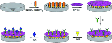
A sensitive label-free immunosensor adopting a novel signal amplification strategy was proposed for the electrochemical detection of human chorionic gonadotropin (hCG). Firstly, a novel composite film consisting of multi-walled carbon nanotubes (MWCNTs) and a room temperature ionic liquid (RTIL) of 1-butyl-3-methylimidazolium hexafluorophosphate (BMIMPF6), which combined the advantages of MWCNTs and RTILs, was fabricated on a glassy carbon electrode surface. The mechanism of the synergy between the MWCNTs and RTIL has been discussed. Secondly, the first film was modified with nanoporous Pd (NP-Pd) prepared by a simple dealloying method. The structure of NP-Pd has been confirmed by EDS, XRD, SEM, TEM and BET analysis. Due to the large specific surface area and excellent electrical conductivity of NP-Pd, electron transfer was promoted and the amount of hCG antibody was enhanced significantly. The results showed that MWCNTs–BMIMPF6/NP-Pd composites were successfully designed as a sensitive immunosensor platform for hCG determination. Under the optimum conditions, the immunosensor exhibited high sensitivity and a wide linear range for hCG from 0.05 to 50 ng mL−1 with a detection limit of 3.2 pg mL−1. The prepared immunosensor showed high sensitivity, reproducibility and stability. This immunosensor preparation strategy presents a promising platform for clinical application.
Introduction
Human chorionic gonadotropin (hCG) exists in the blood and urine of pregnant women. It can be used as a tumor marker, especially in gestational trophoblastic disease. The early detection of hCG produced by the placenta is very important in preventing the spread of pregnancy complications.1–3 Many methods such as fluoroimmunoassays,4,5 resonance scattering spectral assays,6 spectrometry,7 and electrochemical immunosensing8–10 have been developed to detect hCG. Among the techniques mentioned above, electrochemical sensing11,12 has continued to be popular in the last decade due to its high sensitivity, inherent simplicity, low cost, and rapid detection. The modifying technology of the electrode is the crucial step, which affects the signal intensity of the electrochemical detection and the immobilization of the biological molecule. Thus, it is necessary to develop a simple immobilization method to form the efficient matrices to improve the performance of electrochemical immunosensors.
Among the various immobilization methods, the traditional method was to form covalent couplings, which provides stable and strong bindings between the desired biomolecule and the electrode substrate. In this method, molecules such as 1-ethyl-3-(3-dimethylaminopropyl)-carbodiimide (EDC) and N-hydroxysuccinimide (NHS), were usually applied to make the functional groups of the substrate or the desired biomolecule active.13–15 The complicated chemical reaction had some drawbacks,16 such as being cumbersome and time-consuming in practice. Therefore, looking for a new fabrication technology has become a hot research topic.
With the development of nanoporous metallic materials, such as Au, Pt and Pd-based metals, it has been proven that nanoporous metals are ideal materials which can form immobilization matrices easily and amplify signals.17–19 Research work in the field of nanotechnology20,21 has proven that nanoporous nanomaterials are excellent materials for use in electrochemical sensors.22–25 Nanoporous metals26,27 have received considerable attention due to their high surface area and three-dimensional (3D) interconnected network structure. In this work, nanoporous Pd (NP-Pd), prepared by a simple dealloying method, was applied not only to provide a higher surface area for the conjugation of antibodies but also to facilitate the electron transfer. The surface of NP-Pd prepared in a concentrated alkaline solution (without any surfactants) was extremely clean, which was beneficial for its application in further experiments without any purification. Therefore, NP-Pd provides unprecedented promise in electrochemical assays.
Till now, different signal amplification strategies28,29 have been developed to improve the performance of electrochemical immunosensors. Carbon nanotubes (CNTs)30–32 as a typical nanomaterial have attracted wide attention in electroanalysis. CNTs play an important role in improving sensor performance due to their unique structure and extraordinary physical properties, such as large surface area to mass ratio, ultralight weight, high electrical conductivity, and remarkable mechanical strength. Many applications of CNTs in electrochemical sensing and bioelectrochemistry33,34 have been reported. Nevertheless, the development of CNT-based sensing devices is encumbered by the low solubility of CNTs in most solvents. Therefore, there is a pressing need to find pathways that can overcome the above obstacle. Fortunately, it is found that CNTs could be considerably untangled into much finer bundles in room temperature ionic liquids (RTILs). RTILs possess unique physicochemical characteristics, such as high ionic conductivity, wide electrochemical windows, good chemical and thermal stability, and negligible vapour pressure,35–37 which make them suitable for use as the supporting electrolyte or the modifier in electroanalysis. More interestingly, the capability of RTILs to combine with carbon materials to form novel conductive composites makes them very attractive for the preparation of various electrodes with good conductivity.38
In this work, a novel label-free immunosensor for hCG was fabricated by utilizing multi-walled carbon nanotubes (MWCNTs)–1-butyl-3-methylimidazolium hexafluorophosphate (BMIMPF6) (MWCNTs–BMIMPF6) and NP-Pd as the matrix. MWCNTs–BMIMPF6 composites were used as an effective load matrix for NP-Pd and the electrochemical signal was enhanced greatly by the synergistic amplification effect of the two kinds of excellent nanomaterials, NP-Pd and MWCNTs. The modified electrode exhibited excellent sensitivity, selectivity and stability for the determination of hCG. Most importantly, MWCNTs–BMIMPF6/NP-Pd has been used in a label-free immunosensor for the selective detection of hCG for the first time. Under the optimum conditions, the as-prepared immunosensor showed a wide linear range and a low detection limit. The practical application of the as-prepared immunosensor for the determination of hCG in human serum was also fulfilled. The as-prepared immunosensor may have potential applications for the ultrasensitive detection of different biomolecules.
Experimental
Reagents and materials
MWCNTs were purchased from Beijing Dekedaojin Technology Co., Ltd (China). BMIMPF6 was purchased from Sigma-Aldrich (St. Louis, MO, USA). Ag, hCG antibody (Ab), carcinoembryonic antigen (CEA) and alfa fetoprotein (AFP) were purchased from Shanghai Lingcao Biotechnology Co., Ltd (China). N,N-Dimethylformamide (DMF), bovine serum albumin (BSA), urea, L-tyrosine sulfate, methionine, DL-tryptophan, ascorbic acid (Vc), L-glutamic acid, folic acid and sucrose were purchased from Sinopharm Chemical Reagent Co., Ltd (China). Other chemicals and solvents were of guaranteed analytical grade. Ultrapure water was used in all experiments.
Apparatus
Electrochemical measurements were performed on a CHI 760E electrochemical working station (Shanghai Chenhua Instrument, China). Electrochemical impedance measurements (EIS) were performed on a Zennium electrochemical workstation (Zahner, Germany). All electrochemical measurements were performed in an unstirred electrochemical cell at room temperature. X-ray diffraction (XRD) analysis was carried out on a Bruker D8 advanced X-ray diffractometer. The NP-Pd sample was used as the tablet for the scanning electron microscopy (SEM) images obtained using a QUANTA PEG 250 with energy dispersive X-ray spectroscopy (EDS), and the sample was dispersed in solvent for transmission electron microscopy (TEM) performed on a JEM-2100. The Barunauer–Emmett–Teller (BET) analysis was carried out using a nitrogen adsorption instrument (America, Quantachrome).
Synthesis of NP-Pd
NP-Pd was prepared according to the literature.39 PdAl alloy foils were made by refining pure (>99.9%) Pd and Al under the protection of an argon atmosphere in a furnace, followed by melt-spinning. Then, NP-Pd was gained by dipping the PdAl alloy foils in 1 M NaOH solution at room temperature for 24 h. During the dealloying process, Pd atoms left behind self-organized into an interconnected network of pores and ligaments, in terms of effective predetermination of the metallic composition, and the foils became brittle. After dealloying, the foils were crushed to uniform grains using a pestle and mortar prior to characterization. 5 mg of NP-Pd was dispersed in 1 mL of 1 wt% chitosan (CS) under sonication to obtain a black suspension.
Preparation of MWCNTs–BMIMPF6
4 mg of MWCNTs was dispersed in 1.5 mL CS. 0.05 g of BMIMPF6 was added into 0.5 mL DMF. MWCNTs–BMIMPF6 composite was prepared by mixing the above mixture under sonication to obtain a homogeneous dispersion.
Fabrication of the immunosensor
Scheme 1 displays the stepwise procedure for the synthesis of the immunosensor. The GCE was polished to a mirror finish with 0.3 and 0.05 μm alumina slurry, and then thoroughly washed ultrasonically in ethanol and ultrapure water in turn. 5 μL of the mixture of MWCNTs–BMIMPF6 was placed on the surface of the GCE. Next, 5 μL of NP-Pd (5 mg mL−1) was dropped onto the electrode surface to immobilize the Ab. The electrode was thoroughly rinsed with PBS to remove unbound particles. Subsequently, 10 μL of a prepared excessive Ab solution (10 μg mL−1) was added onto the modified working electrode and incubated for 12 h. The modified working electrode was then washed with PBS to remove unbounded biomolecules and immersed in 1% BSA solution for 1 h at 4 °C to block nonspecific binding sites between Ab and electrode surface. Finally, the electrode was incubated in varying concentrations of hCG solution for 2 h. In each step, the working electrode was washed by PBS thoroughly several times. The electrode was stored in the refrigerator prior to use. Cyclic voltammetry (CV) was performed in PBS. After the background current was stabilized, the change in the current response (I) before and after antigen–antibody reaction was recorded.
 |
| Scheme 1 Illustration of the stepwise immunosensor fabrication process. | |
Detection of hCG
Electrochemical measurements were performed with a conventional three-electrode system composed of a glassy carbon working electrode (GCE), a platinum wire counter electrode, and an Ag/AgCl reference electrode. Cyclic voltammetry (CV) was carried out at a scan rate of 100 mV s−1 in PBS. After the background current was stabilized, the change in the current response (I) before and after antigen–antibody reaction was recorded.
Results and discussion
Characterization of MWCNTs–BMIMPF6
SEM was used to examine the morphologies of the MWCNTs and MWCNTs–BMIMPF6. As shown in Fig. 1a, the MWCNTs were highly entangled with one another, which could be attributed to π–π stacking interactions between the MWCNTs. In the bundles, the MWCNTs adhere tightly to one another according to previous studies.40,41 Thus, MWCNTs are difficult to disperse by ordinary solvents. In Fig. 1b, in the case of MWCNTs–BMIMPF6, the surface morphology was distinctly different from the MWCNTs. The heavily entangled MWCNT bundles were found to be untangled in BMIMPF6. This might be ascribed to strong π–π stacking interactions41,42 and weak “cation–π” interactions41,43,44 between the MWCNTs and RTIL. [BMIM+] (Fig. 2) consists of an imidazole ring and alkyl chain. The imidazole ring possesses a π-conjugated structure, and the positive charge is mainly localized on the imidazole ring. π-Electrons and cations in BMIMPF6 could interact with the π-electrons in the MWCNTs. Additionally, the dielectric constants of RTILs are normally very large. When the MWCNTs and BMIMPF6 were mixed with CS and DMF by sonication, the MWCNTs were detached from the bundles by the shear force. BMIMPF6 could prevent the detached MWCNTs from rebinding by shielding the π–π stacking interactions between the MWCNTs. The high surface energy of the detached MWCNTs was effectively appeased since they were enveloped by the RTIL via strong π–π stacking interactions and weak “cation–π” interactions between the MWCNTs and BMIMPF6. Therefore, it could be deduced that BMIMPF6 played an important role in dispersing the MWCNTs.
 |
| Fig. 1 SEM images of (a) MWCNTs and (b) MWCNTs–BMIMPF6 dispersed in CS and DMF by sonication. | |
 |
| Fig. 2 Structure of BMIMPF6. | |
Characterization of the prepared NP-Pd
The composition of the NP-Pd sample was monitored by EDS (Fig. 3a). The results proved that the nanoporous material was composed of Pd with few Al atoms. XRD analysis was also used to characterize the dealloyed sample. As shown in Fig. 3b, it was observed that four diffraction peaks emerged around 40.118°, 46.658°, 68.119°, 82.098° (2q), which could be assigned to diffractions from the (111), (200), (220) and (311) planes of the Pd alloy structure.
 |
| Fig. 3 EDS spectrum (a), XRD patterns (b), SEM image (c), TEM images (d) and (e), and pore size distribution (f) of NP-Pd. | |
Fig. 3c shows the SEM image of the dealloyed sample. It was observed that the dealloyed sample was composed of a number of ultrafine nanopores, and had an open bicontinuous sponge-like morphology. The TEM images (Fig. 3d and e) provide more details on the structure. The clear contrast between the bright pores and the dark ligaments further confirmed the formation of a 3D bicontinuous pore–ligament structure, which is beneficial for mass and electron transport during electrochemical sensing. It is consistent with the SEM observation.
Pore size and pore size distributions are critical factors for porous materials. The pore size distributions of the sample were confirmed by BET analysis. It could be observed that the pore size of the materials has a relatively narrow distribution from Fig. 3f. The pore size was 5.1 nm.
Based on the above results, a Pd alloy with a 3D bicontinuous pore-ligament structure was straightforwardly obtained by a simple dealloying procedure. The dealloying of alloys is in sharp contrast to the traditional approach of chemical synthesis, where the feeding ratio of metal salts does not guarantee the same nominal composition in the final alloy sample, mainly due to the different reducing rates of individual metal salts.45 Furthermore, this method can achieve almost 100% yield.
Electrochemical characteristics of the modified electrodes
The electrochemical behaviors of the bare GCE and modified GCE were studied by taking potassium ferricyanide as the electrochemical probe (Fig. 4). A couple of reversible redox peaks of the probe could be observed on the bare GCE, indicating a reversible electrochemical process. It is noticeable that the peak current was larger than that of the bare GCE after coating with MWCNTs or NP-Pd, suggesting that the introduction of MWCNTs or NP-Pd played an important role in the enhancement of its conductivity and active electrode area. Moreover, it could be clearly seen that the current of the NP-Pd/GCE was superior to that of the MWCNTs/GCE due to NP-Pd’s higher capability of electron transfer. The electrochemical performance of the NP-Pd/MWCNTs/GCE was also superior to that of the MWCNTs/GCE and NP-Pd/GCE, which could be ascribed to the synergistic amplification effect of the two kinds of nanomaterials, NP-Pd and MWCNTs. The peak current of the MWCNTs–BMIMPF6/GCE showed a higher current in comparison with the bare GCE and MWCNTs/GCE, which indicated that the presence of BMIMPF6 could increase the peak current. Additionally, after modification of the MWCNTs–BMIMPF6/GCE with NP-Pd, the peak current further increased distinctly, suggesting that the NP-Pd/MWCNTs–BMIMPF6 film on the GCE could further increase the electron transfer rate. The NP-Pd/MWCNTs–BMIMPF6/GCE showed an increase in current in contrast with the NP-Pd/MWCNTs/GCE, which implied that the rate of electron transfer was faster, due to the synergic effect of the MWCNTs and BMIMPF6. Based on this observation, the MWCNTs–BMIMPF6 composite film was fit for the effective load matrix for NP-Pd. Therefore, it demonstrated that the combination of BMIMPF6 and MWCNTs could bring about an advanced and sensitive electrode substrate and the introduction of NP-Pd was believed to offer unprecedented benefits in catalysis design, which might be a new idea for the construction of novel and powerful electrochemical sensors.
 |
| Fig. 4 CVs of (a) bare GCE; (b) MWCNTs/GCE; (c) MWCNTs–BMIMPF6/GCE; (d) NP-Pd/GCE; (e) NP-Pd/MWCNTs/GCE; (f) NP-Pd/MWCNTs–BMIMPF6/GCE. | |
In order to further confirm the above speculation, the surface coverage (Γ*) of the six kinds of electrodes were evaluated from the equation:46
where
Q is the quantity of electric charge,
n is the transferring electron number and
A is the electroactive surface area. The average electroactive surface areas could be calculated based on the Randles–Sevcik equation:
47,48Ip = 2.65 × 10⁵n3/2AD1/2ν1/2c |
where
Ip is the peak current,
D is the diffusion coefficient of the redox probe,
ν is the scan rate and
C is the bulk concentration of the oxidized form. For K
3[Fe(CN)
6],
n = 1,
D = 7.0 × 10
−6 cm
2 s
−1. The calculated results are revealed in
Table 1. The surface coverage of the NP-Pd deposited on the MWCNTs–BMIMPF
6/GCE was larger than that of other modified electrodes, which further proved that the modified electrode combined the superior characteristics of MWCNTs and RTILs. This was related to their interaction. MWCNTs bundles could be considerably untangled within BMIMPF
6, greatly increasing the effective area of the electrode to load more NP-Pd. In addition, it might also be because the MWCNTs and BMIMPF
6 could interact with NP-Pd through hydrophobic interactions, so as to facilitate the deposition of NP-Pd.
35
Table 1 The surface coverage (Γ*) of different modified electrodes
Electrode |
A (cm2) |
Γ* (mol cm−2) |
GCE |
0.0663 |
2.546 × 10⁻⁸ |
MWCNTs/GCE |
0.177 |
2.859 × 10⁻⁸ |
MWCNTs–BMIMPF6/GCE |
0.201 |
2.894 × 10⁻⁸ |
NP-Pd/GCE |
0.293 |
2.715 × 10⁻⁸ |
NP-Pd/MWCNTs/GCE |
0.426 |
3.348 × 10⁻⁸ |
NP-Pd/MWCNTs–BMIMPF6/GCE |
0.538 |
3.504 × 10⁻⁸ |
Characterization of the immunosensor fabrication
CV, an effective and convenient method for probing the features of the modified electrode, was used to characterize the fabrication of the proposed immunosensor. After treatment with MWCNTs–BMIMPF6, an obvious increase in the peak current occurred (Fig. 5A(b)), indicating that MWCNTs–BMIMPF6 made the electron transfer easier. The magnitude of the peak current increased (Fig. 5A(c)) due to the increment of electrode surface area when NP-Pd was coated on the MWCNTs–BMIMPF6 matrix. Nevertheless, after the Ab was loaded into the channel of NP-Pd through adsorption, an obvious decrease in amperometric response was obtained (Fig. 5A(d)). This is because Ab, as a biomacromolecule, has weak conductivity, so as to generate the insulating layer and hinder the diffusion of the redox marker toward the electrode surface and hinder electron transfer. After BSA was employed to block the non-specific binding sites, a successive decrease in the current was observed (Fig. 5A(e)) due to the hindering effect of the protein on electron transfer. Subsequently, the reaction between the Ab/NP-Pd/MWCNTs–BMIMPF6/GCE and hCG led to a further reduction in the peak current (Fig. 5A(f)), which could be attributed to the formation of an immunocomplex blocking layer.
 |
| Fig. 5 (A): CVs of (a) bare GCE; (b) Ag/BSA/Ab/NP-Pd/MWCNTs–BMIMPF6/GCE; (c) MWCNTs–BMIMPF6/GCE; (d) BSA/Ab/NP-Pd/MWCNTs–BMIMPF6/GCE; (e) Ab/NP-Pd/MWCNTs–BMIMPF6/GCE; (f) NP-Pd/MWCNTs–BMIMPF6/GCE; (B): EIS of (a) NP-Pd/MWCNTs–BMIMPF6/GCE; (b) Ab/NP-Pd/MWCNTs–BMIMPF6/GCE; (c) BSA/Ab/NP-Pd/MWCNTs–BMIMPF6/GCE; (d) MWCNTs–BMIMPF6/GCE; (e) Ag/BSA/Ab/NP-Pd/MWCNTs–BMIMPF6/GCE; (f) bare GCE. Conditions: PBS (pH 7.2) containing 0.2 M KCl and 5 mM K3[Fe(CN)6]. | |
In order to gain insight into the fabrication process of the immunosensor, Fig. 5B represents the Nyquist plots from the electrochemical impedance spectroscopy (EIS) of the various modified electrodes. One of the important advantages of EIS over other electrochemical techniques is the small amplitude perturbation from steady state, which makes it possible to treat the response theoretically by linearised or otherwise simplified current–potential characteristics. EIS was performed in a background solution of 5 mM K3[Fe(CN)6] containing 0.2 M KCl. In EIS, the electron transfer resistance (Ret) could be estimated from the semicircle diameter which was at the high frequencies corresponding to the electron transfer-limited process. It is observed that the bare GCE showed a relatively large resistance. After the deposition of the NP-Pd/MWCNTs–BMIMPF6 composites, a remarkable decrease in the Ret value was observed (Fig. 5B(a–c)), implying that the NP-Pd/MWCNTs–BMIMPF6 composites could promote electron transfer. However, Ab with weak conductivity which could resist the electron transfer kinetics of the redox probe at the interface of the electrode were assembled onto the NP-Pd/MWCNTs–BMIMPF6/GCE (Fig. 5B(d)), so as to increase the resistance. Similarly, the capture of BSA and hCG resulted in the increase of impedance of the electrode (Fig. 5B(e and f)). The results showed that BSA and hCG were successively assembled onto the GCE.
Based on the above results, it was confirmed that the fabrication program was feasible and the proposed immunosensor was successfully fabricated.
Optimization of the immunoassay procedure
The amperometric signal will be affected by the mass ratio of the MWCNTs and BMIMPF6, and further influence the electrochemical performance of the immunosensor. As shown in Fig. 6a, the highest value of electrochemical response was achieved at 2.0
:
25 among different ratios ranging from 1.0
:
25 to 3.0
:
25. The reason might be put down to the solvent effect of the RTIL. If there was too much BMIMPF6, the MWCNTs could be wrapped up by BMIMPF6, so as to counteract the advantage of the MWCNTs.
 |
| Fig. 6 Effect of (a) the mass ratio of MWCNTs and BMIMPF6, (b) the pH value, (c) the incubation time of the antibodies and antigens on the electrochemical signal of the immunosensor. | |
Solution pH has great effects on both the bioactivity of immobilized immunoproteins and the electrochemical performance of the GCE. In order to optimize the pH, a series of PBS buffers with pH values from 5.8 to 8.0 were prepared. The amperometric signal decreased in strong acidic and alkaline solutions. It might be on account of the irreversible behavior of the denaturation of proteins involved in the process which was caused by the pH. The optimal response was obtained at pH 7.2 (Fig. 6b), which indicated that the weak alkaline environment was more conducive for the antibody to be in operation. Thus, pH 7.2 was chosen as the optimal pH value for the determination of hCG.
The reaction of the antibodies and antigens depends on the incubation temperature and incubation time. Considering the convenience of the immunoassays in future applications, room temperature was selected throughout the experiment. As shown in Fig. 6c, the results demonstrated that with increasing incubation time, the amperometric signal increased during the first 80 min, and then tended to level off due to the saturated formation of antigen–antibody complexes. Therefore, an incubation time of 2 h was chosen for the determination of hCG.
Sensitivity of the immunosensor
Under optimal conditions, the current change increased with the increment of hCG concentration. As expected, the amperometric signal increased linearly with hCG concentration over the range of 0.05–50 ng mL−1 with a detection limit of 3.2 pg mL−1 at a signal-to-noise ratio of 3 (Fig. 7). Therefore, the immunosensor exhibited higher sensitivity. Some possible explanations might be considered as followed: (1) the excellent film-forming ability, adsorption capabilities and biocompatibility of CS might help the detection of hCG; (2) BMIMPF6 may not only be a good solvent but could also have acted as a suitable charge-transfer bridge to facilitate the electrode transfer rate; (3) MWCNTs–BMIMPF6 composites were used as an effective load matrix for the deposition of NP-Pd; (4) NP-Pd with lots of pores could display a high surface-to-volume ratio and could enhance the immobilized quantity of Ab, which improved the amperometric signal greatly; (5) the good electron transfer ability of MWCNTs and NP-Pd resulted in the dual-amplification effects; additionally, the proposed method for the determination of hCG is compared with the previously reported methods in Table 2. The proposed method had a relatively high sensitivity and low detection limit, which indicated a promising sensitive method to quantify hCG.
 |
| Fig. 7 Calibration curve for hCG determination (n = 3). | |
Table 2 Comparison of hCG determinations using the proposed and reference methods
Methods |
Reagents or condition |
Linear range |
Detection limit |
Reference |
Fluoroimmunoassay |
Magnetic particles |
0.16–450 ng mL−1 |
0.08 ng mL−1 |
5 |
Resonance scattering spectral assay |
Gold nanoparticles |
2.5–208.3 mIU mL−1 |
0.83 mIU mL−1 |
6 |
Spectrometry |
An inductively coupled plasma mass spectrometry |
5.0–170 μg L−1 |
1.7 μg L−1 |
7 |
Label-free electrochemical immunosensor |
Pt–Au alloy nanotube |
25–400 mIU mL−1 |
12 mIU mL−1 |
8 |
Automated flow immunosensor |
Flow kinetic-exclusion analytical technology |
0.2–50 ng mL−1 |
0.2 ng mL−1 |
9 |
Immunosensor |
Epitaxial graphene |
0.62–5.62 ng mL−1 |
0.62 ng mL−1 |
10 |
Label-free electrochemical immunosensor |
MWCNTs–BMIMPF6, NP-Pd |
0.05–50 ng mL−1, (0.5–500 mIU mL−1) |
3.2 pg mL−1, (0.32 mIU mL−1) |
Present work |
Specificity, reproducibility and stability
Specificity is an important criterion for immunosensors. Other possible interferents such as CEA, AFP, BSA, urea, L-tyrosine sulfate, DL-tryptophan and Vc, etc., were investigated. The immunosensors were separately exposed to 10 ng mL−1 hCG solutions with and without 50 ng mL−1 interference. As shown in Fig. 8, no remarkable changes were observed. The results indicated that the specificity of the present immunoassay protocol was satisfactory.
 |
| Fig. 8 Amperometric response of the immunosensor to interferents. | |
Reproducibility is a key factor for developing a practical immunosensor. The intra-assay precision was evaluated by analyzing three hCG levels for five replicate measurements. The relative standard deviations (RSDs) were 4.9%, 3.8% and 5.1% for 5.0, 10.0 and 20.0 ng mL−1 hCG, respectively. Similarly, the inter-assay precision was evaluated by analyzing the same hCG level with five immunosensors made using the same conditions independently. The RSDs were 3.8%, 4.5%, and 4.2% for 5.0, 10.0 and 20.0 ng mL−1 hCG, respectively. The results obtained were acceptable.
The stability of the immunosensor was also examined. When it was not in use, it was stored at 4 °C. The response of the immunosensor was retained at 85% of the initial response after 2 weeks. The immunosensor kept long-term stability, which might be on account of the biocompatibility of NP-Pd.
Application of hCG immunosensor in human serum samples
The feasibility of the immunoassay for clinical applications was investigated by standard addition methods in human serum. The analytical results are shown in Table 3. The recovery efficiency was in the range of 96–104.55%. Therefore, the proposed immunosensor could be satisfactorily applied to the clinical determination of hCG in real samples.
Table 3 hCG determination in serum by the proposed immunosensor (n = 5)
Amount added (ng mL−1) |
This method (ng mL−1 ± RSD%) |
Recovery (%) |
0.5 |
0.48 ± 2.47 |
96 |
1.0 |
1.05 ± 3.89 |
105 |
5.0 |
5.23 ± 4.13 |
104.6 |
10.0 |
9.87 ± 3.43 |
98.7 |
20.0 |
20.91 ± 2.18 |
104.55 |
Conclusions
In this work, a simple and sensitive electrochemical immunosensor using MWCNTs–BMIMPF6/NP-Pd as a sensor platform to sequentially immobilize hCG antibody was developed for hCG detection. The structures of MWCNTs–BMIMPF6 and NP-Pd were characterized by EDS, XRD, SEM, TEM and BET analysis. The synergy of the MWCNTs and RTIL greatly enhanced the conductivity and the MWCNTs–BMIMPF6 composites acted as an effective load matrix for NP-Pd. Moreover, NP-Pd with good biocompatibility could display a high surface-to-volume ratio, which could enhance the immobilized quantity of Ab. The proposed immunosensor showed high sensitivity, wide linear range, good reproducibility, acceptable precision and accuracy. The presented strategy was demonstrated to be simple and specific, which provides a novel promising platform for a clinical immunoassay for hCG.
Acknowledgements
This project was financially supported by the Shandong Provincial Natural Science Foundation, China (Grant no. ZR2012BL11), the Shandong Provincial Science and Technology Development Plan Project, China (Grant no. 2013GGX10705) and the National Natural Science Foundation of China (Grant no. 51003040 and 51102114).
References
- G. G. Yang, X. Y. Yang, C. Y. Yang and Y. H. Yang, Colloids Surf., A, 2011, 389, 195–200 CrossRef CAS PubMed.
- X. K. Ding and K. L. Yang, Anal. Chem., 2013, 85, 10710–10716 CrossRef CAS PubMed.
- J. F. Wang, R. Yuan, Y. Q. Chai, S. R. Cao, S. Guan, P. Fu and L. G. Min, Biochem. Eng. J., 2010, 51, 95–101 CrossRef CAS PubMed.
- Y. H. He, Y. P. Li and X. Hun, Microchim. Acta, 2010, 171, 393–395 CrossRef CAS.
- J. Y. Hou, T. C. Liu, Z. Q. Ren, M. J. Chen, G. F. Lin and Y. S. Wu, Analyst, 2013, 138, 3697–3704 RSC.
- A. H. Liang, M. J. Zou and Z. L. Jiang, Talanta, 2008, 75, 1214–1220 CrossRef CAS PubMed.
- S. C. Zhang, C. Z. Zhang, Z. Xing and X. R. Zhang, Clin. Chem., 2004, 50, 1214–1221 CAS.
- M. L. Tao, X. F. Li, Z. S. Wu, M. Wang, M. Hua and Y. H. Yang, Clin. Chim. Acta, 2011, 412, 550–555 CrossRef CAS PubMed.
- T. A. Wani and I. A. Darwish, New J. Chem., 2012, 36, 1114–1120 RSC.
- S. Teixeira, G. Burwell, A. Castaing, D. Gonzalezb, R. S. Conlan and O. J. Guy, Sens. Actuators, B, 2014, 190, 723–729 CrossRef CAS PubMed.
- K. S. Arya and M. K. Park, Biosens. Bioelectron., 2014, 61, 260–265 CrossRef PubMed.
- S. F. Liu, Y. Lin, T. Liu, C. B. Cheng, W. J. Wei, L. Wang and F. Li, Biosens. Bioelectron., 2014, 56, 12–18 CrossRef CAS PubMed.
- R. Genc, D. Murphy, A. Fragoso, M. Ortiz and C. K. O’Sullivan, Anal. Chem., 2011, 83, 563–570 CrossRef CAS PubMed.
- U. Jarocka, R. Sawicka, A. Góra-Sochacka and A. Sirko, et al., Biosens. Bioelectron., 2014, 55, 301–306 CrossRef CAS PubMed.
- L. T. Truong, M. Chikae, Y. Ukitaa and Y. Takamura, Talanta, 2011, 85, 2576–2580 CrossRef CAS PubMed.
- X. Y. Zhang, G. Y. Shen, S. Y. Sun, Y. M. Shen, C. X. Zhang and A. G. Xiao, Sens. Actuators, B, 2014, 200, 304–309 CrossRef CAS PubMed.
- R. Li, D. Wu, H. Li and C. X. Xu, et al., Anal. Biochem., 2011, 414, 196–201 CrossRef CAS PubMed.
- T. T. Qi, J. F. Liao, Y. S. Li and J. R. Peng, et al., Biosens. Bioelectron., 2014, 61, 245–250 CrossRef CAS PubMed.
- R. Feng, Y. Zhang, H. Q. Yu, D. Wu and H. M. Ma, et al., Biosens. Bioelectron., 2013, 42, 367–372 CrossRef CAS PubMed.
- E. Turkmen, S. Z. Bas, H. Gulceb and S. Yildiz, Electrochim. Acta, 2014, 123, 93–102 CrossRef CAS PubMed.
- K. K. Liu, J. R. Zhang, Q. Liu and H. P. Huang, Electrochim. Acta, 2013, 114, 448–454 CrossRef CAS PubMed.
- M. Li, M. Zhang, S. G. Ge, M. Yan, J. H. Yu, J. D. Huang and S. Liu, Sens. Actuators, B, 2013, 181, 50–56 CrossRef CAS PubMed.
- C. X. Xu, Q. Li, Y. Q. Liu, J. P. Wang and H. R. Geng, Langmuir, 2012, 28, 1886–1892 CrossRef CAS PubMed.
- C. X. Xu, Y. Q. Liu, J. P. Wang and H. R. Geng, J. Power Sources, 2012, 199, 124–131 CrossRef CAS PubMed.
- F. L. Jia, C. F. Yu and L. Z. Zhang, Electrochem. Commun., 2009, 11, 1944–1946 CrossRef CAS PubMed.
- L. F. Liu, O. Albrecht and U. Gosele, Electrochem. Commun., 2010, 12, 835–838 CrossRef CAS PubMed.
- J. S. Yu, Y. Ding and M. W. Chen, Chem. Mater., 2008, 20, 4548–4550 CrossRef CAS.
- S. Zang, Y. J. Liu, M. H. Lin, J. L. Kang, Y. M. Sun and H. T. Lei, Electrochim. Acta, 2013, 90, 246–253 CrossRef CAS PubMed.
- L. Hou, Y. L. Cui, M. D. Xu, Z. Q. Gao, J. X. Huang and D. P. Tang, Biosens. Bioelectron., 2013, 47, 148–156 CrossRef PubMed.
- E. Mojdeh, M. A. Khalilzadeh and K. M. Hassan, Food Chem., 2013, 141, 4311–4317 CrossRef PubMed.
- J. J. Lu, S. Q. Liu, S. G. Ge, M. Yan, J. H. Yu and X. T. Hu, Biosens. Bioelectron., 2012, 33, 29–35 CrossRef CAS PubMed.
- P. Yanez-Sedeno, J. M. Pingarron, J. Riu and F. Rius, Anal. Chem., 2010, 29, 939–953 CAS.
- L. Zhou, J. P. Wang, L. Gaia, D. J. Li and Y. B. Li, Sens. Actuators, B, 2013, 181, 65–70 CrossRef CAS PubMed.
- E. Lahiff, C. Lynam, N. Gilmartin, R. O’Kennedy and D. Diamond, Anal. Bioanal. Chem., 2010, 39, 1575–1589 CrossRef PubMed.
- F. Xiao, F. Q. Zhao, J. W. Li, L. Q. Liu and B. Z. Zeng, Electrochim. Acta, 2008, 53, 7781–7788 CrossRef CAS PubMed.
- B. Haghighi, H. Hamidi and L. Gorton, Electrochim. Acta, 2010, 55, 4750–4757 CrossRef CAS PubMed.
- X. L. Niu, W. Yang, H. Guo, J. Ren and J. Z. Gao, Biosens. Bioelectron., 2013, 41, 225–231 CrossRef CAS PubMed.
- X. H. Liu, Z. Ding, Y. H. He, Z. H. Xue, X. P. Zhao and X. Q. Lu, Colloids Surf., B, 2010, 79, 27–32 CrossRef CAS PubMed.
- C. X. Xu, A. H. Liu, H. J. Qiu and Y. Q. Liu, Electrochem. Commun., 2011, 13, 766–769 CrossRef CAS PubMed.
- J. Y. Wang, H. B. Chu and Y. Li, ACS Nano, 2008, 2, 2540–2546 CrossRef CAS PubMed.
- T. Fukushima, A. Kosaka, Y. J. Ishimura, T. Yamamoto, T. Takigawa, N. Ishii and T. Aida, Science, 2003, 2072–2074 CrossRef CAS PubMed.
- J. C. Ma and D. A. Dougherty, Chem. Rev., 1997, 97, 1303–1324 CrossRef CAS PubMed.
- T. Fukushima and T. Aida, Chem.–Eur. J., 2007, 13, 5048–5058 CrossRef CAS PubMed.
- S. Bellayer, J. W. Gilman, N. Eidelman, S. Bourbigot and X. Flambard, et al., Adv. Funct. Mater., 2005, 15, 910–916 CrossRef CAS.
- C. X. Xu, J. P. Wang and J. H. Zhou, Sens. Actuators, B, 2013, 182, 408–415 CrossRef CAS PubMed.
- X. Y. Li, Y. X. Liu, L. C. Zheng, M. J. Dong, Z. H. Xue, X. Q. Lu and X. H. Liu, Electrochim. Acta, 2013, 113, 170–175 CrossRef CAS PubMed.
- S. MansouriMajda, H. Teymourian, A. Salimi and R. Hallaj, Electrochim. Acta, 2013, 108, 707–716 CrossRef PubMed.
- M. Elyasi, M. A. Khalilzadeh and H. Karimi-Maleh, Food Chem., 2013, 141, 4311–4317 CrossRef CAS PubMed.
|
This journal is © The Royal Society of Chemistry 2014 |
Click here to see how this site uses Cookies. View our privacy policy here.