DOI:
10.1039/C4RA09790D
(Paper)
RSC Adv., 2014,
4, 48004-48011
Fluorometric appraisal of HSO4− in aqueous media and daily utilities using organic–inorganic nanohybrids†
Received
4th September 2014
, Accepted 17th September 2014
First published on 17th September 2014
Abstract
Receptor 1 based on rhodamine dye was designed and characterized using 1H, 13C NMR and mass spectroscopy like spectroscopic techniques. Re-precipitation methods have been used for developing nano-aggregates of 1 (N1) in aqueous medium, which were further used for preparation of inorganic–organic hybrid nanomaterial (H1) i.e. by coating organic nanoparticles with gold nanoparticles. Changes in the photophysical profile by anion binding of N1 and H1 were evaluated using fluorescence spectroscopy. No selectivity was perceived with nano-aggregates (N1) for any anion. However, the hybrid nanomaterial (H1) selectively recognized HSO4− through enhancement of fluorescence emission intensity in aqueous medium. Thus, the hybrid nanomaterial (H1) has been established for sensing of HSO4− and was considered for various parameters like rapid response, good stability in water and is validated for determination of HSO4− ions in daily use items.
Introduction
Anions play a crucial role in various biological and chemical processes.1–4 Among various anions cyanide, halides, phosphates, nitrates and sulphates have received special interest as they have been used extensively in various industries, for example fluoride has been found in toothpastes and medicines,5 chlorine has been used for disinfecting drinking water,6 phosphate anions play an important role in energy transfer and information storage,7 nitrates and phosphates have been used in various fertilizers8 and used as counter ions for various metallic cations. Beside these enormous uses, the flip side of anions is also present, for example excess of fluoride anion causes fluorosis,9 phosphate containing fertilizers cause eutrophication of rivers due to over-use,10 various metabolites of nitrate cause cancer,11 anionic nuclear wastes, etc., cause deleterious and irreversible effects in the environment.12,13 Thus, their determination in various samples of analytical and environmental significance becomes of extreme prominence. But, their determination pose a greater challenge in comparison with their counter parts, because of their large size as compared to isoelectronic cations, pH sensitivity, wide range of geometries, competition between solvent molecule and receptor etc.14 Hence, pursuit of methods for sensitive and selective monitoring of anions that are rapid, easy and economical has risen alarmingly in analytical chemistry.15–17 Various techniques like fluorescence have been employed for determination of anions in various samples of significance18–20 using various synthetic sensors of inorganic21 and organic origin.22 Recently, various organic dyes like BODIPY, rhodamine, fluorescein, coumarin derivatives have been used in sensing of anions attributable to their incredible spectroscopic properties like high absorption coefficient, high fluorescence quantum yield, sharp absorption and emission peaks.23–27 To improve the sensitivity and selectivity of such sensors, nanomaterials have come up as one of the potential candidate, because of their shape and size dependent chemical and physical properties28 and qualities like high surface area, volume ratio, exceptional surface chemistry, thermal and electrical properties.29 Among various anions, the hydrogen sulfate is of great significance as it is used in various industries like in production of fertilizers, chemicals, dyes, glass, paper, soaps, textiles, fungicides, insecticides, astringents, cosmetics and in leather industries.30,31 It dissociates to sulfate32 that cause acid rain which is very toxic to flora and fauna.33 Drinking of sulphate containing water in concentration less than 600 mg litre−1 causes cathartic effect34 like dehydration as side effect and cause diseases like catharsis in adult males. Hence, its determination in trace level from samples of analytical and environmental prominence is of prodigious significance.
In the present work, we have tried to collaborate between two branches of chemistry i.e. photophysical chemistry of organic compounds with recent branch of nanomaterials to increase the sensitivity and selectivity of prepared sensor for analyte of interest in aqueous samples of various origins. We synthesized a rhodamine based receptor and its nanoaggregates in aqueous medium. Nanoaggregates were further used for fabrication of organic–inorganic hybrid with gold nanoparticles. Gold nanoparticles (AuNPs) have received much attention in chemical and biological sensors because they are quite convenient to functionalize with organic ligand.35–37 A well-disseminated AuNPs of 10–50 nm in diameter in solution unveils a pink colour. Whereas, aggregated AuNPs impart purple or blue38,39 colour for the solution. The hybrid nanomaterial showed a great deal of selectivity for determination of HSO4− over other anions, as no such sensitivity and selectivity was observed in case of organic nanoparticles of the receptor. Thus, it was perceived that nano-hybrids present a new class of materials, which have improved response and selectivity for analyte of environmental and analytical importance, when compared with either of the components they are produced from in aqueous medium.
Results and discussion
Synthesis
Receptor 1 (Fig. 1) was synthesized by reacting rhodamine 6G with diethylenetriamine, which was further reacted with 2-hydroxy-5-nitrobenzaldehyde to form receptor 1 (Fig. 1). The receptor 1 was copiously characterized with 1H, 13C NMR, mass spectroscopy (Fig. S1–S3†) and elemental analysis.
 |
| | Fig. 1 Design of receptor 1. | |
Geometry optimization
The geometry of synthesized receptor 1 was optimized (Fig. 2a) using DMol 3 package with DFT (density functional theory) calculations run through GGA with basis set DNP (double numeric plus polarization). HOMO (highest occupied molecular orbital) and LUMO (lowest unoccupied molecular orbitals) were also calculated using the same (Fig. 2b). It is quite evident from the HOMO and LUMO orbitals that HOMO is dispersed over rhodamine moiety and LUMO is highly concentrated on 2-hydroxy-5-nitrobenzaldehyde part of the receptor 1. It can be established on the basis of HOMO and LUMO energies of receptor 1 and receptor 1–HSO4− complex that after complexation with HSO4− both HOMO and LUMO get stabled, when compared with their parent counterparts indicated by decrease in the energies for both HOMO and LUMO in Fig. 2b.
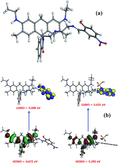 |
| | Fig. 2 (a) Optimized geometry of receptor 1 using DMol 3 package (b) HOMO, LUMO diagrams as per calculated theoretically using optimized structure of receptor 1 and its interaction with HSO4− ions. | |
It is quite apparent from HOMO and LUMO energy gaps before and after the complexation that, the gap remains almost the same, with a negligible difference in energy gap. It is also established on the basis of above studies that, no shift in photo physical peaks of receptor 1 on complexation with HSO4− ions is expected. However, such system can offer “switch on”/“switch off” behaviour fluorescence intensity of receptor 1.
Fabrication of nano-aggregates (N1) of receptor 1
Organic nano-aggregates are fine suspensions of organic compounds in aqueous medium ranging from 10 nm to 1 μm in diameter.40 Exploration of various methods of nano-aggregate formation has expanded exponentially due to their developing demands in wide spectrum of fields ranging from electronics to photonics, conducting materials to sensors, etc. Workability of nano-aggregates in various applications depend upon various physio-chemical parameters and properties like binding sites, solubility of compound and environmental factors such as pH, temperature, salt strength41 of the organic compound. From design and structure of receptor 1, it was established that the material can be easily changed to nano-aggregates using single step re-precipitation method instead of other two steps techniques. In re-precipitation approach, organic compound solution prepared in organic solvent is injected slowly to the bulk of aqueous medium without prior emulsification. The N1 of receptor 1 was synthesized by slow injection of solution of 1 (0.1 mL) in DMF to water (200 mL). Influence of water on the photophysical properties of receptor 1 was studied using UV-visible and fluorescence spectroscopy. It was observed that receptor one gave two absorption peaks at 370 and 419 nm, when studied using UV-visible spectroscopy. On shifting the solvent system from THF to water i.e. formation of nano-aggregates (N1) the two peaks of the receptor 1 gets diminished and another peak at 530 nm was observed. On formation of nano-hybrid i.e. decoration of gold nano particles on the surface of nano-aggregates of receptor 1 the peak of the receptor shows further red shift to 542 nm (Fig. 3).
 |
| | Fig. 3 Diagram showing UV-visible profile of receptor 1, nano-aggregates (N1) and hybrid nanomaterial (H1). | |
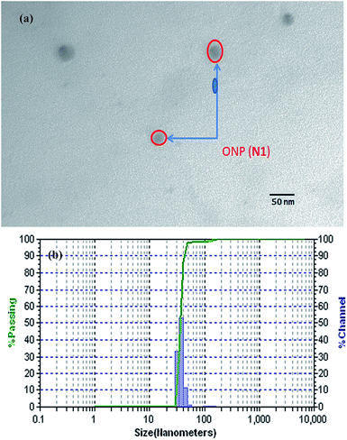
On exciting at a wavelength of 370 nm for fluorescence spectroscopy, receptor 1 showed peaks in the region 510–520 nm and 545–555 nm with DMF as solvent. As the solvent system was changed from DMF to water i.e. nano-aggregates were formed, a solvatochromic change in peak was observed as the peaks perceived in DMF system got merged and showed a slight bathochromic shift to 550–560 nm for N1 (Fig. 4), on excitation at a wavelength of 530 nm. Analysis of N1 particle size was performed using DLS (Dynamic Light Scattering) and TEM (transmission electron micrograph). TEM analysis has confirmed the formation of spherical nano-aggregates of receptor 1 with a spherical size of 25–30 nm (Fig. 5a), while DLS analysis has revealed a narrow size distribution of 35 nm (Fig. 5b).
 |
| | Fig. 4 Emission profile of N1 in water and receptor 1 in DMF. | |
 |
| | Fig. 5 (a) TEM Image of nano-aggregate (N1) (b) DLS histogram of N1 showing average particle size of 35 nm. | |
Fabrication of hybrid nanomaterial (H1)
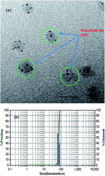
The H1 was developed by simultaneous mixing of N1 and HAuCl4 in 4![[thin space (1/6-em)]](https://www.rsc.org/images/entities/char_2009.gif) :1 ratio under constant sonication. Addition of ascorbic acid causes formation of Au0 by completely reducing Au+ and result in gold nanoparticle formation with advent of pink colour in solution. The formation of H1 was confirmed by TEM analysis as TEM images showed appearance of black heads over nano-aggregates with a size of 60–65 nm (Fig. 6a) and DLS studies also confirmed same sort of size distribution for H1 as in N1, with peak centred at 70 nm (Fig. 6b).
:1 ratio under constant sonication. Addition of ascorbic acid causes formation of Au0 by completely reducing Au+ and result in gold nanoparticle formation with advent of pink colour in solution. The formation of H1 was confirmed by TEM analysis as TEM images showed appearance of black heads over nano-aggregates with a size of 60–65 nm (Fig. 6a) and DLS studies also confirmed same sort of size distribution for H1 as in N1, with peak centred at 70 nm (Fig. 6b).
 |
| | Fig. 6 (a) TEM Image of nano-hybrid (H1), (b) DLS histogram of H1 showing average particle size of 70 nm. | |
The emission profile of H1 was studied by exciting at a wavelength of 541 nm. It was observed that H1 was having same emission peak as that of N1, with a considerable enhancement in the intensity (Fig. 7). It is quite evident from the emission profile that there is considerable enhancement in sensitivity of the receptor 1 in H1 form as compared to N1. Although metal nanoparticles cause quenching of fluorescence of various dyes, when added to their solutions but sharp enhancement in fluorescence of N1, when coupled with gold nanoparticles to form H1 is observed. The enhancement in the fluorescence intensity can be attributed to the reason that surface plasma resonance (SPR) of metal nanoparticles overlap with emission band of the dye.42
 |
| | Fig. 7 Emission profile of N1 and H1 showing enhancement in intensity of emission peak on formation of hybrid from nano-aggregate. | |
Recognition studies
Anion binding with nano-aggregates (N1). For the evaluation of anion recognition behaviour of N1 of receptor 1 in aqueous medium, tetrabutylammonium (TBA) salts of various anions such as F−, Cl−, Br−, I−, NO3−, CN−, CH3COO−, H2PO4−, HSO4−, ClO4− (10 μM) were added to fixed concentration of (385 nM) of N1. The fluorescence spectrum of N1 has shown emission profile at 555 nm and no variation in emission profile of N1 was observed on addition of various anions, even with such high concentration (Fig. S4†). Thus, it can be established that N1 had no selectivity or sensitivity profile for any of the anions.
Anion binding of hybrid nanomaterial (H1). On similar terms the anion binding ability of H1 was also evaluated by observing the change in the fluorescence spectral profile of H1 upon addition of a various anion solutions (F−, Cl−, Br−, I−, NO3−, CN−, CH3COO−, H2PO4−, HSO4−, ClO4−) (10 μM) to its buffered solution in aqueous medium. The emission profile of nano-hybrid (H1) comprise of a band at 555 nm, Anions such as F−, Cl−, Br−, I−, NO3−, CN−, CH3COO−, H2PO4−, ClO4− showed slight quenching in emission profile of H1. On the contrary, HSO4− ions showed significant enhancement (2-fold) in fluorescence intensity at 555 nm (Fig. 8), which has been further utilized in projecting H1 as selective sensor for HSO4− anions.
 |
| | Fig. 8 Changes in emission profile of hybrid nanomaterial H1 (100 μM) in aqueous medium upon addition of 10 μM of TBA salt of particular anion under buffered conditions. | |
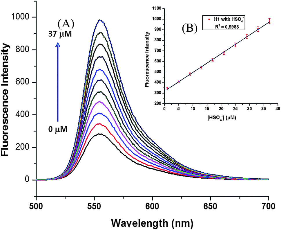
To explore more about the sensing ability of H1 as a sensor for HSO4− anion, titration was carried out with addition of small aliquots of HSO4− ions (0–37 μM) (Fig. 9A) to hybrid H1 (100 μM). Upon successive addition of TBA salt of HSO4−, there was a regular increase in fluorescence intensity profile of H1. The successive addition was carried out till the saturation point for the instrument was achieved and it was observed that after 37 μM addition the intensity of the peak goes beyond detection and hence, no further additions were made.
 |
| | Fig. 9 (A) Changes in emission profile of H1 (100 μM) upon successive addition of HSO4− (0–37 μM), (B) calibration plot for successive additions of HSO4− to H1. | |
Calibration curve was plotted on the basis of emission profile change of H1 on successive additions of HSO4− ions. The calibration plot has showed an excellent linear regression for concentration range upto 37 μM, with r2 value of 0.9988 (Fig. 9B).
To establish the prepared receptor nano hybrid as a potential sensor for HSO4− ions, a competitive binding study was performed to check potential interference with other anions in determination of HSO4− ions by estimating it (10 μM) in the presence of same concentration of other anions like F−, Cl−, Br−, I−, NO3−, CN−, CH3COO−, H2PO4−, ClO4−. Barely any change in the fluorescence spectrum of HSO4−–H1 complex was observed, when compared with its profile in presence of various anions (Fig. 10). It clearly indicates that H1 selectively recognize HSO4− ions and have no interference from any of the potential interfering anions. Thus, the proposed sensor can be used for determination of HSO4− ions with a dynamic range of 0–37 μM and a detection limit of 108 nm.
 |
| | Fig. 10 Influence of other anions on the HSO4− based changes in the emission profile of hybrid H1. | |
The effect of variation in solution pH, on performance of H1 was checked by studying the emission spectra at different pH in water. It was observed that on moving towards acidic range, there was hardly any change in the intensity of emission band. However, as the pH goes below 4, it causes slight enhancement in the emission profile. On the other hand fluorescence intensity of emission band gets quenched as the pH of the solution was changed to basic from neutral (Fig. S5†). So from the above experimentation it can be concluded that the proposed sensors works best with in pH range of 4–7 with negligible change in spectral behaviour.
The effect of ionic strength on H1 emission spectra is studied by adding TBA salt of perchlorate anion (0–100 equiv.) to H1 and was compared with fluorescence spectra of H1 (Fig. S6†). It is quite evident from the spectral behaviour of H1 that there is very negligible change in fluorescence intensity of H1 with additions upto 40 equiv. and very slight decrease on addition upto 100 equiv. So it can be established that the proposed sensor remains stable even in high concentration of salt and shows no effect in its spectral behaviour even in presence of high concentration of salts. Further, the effect of time on response of H1 for HSO4− was studied by monitoring the changes in the emission spectra as function of time. Different concentrations of HSO4− (5, 10, 20 and 40 μM) were added to a fixed concentration (100 μM) of H1 and emission spectra were recorded after small intervals of time (Fig. 11). From the result it is clear that the interaction of HSO4− ion with H1 occurs within the first minute and thereafter no change in the fluorescence intensity was observed.
 |
| | Fig. 11 Plot of fluorescence intensity of H1 and HSO4− at different concentrations (5, 10, 20 and 40 μM) as a function of time (minutes). | |
Real sample analysis
Various samples having sulphate were identified from our daily chores like toothpaste, shampoo etc. and were tested using the proposed sensors and results are shown in Fig. 12 and the results were compared with the calibration curve for estimation of the concentration present in the samples (Table 1). The proposed sensor performed exceptionally well with all the samples and showed enhancement in the emission profile of H1 in presence of HSO4− ions, as was expected from the sensor. Hence it can be concluded that the proposed sensor can be used for selective determination of HSO4− ions in all kind of aqueous samples, without having any interference from any of its potential interfering anions.
 |
| | Fig. 12 Real sample analysis of various commercially available articles of daily chores and their interaction with H1. | |
Table 1 Real sample analysis of commercially available articles of daily chores and their estimated concentrations
| S. No. |
Sample name |
Concentration using turbidimetric method47 (μM) |
Concentration using proposed method (μM) |
| 1 |
S1 |
27.68 ± 0.05 |
27.71 ± 0.03 |
| 2 |
S2 |
26.52 ± 0.12 |
26.75 ± 0.07 |
| 3 |
T1 |
23.39 ± 0.02 |
23.25 ± 0.08 |
| 4 |
T2 |
21.7 ± 0.05 |
21.71 ± 0.06 |
| 5 |
D1 |
12.61 ± 0.01 |
12.34 ± 0.13 |
| 6 |
TS1 |
4.97 ± 0.02 |
4.95 ± 0.02 |
Conclusion
Rhodamine dye based receptor synthesized and characterized using various spectroscopic techniques was suspended in aqueous media as nano-aggregates using single step re-precipitation method. The prepared nano-aggregates were tested for their binding ability with various anions, but no change in fluorescence intensity of the receptor 1 was found. Although formation of inorganic–organic nano-hybrids by decorating gold nanoparticles on the surface of nano-aggregates caused the change is selectivity, as they responded selectively for HSO4− ions, with a detection limit of 37 μM. The proposed chemical sensor had no interference from any of the possible interferent anions and provided with excellent, rapid and stable response towards HSO4− ions. The proposed sensor was also used for determination of HSO4− ions in daily utility items and validated the results using standard reported method.
Experimental
General information
All chemicals used were of analytical grade and were purchased from Sigma-Aldrich Co. 1H and 13C NMR spectra were recorded on Avance-II (Bruker) instrument, which operated at 400 MHz for 1H NMR and 100 MHz for 13C NMR (chemical shifts are expressed in ppm). The fluorescence measurements were performed on a Perkin Elmer LS55 Fluorescence. TEM images were recorded on Hitachi (H-7500) instrument worked at 120 kV. This instrument has the resolution of 0.36 nm (point to point) with 40–120 kV operating voltage. A 400-mesh formvar carbon-coated copper grid was used for sample preparation. The particle size of nano-aggregates was determined with Dynamic Light Scattering (DLS) using external probe feature of Metrohm Microtrac Ultra Nanotrac Particle Size Analyser.
Synthesis of compound 1
Rhodamine 6G (0.5 g, 1.04 mmol) was dissolved in 50 mL of ethanol and then diethylenetriamine (562 μL, excess) was added drop wise to the solution and refluxed overnight (24 h) until the solution lost its red colour. The solvent was removed under reduced pressure. To the obtained orange oily liquid residue water was added and extraction was done with DCM. Water washing of the organic phase was done and dried over Na2SO4. The solvent was removed under reduced pressure and compound 1a recrystallized using hexane was obtained with 81% yield (Rf value = 0.54). Further this product (0.2 g, 0.4 mmol) was dissolved in 10 mL of methanol and its reaction was carried out with 2-hydroxy-5-nitrobenzaldehyde (86.9 mg, 0.52 mmol) for overnight under refluxing condition. The solvent was removed under reduced pressure followed by recrystallization using diethyl ether to get final product 1 with 74% yield. 1H NMR (400 MHz, CDCl3) δ: 1.29–1.33 (t, 6H, –CH3), 1.88 (s, 3H, Ar–CH3), 2.17 (s, 3H, Ar–CH3), 2.59 (broad s, 1H, Ar–NH), 2.98 (broad s, 1H, Ar–NH), 3.17–3.28 (m, 12H, –CH2), 3.54 (m, 1H, –NH), 6.16 (s, 2H, Ar–H), 6.33 (s, 2H, Ar–H), 6.94–6.97 (d, 1H, Ar–H), 7.02–7.04 (m, 1H, Ar–H), 7.11–7.14 (d, 1H, Ar–H), 7.45–7.47 (m, 1H, Ar–H), 8.19–8.21 (d, 1H, Ar–H), 8.29 (s, 1H, –N![[double bond, length as m-dash]](https://www.rsc.org/images/entities/char_e001.gif) CH), 8.39–8.42 (dd, 1H, Ar–H), 8.56–8.57 (d, 1H, Ar–H), 10 (s, 1H, –OH); 13C NMR (100 MHz, DMSO-d6) δ: 14.8, 16.8, 38.4, 39.6, 48.1, 48.5, 65.7, 96.6, 105.4, 118.2, 119.6, 122.9, 123.9, 128.2, 128.3, 128.4, 128.4, 129, 133, 147.7, 151.8, 153.5, 169.2, 195.4. ESI-MS m/z = 649.6 [M + H]+. CHN analysis calculated for C37H40N6O5: C, 68.5; H, 6.21; N, 12.95; O, 12.33; found: C, 67.83; H, 6.31; N, 12.65; O, 12.15.
CH), 8.39–8.42 (dd, 1H, Ar–H), 8.56–8.57 (d, 1H, Ar–H), 10 (s, 1H, –OH); 13C NMR (100 MHz, DMSO-d6) δ: 14.8, 16.8, 38.4, 39.6, 48.1, 48.5, 65.7, 96.6, 105.4, 118.2, 119.6, 122.9, 123.9, 128.2, 128.3, 128.4, 128.4, 129, 133, 147.7, 151.8, 153.5, 169.2, 195.4. ESI-MS m/z = 649.6 [M + H]+. CHN analysis calculated for C37H40N6O5: C, 68.5; H, 6.21; N, 12.95; O, 12.33; found: C, 67.83; H, 6.31; N, 12.65; O, 12.15.
Synthesis of nano-aggregates (N1)
The N1 of receptor 1 was synthesized by re-precipitation single step method. Solution of the ligand was dissolved in various concentrations using DMF. The working solution was slowly injected into water (200 mL) under sonication and size of the nano-aggregates formed were analysed using DLS probe after every injection. It was observed after using various concentrations of ligand that concentration having 771 nM of ligand in 1 mL of DMF gave the optimum sized nano-aggregates. Prepared nano-aggregates were sonicated again for 10 minutes, making sure the temperature of the solution containing nano-aggregates does not rise above 10 °C and was later again observed under DLS for particle size to confirm stability and size of the nano-aggregates. It was perceived that at concentration higher than this triggered agglomeration causing increase in particle size considerably and suspension settled down in the beaker even after sonication, while at concentration lower than the optimum concentration, no nano-aggregates were observed as the material simply settles down without giving any suspension.
Synthesis of hybrid nanomaterial (H1)
For H1 formation, various combinations of gold solution, ascorbic acid and nano-aggregates were tried using various concentrations of the three. Gold solution was prepared by dissolving HAuCl4 (0.1 mM) in water; ascorbic acid solution was prepared by dissolving ascorbic acid (0.1 mM) in water, nano-aggregates were formed as explained earlier. The three solutions were mixed under sonication and were observed under DLS probe for particle size distribution. It was also observed that hybrid nanomaterial (H1) was prepared by mixing the gold solution, ONP and ascorbic acid in the ratio of 4:1:4 respectively, the formation of pink colour had confirmed the development of H1. Any combination above or below this concentration have not yielded the hybrid. With solution having higher concentration of either of three resulted in increase in particle size indicated by change in colour of the solution from pink to blue to dark blue, while no colour change was observed in the solution, if the concentration of either of three is less than the optimized above confirmed by the DLS probe inserted into solution after each sonication. Temperature of the solution was maintained at 25 ± 5 °C during the whole process and during sonication. The nano-hybrid thus was centrifuged and re-suspended under constant stirring and sonication. The particle size of re-suspended nano-hybrid material was again measured and confirmed using DLS.
Recognition studies
The recognition studies were performed at 25 ± 1 °C, and the solutions were shaken for a sufficient time before recording the spectrum. The binding ability of N1 (385 nM) and of H1 (100 μM) of receptor 1 in aqueous medium was determined by adding 10 μM of tetrabutylammonium salt of anions to 5 mL solution of N1 and H1 taken in volumetric flasks. The pH of the solution was stabilized in range of 6 to 7 using TRIS buffer. The volumetric flasks were allowed to stand for 30 minutes before spectra were recorded. For HSO4− titrations, the TBA salt of HSO4− was added to volumetric flasks containing H1 in aqueous medium. To evaluate any possible interference due to different anions for the estimation of HSO4−, solutions of H1 were prepared along with both with and without other interfering anions (10 μM). The effect of ionic strength was explored by recording the spectra at different concentration of tetrabutylammonium perchlorate (0–100 equivalent). The pH titrations were performed to understand the effect of pH on the recognition profile of H1.
Theoretical studies
The geometry of the complex was optimized using DMol3 package43,44 with GGA-DFT, using double numerical plus polarization (DNP) as basic set. All electrons of the system were treated with BLYP45,46 local functions for the exchange-correlation potential.
Real sample analysis
Real time application of the proposed sensor was tested using commercially available samples of HSO4− ions i.e. S1 and S2 are taken from commercially available shampoos, T1 and T2 are taken from toothpastes, D is taken from laboratory drain, while ES is Epsom salt used in kitchen are being used. These are some of the items which are used daily in various chores of our routine and household needs. The results of the proposed chemical sensor are validated using easy to perform turbidimetric method using barium sulfate.47
Acknowledgements
This work was supported with research grant (SR/FT/CS-97/2010(G) from Department of Science and Technology (DST), Government of India. R.K. acknowledges CSIR for fellowship.
References
- Y. M. Hijji, B. Barare, A. P. Kennedy and R. Butcher, Sens. Actuators, B, 2009, 136, 297–302 CrossRef CAS PubMed.
- Q.-Y. Cao, Y.-M. Han, P.-S. Yao, W.-F. Fu, Y. Xie and J.-H. Liu, Tetrahedron Lett., 2014, 55, 248–251 CrossRef CAS PubMed.
- P. A. Gale, N. Busschaert, C. J. E. Haynes, L. E. Karagiannidis and I. L. Kirby, Chem. Soc. Rev., 2014, 43, 205–241 RSC.
- X. Lou, D. Ou, Q. Li and Z. Li, Chem. Commun., 2012, 48, 8462–8477 RSC.
- Q. Li, Y. Yue, Y. Guo and S. Shao, Sens. Actuators, B, 2012, 173, 797–801 CrossRef CAS PubMed.
- S. Madhu, R. Kalaiyarasi, S. K. Basu, S. Jadhav and M. Ravikanth, J. Mater. Chem. C, 2014, 2, 2534–2544 RSC.
- D. Zhang, X. Jiang, Z. Dong, H. Yang, A. Martinez and G. Gao, Tetrahedron, 2013, 69, 10457–10462 CrossRef CAS PubMed.
- K. H. Hovland, Y. Thomassen, N. P. Skaugset, K. Skyberg, M. Skogstad and B. Bakke, J. Environ. Monit., 2012, 14, 2092–2099 RSC.
- B. Hu, P. Lu and Y. Wang, Sens. Actuators, B, 2014, 195, 320–323 CrossRef CAS PubMed.
- S. Hamoudi and K. Belkacemi, Fuel, 2013, 110, 107–113 CrossRef CAS PubMed.
- D. Kim, I. B. Goldberg and J. W. Judy, Analyst, 2007, 132, 350–357 RSC.
- S. Su, W. Wu, J. Gao, J. Lu and C. Fan, J. Mater. Chem., 2012, 22, 18101–18110 RSC.
- P. Kaur, H. Kaur and K. Singh, Analyst, 2013, 138, 425–428 RSC.
- P. D. Beer and P. A. Gale, Angew. Chem., Int. Ed., 2001, 40, 486–516 CrossRef CAS.
- A. Satheshkumar, R. Manivannan and K. P. Elango, J. Organomet. Chem., 2014, 750, 98–106 CrossRef CAS PubMed.
- H. Tavallali, G. D. Rad, A. Parhami and E. Abbasiyan, Dyes Pigm., 2012, 94, 541–547 CrossRef CAS PubMed.
- D. Sharma, S. K. A. Kumar and S. K. Sahoo, Tetrahedron Lett., 2014, 55, 927–930 CrossRef CAS PubMed.
- C. He, W. Zhu, Y. Xu, Y. Zhong, J. Zhou and X. Qian, J. Mater. Chem., 2010, 20, 10755–10764 RSC.
- L. Bau, P. Tecilla and F. Mancin, Nanoscale, 2011, 3, 121–133 RSC.
- B. Valeur and I. Leray, Coord. Chem. Rev., 2000, 205, 3–40 CrossRef CAS.
- N. Singh, R. C. Mulrooney, N. Kaur and J. F. Callan, Chem. Commun., 2008, 4900–4902 RSC.
- D. Y. Lee, N. Singh, M. J. Kim and D. O. Jang, Org. Lett., 2011, 13, 3024–3027 CrossRef CAS PubMed.
- J. Fan, M. Hu, P. Zhan and X. Peng, Chem. Soc. Rev., 2013, 42, 29–43 RSC.
- M. J. Culzoni, A. M. de la Pena, A. Machuca, H. C. Goicoechea and R. Babiano, Anal. Methods, 2013, 5, 30–49 RSC.
- C. Kaewtong, B. Wanno, Y. Uppa, N. Morakot, B. Pulpoka and T. Tuntulani, Dalton Trans., 2011, 40, 12578–12583 RSC.
- B. Bag and A. Pal, Org. Biomol. Chem., 2011, 9, 4467–4480 CAS.
- V. K. Bhardwaj, H. Sharma, N. Kaur and N. Singh, New J. Chem., 2013, 37, 4192–4198 RSC.
- T. Asefa, C. T. Duncan and K. K. Sharma, Analyst, 2009, 134, 1980–1990 RSC.
- A. Chen and S. Chatterjee, Chem. Soc. Rev., 2013, 42, 5425–5438 RSC.
- WHO/SDE/WSH/03.04/114, Sulfate in Drinking water, Background document for development of WHO Guidelines for Drinking-water Quality.
- N. N. Greenwood and A. Earnshaw, Chemistry of the elements, Pergamon Press, Oxford, 1984 Search PubMed.
- Q. Li, Y. Guo and S. Shao, Analyst, 2012, 137, 4497–4501 RSC.
- J. G. M. Roelofs, E. Brouwer and R. Bobbink, Hydrobiologia, 2002, 478, 171–180 CrossRef.
- L. Chien, H. Robertson and J. W. Gerrard, Can. Med. Assoc. J., 1968, 99, 102–104 CAS.
- S. Zeng, D. Baillargeat, H.-P. Ho and K.-T. Yong, Chem. Soc. Rev., 2014, 43, 3426–3452 RSC.
- A. Feis, C. Gellini, P. R. Salvi and M. Becucci, Photoacoustics, 2014, 2, 47–53 CrossRef PubMed.
- Y. Zhou, H. Dong, L. Liu, M. Li, K. Xiao and M. Xu, Sens. Actuators, B, 2014, 196, 106–111 CrossRef CAS PubMed.
- H. Chen, L. Shao, Q. Li and J. Wang, Chem. Soc. Rev., 2013, 42, 2679–2724 RSC.
- H. Guan, W. Wang, X. Liu and J. Liang, Colloids Surf., A, 2014, 448, 147–153 CrossRef CAS PubMed.
- V. J. Mohanraj and Y. Chen, Trop. J. Pharm. Res., 2006, 1, 562 Search PubMed.
- H. Yao and T. Funada, Chem. Commun., 2014, 50, 2748 RSC.
- J. Zhu, K. Zhu and L. Huang, Phys. Lett. A, 2008, 372, 3283 CrossRef CAS PubMed; M. Iosin, P. Baldeck and S. Astilean, Nucl. Instrum. Methods Phys. Res., Sect. B, 2009, 267, 403 CrossRef PubMed; E. G. Matveeva, T. Shtoykob, I. Gryczynski, I. Akopova and Z. Gryczynskia, Chem. Phys. Lett., 2008, 454, 85 CrossRef PubMed.
- X. Fang, Y. Huo, Z. Wei, G. Yuan, B. Huang and S. Zhu, Tetrahedron, 2013, 69, 10052 CrossRef CAS PubMed.
- L. Wang, S. Jiao, W. Zhang, Y. Liu and G. Yu, Chin. J. Chem., 2013, 58, 2733 CAS.
- C. Lee, W. Yang and R. G. Parr, Phys. Rev. B: Condens. Matter Mater. Phys., 1988, 37, 786 Search PubMed.
- A. D. J. Becke, Chem. Phys., 1988, 88, 2547 CAS.
- M. A. Tabatabai, A Rapid Method for Determination of Sulfate in Water Samples, Environ. Lett., 1974, 7, 237 CrossRef CAS.
Footnotes |
| † Electronic supplementary information (ESI) available: Experimental details, UV-vis absorption spectra, fluorescence spectra, NMR spectra, mass spectra. See DOI: 10.1039/c4ra09790d |
| ‡ Both authors contributed equally. |
|
| This journal is © The Royal Society of Chemistry 2014 |
Click here to see how this site uses Cookies. View our privacy policy here.