Rapid synthesis of a flower-like ZnO/rGO/Ag micro/nano-composite with enhanced photocatalytic performance by a one-step microwave method

Alan Menga, Jian Shaoa, Xiaoyan Fanc, Junhu Wangd and Zhenjiang Li*b
aState Key Laboratory Base of Eco-chemical Engineering, College of Chemistry and Molecular Engineering, Qingdao University of Science and Technology, Qingdao 266042, Shandong, P. R. China
bKey Laboratory of Polymer Material Advanced Manufacturings Technology of Shandong Provincial, Qingdao University of Science and Technology, Qingdao 266061, Shandong, P. R. China. E-mail: zhenjiangli@qust.edu.cn; Fax: +86 532 88956118; Tel: +86 532 88958602
cSchool of Mathematics and Physics, Qingdao University of Science and Technology, Qingdao 266061, Shandong, P. R. China
dCollege of Materials Science and Engineering, Qingdao University of Science and Technology, Qingdao 266042, Shandong, P. R. China

Received 3rd September 2014 , Accepted 31st October 2014

First published on 31st October 2014


Abstract

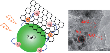
Flower-like ZnO/rGO/Ag micro/nano-composites (MNCs) have been engineered by a one-step microwave technique using graphene oxide, AgNO3 and Zn(CH3COO)2 as raw materials without adding any external toxic reagent. This is a facile and rapid process requiring only low power microwave irradiation (120 W). Various characterization results showed that the flower-like ZnO/rGO/Ag MNCs consisted of a ZnO nanosheet and Ag nanoparticles and reduced graphene oxide (rGO) deposited on ZnO nanosheet surface. The composite shows an enhanced and faster ultraviolet and simulated daylight photocatalytic property, i.e. 92.73% and 70.43% degradation of methyl orange in 20 minutes as compared to the values of 70.91% and 60.82%, 55.48% and 50.61% by bare ZnO and rGO/ZnO, respectively. The enhanced photocatalytic property is attributed to an efficient charge transfer process from ZnO to both Ag and rGO. This method would be beneficial for synthesizing efficient ZnO-based ternary photocatalysts with a combination of metal and rGO.


1. Introduction

ZnO is inexpensive and environmentally friendly. Because of the strong photoinduced hole oxidation ability, it has been a material of considerable interest for photocatalysis applications. In addition, it is widely used in solar cells, photodetection, photoluminescence, etc. However, ZnO as a photocatalyst suffers from an inherent limitation of fast recombination of photoinduced electrons and holes which would reduce the photocatalytic activity.1 To overcome this, considerable research has been carried out by a combination of other materials to transfer the photoinduced electrons. Typically, it has been found that the combination of Ag can increase the rate of the electron-transfer process to improve the photocatalytic performance of ZnO.2–4 However, even then, a complete and faster degradation rate is not yet achieved. Therefore, exploring a highly efficient ZnO-based photocatalyst through a tactful design is needed.

Graphene, with a two-dimensional honeycomb structure, is a new material developed in recent years. Because of the good electron conductivity, high specific surface area and adsorption, it is often used in combination with ZnO to improve the photocatalytic performance through improving the electron–hole pair separation efficiency.5–9 In order to achieve more efficient charge separation, synthesizing a ternary hybrid system is the effective way. By far, few methods have been reported to synthesize ZnO/rGO/Ag MNCs such as photoreduction method,10 hydrothermal method,11 and so on. However, most of these methods are limited for research purpose because of high temperature, high pressure, expensive equipment, or long reaction time. Thus, a simple and fast route for the synthesis of a novel morphology ZnO/rGO/Ag MNCs with excellent properties is still need to be explored to meet economic and industrial needs.

Microwave-assisted synthesis is an effective tool in synthetic organic chemistry due to its unique features such as rapid and selective heating, higher reaction rates, increased product yields, and energy saving.12 In this work, flower-like ZnO/rGO/Ag MNCs have been synthesized via microwave method at the first time. The structures and morphologies of the constructed materials were analyzed by Raman, XRD, SEM, and TEM. Photocatalytic activity was assessed by analyzing the decomposition of MO solution. The significant enhancement of photocatalytic properties was observed. The presented method needs no surfactant or post-annealing treatment, which can reduce cost. Thus, it is suitable for industrial production of ZnO/rGO/Ag MNCs.

2. Experimental methods

2.1 Preparation of the rGO/ZnO composite materials

Graphene oxide was prepared by the Hummer method.13–15 0.05 M of zinc acetate solution was prepared by dissolving zinc acetate dihydrate (Zn(CH3COO)2·2H2O) in distilled water. 8 mL of 1.7 mg mL−1 GO gel was sonicated in distilled water to get uniform GO suspension. Under stirring, GO solution was added into the mixed solution of zinc acetate. Then, 0.5 M NaOH was added into the mixed solution at a rate of a drop per 5 s. The mixture was then placed in a microwave reactor (XH-300UL Beijing Xiang Hu Science and Technology Development Co. Ltd) and treated at 100 °C for 30 min with a power of 120 W under atmosphere. After completion of the reaction, the precipitates were washed three times with distilled water and finally dried in oven at 60 °C for 24 h.

2.2 Preparation of the ZnO/rGO/Ag composite materials

A mixed solution of zinc acetate and silver nitrate was prepared by dissolving zinc acetate (Zn(CH3CH2COO)2·2H2O) and silver nitrate (AgNO3) in distilled water. 8 mL of 1.7 mg mL−1 GO gel was sonicated in distilled water to get uniform GO suspension. Under stirring, GO solution was added into the mixed solution of zinc acetate and silver nitrate, then 0.5 M NaOH was added into the mixed solution at a rate of a drop per 5 s. The precipitation of silver oxide occurred because of the reaction between silver ions and hydroxyl ions with the adding of NaOH. In order to eliminated the precipitation, 10 mL ethanol was also adding by which the silver oxide was reduced to silver. The mixture was then placed in a microwave reactor and treated at 100 °C for 30 min with a power of 120 W under atmosphere. The precipitates were washed three times with distilled water and finally dried in oven at 60 °C for 24 h.

2.3 Analyses of material properties

The structures of the samples were investigated by X-ray diffraction (XRD, Philips PW3710) in 2θ mode and Raman spectra (invia-reflex). Scanning electron microscope (SEM, Hitachi S-4200) and transmission electron microscope (TEM, JEOL JEM-2100F) were used to examine the morphologies and constituent of the ZnO/rGO/Ag MNCs. Room temperature photoluminescence (PL) spectrum was recorded by Perkin Elmer Instruments (LS-55). To measure the photocatalytic property of the hybrid, MO has been taken as a representative organic pollutant in water since most of the fabric industries use MO as the coloring agent. The photo degradation experiment of MO is performed by taking 10 mL of 1 × 10⁻³ g L−1 MO solution in a cylindrical vessel. 0.01 g prepared photocatalysts was added to 10 mL MO with a concentration of 10 mg L−1. After the dark adsorption in 30 min, the measurement was done (see ESI). The solution was stirred while being irradiated by UV lamps of 100 W (Xu Jiang Nanjing) and Xenon lamps of 800 W (Xu Jiang Nanjing) (The light intensities of 100 W Hg lamp is about 63[thin space (1/6-em)]000 lux and 800 W Xenon lamp is about 4000 lux). After every 5 min for UV and 15 min for simulate daylight light irradiation, the suspensions were collected and then centrifuged (10[thin space (1/6-em)]000 rpm, 5 min) to remove the photocatalyst particles. The absorbance property of the dye-containing solution was measured by UV-vis spectrophotometer. The photocatalyst was recycled by the centrifugation of the solution after dye degradation and the photocatalyst was used for the next cycle of the MO degradation after sufficiently drying. The times of the photocatalyst recycling are 5.

3. Results and discussion

XRD was employed to determine the crystal phases of the synthesized samples. As shown in Fig. 1, the characteristic diffraction peaks at 2θ of 31.7°, 34.4°, 36.3°, 47.4°, 56.5°, 62.7°, 66.2°, 67.8° and 68.9° are assigned to (100), (002), (101), (102), (110), (103), (200), (112) and (201) plane of ZnO with a hexagonal (wurtzite) ZnO phase (JCPDS 36-1451). And, two small peaks at 2θ of 38.1° and 44.3° correspond to (111) and (200) diffraction planes of face-centered-cubic (FCC) metallic Ag (JCPDS 04-0783). Diffraction peaks for rGO are not observed, which may be related to the low amount and low diffraction intensity of rGO. In addition, The XRD data showed a main peak in the [111] direction for synthesized Ag nanoparticles, the property is important to the enhanced photocatalytic activity of our ZnO/rGO/Ag sample that will be discussed below.
image file: c4ra09695a-f1.tif
Fig. 1 XRD pattern of ZnO/rGO/Ag.

Fig. 2 shows the Raman spectra of as-synthesized GO and ZnO/rGO/Ag. It is well known that GO exhibits two characteristic main peaks of D band at 1350 cm−1 and G bands at 1594 cm−1. The G band is attributed to the in-plane vibration of sp2 bonded carbon atoms while the D band suggests the presence of defects within the hexagonal graphite structure.16 The ID/IG value of ZnO/rGO/Ag is about 0.9835 and the ID/IG value of GO is about 0.8583. It was found that the composite showed relatively higher intensity of D to G band where the intensity ratio of D/G (ID/IG) was increased in rGO composite (after microwave) compared with GO (before microwave) which confirmed that the GO sheets were restored during microwave treatment.17


image file: c4ra09695a-f2.tif
Fig. 2 Raman spectra of ZnO/rGO/Ag and GO powder.

Fig. 3a and b shows SEM images of ZnO/rGO/Ag MNCs. As can be seen from SEM, the flower-like ZnO microstructures are assembled by many interleaving nanosheets which have the uniform thickness of about 10 nm. The diameter of the flower-like ZnO is about 1 μm. The surfaces of ZnO sheets are not very smooth, there are some particles and pits on the surface of ZnO sheets, the particles may be Ag nanoparticles. From TEM (showed in Fig. 3c), it can be identified that rGO sheets, ZnO sheets and Ag nanoparticles all exist in ZnO/rGO/Ag MNCs. We can find that Ag nanoparticles are evenly deposited on the whole ZnO nanosheets and the size of Ag nanoparticles ranges from 10 nm to 30 nm. Obviously, the rGO sheets and Ag nanoparticles are attached on the surface of ZnO sheets, which confirms the perfect preparation of this ternary composite. Furthermore, the ZnO sheets and Ag nanoparticles of ZnO/rGO/Ag synthesized in this work are single-crystalline structure, which is demonstrated by the selected area electron diffraction (SAED) pattern as shown in Fig. 3d and e.


image file: c4ra09695a-f3.tif
Fig. 3 TEM and SEM images of ZnO/rGO/Ag composites.

The photocatalytic activity of ZnO/rGO/Ag samples was studied by the photodegradation of methyl orange under UV light and simulated daylight, and the results were compared with those of ZnO and rGO/ZnO samples as shown in Fig. 4. The degradation rate of MO solutions in the presence of the ZnO, rGO/ZnO and ZnO/rGO/Ag photocatalysts were about 55.48, 70.91, and 92.73% after under irradiation by UV for 20 min. The degradation rate of MO was about 50.61, 60.82, and 70.43% under irradiation by simulate daylight for 1 hour. ZnO/rGO/Ag samples exhibited better photocatalytic activity than that of pure ZnO and rGO/ZnO. This result demonstrates that the photocatalytic activity of the ZnO was significantly enhanced by rGO and Ag. The results in Fig. 4(c) and (d) indicate that the ZnO/rGO/Ag hybrid catalyst is quite stable. However, the figure shows that the catalyst itself is degraded slightly in the 5th cycle, which may be rather due to loss of catalyst during the catalysis experiments.


image file: c4ra09695a-f4.tif
Fig. 4 The degradation rate of MO under UV (a) and simulated daylight radiation (b). Recyclability of the photocatalyst under UV (c) and simulated daylight radiation (d).

To have a better understanding of the reaction kinetics of the MO degradation, the experimental data were fitted by a first-order model as expressed by eqn (1). The value of the rate constant k commonly gives an indication of the activity of the composite photocatalyst.

 
−ln(C/C0) = kt (1)
Where k is the rate constant (min−1), C0 is the initial concentration and C is the concentration after the MO degradation for time t. Fig. 5 shows a linear relationship between ln(C/C0) and the irradiation time for MO degradation. As can be seen from Fig. 5, the photocatalytic degradation curves in all cases fit well with pseudo-first-order kinetics. Furthermore, the corresponding first-order kinetics plot shown in Fig. 6 indicates that the ZnO/rGO/Ag exhibit the highest degradation rate, which is the best catalyst of all three samples.


image file: c4ra09695a-f5.tif
Fig. 5 First-order kinetics plot for the photodegradation of MO under (1) UV light (2) simulated daylight.

image file: c4ra09695a-f6.tif
Fig. 6 Degradation rate constant k (min−1) for the photodegradation of MO under (1) UV light (2) simulated daylight.

Fig. 7 shows UV-vis absorption spectra of ZnO, rGO/ZnO and ZnO/rGO/Ag. Pure ZnO shows the characteristic absorption edge at 399 nm, indicating ZnO only can absorb UV light. The wavelength threshold is corresponding to a band gap of 3.11 eV. The wavelength threshold of rGO/ZnO composite photocatalysts is estimated to be 407 nm corresponding to the band gap of 3.05 eV. And, rGO/ZnO shows a broad absorption in visible region (420–800 nm) due to the introduction of rGO sheets, which has also been observed in previous reports.18,19 The wavelength threshold of ZnO/rGO/Ag MNCs photocatalysts is estimated as to be 422 nm, corresponding to the band gap of 2.94 eV. ZnO/rGO/Ag shows the strongest absorption in the whole visible range because of the synergetic effects of rGO sheets and Ag nanoparticles. According to the above results, it can be also inferred that the photocatalytic properties of ZnO are significantly improved by the rGO and Ag nanoparticles.


image file: c4ra09695a-f7.tif
Fig. 7 UV-vis spectra of ZnO, rGO/ZnO and ZnO/rGO/Ag.

The electrons in the excited state would undergo recombination with the holes before diffused to the surface of ZnO, the recombination process cause the photocatalytic activity reduced. The recombination process can be greatly suppressed through constructing a ternary composites of ZnO with rGO and Ag. Based on the fluorescence characteristics of ZnO, the transfer mechanism of photon-induced electrons among ZnO, Ag, and graphene sheets can be monitored by the fluorescence decay of the samples20 which can be measured by PL. Therefore, the PL spectra of all the samples have been measured with an excitation wavelength of 325 nm and are shown in Fig. 8. The ZnO shows two major peaks that a UV emission at around 395 nm due to the recombination of photogenerated electrons and holes21 and a broad visible emission at around 550 nm due to oxygen vacancy.22,23 The intensity of the visible emission is stronger compared with the UV emission. The weak blue emission peak at 448 nm most likely occurs from the donor level of Zn interstitial to acceptor energy level on Zn valency.24 The blue-green band around 470 nm is probably caused by radiative transition of electron from shallow donor levels created by the oxygen vacancy to valence band.25 For the rGO/ZnO, the visible emission and the UV emission are suppressed, which is probably due to charge transfer from the trapped states and CB of ZnO to rGO.26,27 Moreover, the PL intensity of ZnO/rGO/Ag is the weakest among the three samples, indicating the fluorescence of the composite is quenched more efficiently than that of rGO/ZnO. This phenomenon suggests the incorporation of ZnO with Ag and rGO can improve the separation of photoinduced electrons and holes. It is indicated that ZnO/rGO/Ag should possess the best photocatalytic properties which is consistent with the results of photocatalytic activity.


image file: c4ra09695a-f8.tif
Fig. 8 PL spectra of ZnO, rGO/ZnO and ZnO/rGO/Ag.

A schematic of the band alignment between the three components of ZnO/rGO/Ag is shown in Fig. 9. In the ZnO/rGO/Ag sample, one part of the Ag particles is attached to ZnO and the other part is attached to rGO. At the rGO/ZnO interface, the charge transfer from the trapped states and CB of ZnO to rGO can occur which is verified by the fact that the visible emission and the UV emission are both depressed in the PL measurements. At the interface of ZnO/Ag, the photogenerated electron of ZnO will be transferred from the trapped states and CB of ZnO to Ag. This can also be proved by the intensity decreasing of the PL peaks in the visible region and the UV region with the addition of Ag into rGO/ZnO composite. Therefore, there are two electron transfer routes for the ZnO/rGO/Ag heterostructured sample: (i) the transfer of electrons to Ag from ZnO and (ii) the transfer of electrons to rGO. Electrons transferred to rGO have a capacity to transfer again from rGO to Ag, resulting in the observed extra photodegradation effect. The extra photodegradation of the ZnO/rGO/Ag sample is caused by the difference of the work function between Ag and rGO.28 In general, the work function of a material will have different values in different crystal directions, as does Ag. A common value of the work function for a Ag polycrystal is about −4.26 eV. As seen in the XRD results, however, Ag particles prepared for this study were located mainly in the (111) direction, and the work function of Ag at this surface is about −4.74 eV, which is lower than that of rGO (∼−4.5 eV).29 Hence, photoinduced electron transfer to rGO further transfers to Ag from rGO, as shown in Fig. 9. This transfer mechanism is expected to significantly suppress recombination of photoinduced electrons and holes.


image file: c4ra09695a-f9.tif
Fig. 9 Schematics of the band diagram and charge transfer mechanism among ZnO, rGO and Ag.

4. Conclusions

In summary, we have demonstrated a simple method for rapid synthesis of ZnO/rGO/Ag MNCs. The SEM and TEM images show that the Ag nanoparticles were homogeneously anchored on the reduced graphene oxide and ZnO sheets. Flower-like ZnO/rGO/Ag MNCs can be prepared by microwave method, which was confirmed by SEM. The photocatalytic activity was enhanced due to an efficient charge transfer from the trapped states and CB of ZnO to rGO and Ag. The interaction of the ZnO with Ag and the reduced graphene oxide sheet was further investigated by the PL measurements. According the bonding state of the three components and the PL measurement, a reasonable transfer mechanism of photoninduced electrons is proposed among Ag, ZnO and rGO, from which it can be understood why this ternary composites has so high efficient photocatalytic performance. Therefore, it is obvious that our method paves a way to prepare multi-component ZnO-based composites with high photocatalytic efficiency.

Acknowledgements

The work reported here was supported by the National Natural Science Foundation of China under Grant no. 51272117, 51172115, 50972063, the Natural Science Foundation of Shandong Province under Grant no. ZR2011EMZ001, ZR2013EMQ006, the Research Award Fund for Outstanding Young Scientists of Shandong Province Grant no. BS2013CL040, the Specialized Research Fund for the Doctoral Program of Higher Education of China under Grant no. 20123719110003 and the Tackling Key Program of Science and Technology in Shandong Province under Grant no. 2012GGX10218, the Application Foundation Research Program of Qingdao under Grant no. 13-1-4-117-jch, 14-2-4-29-jch. We express our grateful thanks to them for their financial support.

References

  1. Y. Dae-Hwang, V. C. Tran, H. L. Van, T. K. Nguyen, J. K. Eui, H. H. Seung and H. H. Sung, J. Phys. Chem. C, 2012, 116, 7180–7184 Search PubMed.
  2. R. Georgekutty, M. K. Seery and S. C. Pillai, J. Phys. Chem. C, 2008, 112, 13563–13570 CAS.
  3. Y. H. Zheng, L. R. Zheng, Y. Y. Zhan, X. Y. Lin, Q. Zheng and K. M. Wei, Inorg. Chem., 2007, 46, 6980–6986 CrossRef CAS PubMed.
  4. Y. H. Zheng, C. Q. Chen, Y. Y. Zhan, X. Y. Lin, Q. Zheng and K. Wei, et al., J. Phys. Chem. C, 2008, 112, 10773–10777 CAS.
  5. T. Xu, L. Zhang, H. Cheng and Y. Zhu, Appl. Catal., B, 2011, 101, 382–387 CrossRef CAS PubMed.
  6. B. Li and H. Cao, J. Mater. Chem., 2011, 21, 3346–3349 RSC.
  7. J. Wang, T. Tsuzuki, B. Tang, X. Hou, L. Sun and X. Wang, ACS Appl. Mater. Interfaces, 2012, 4, 3084–3090 CAS.
  8. Q. Zhang, C. Tian, A. Wu, T. Tan, L. Sun, L. Wang and H. Fu, J. Mater. Chem., 2012, 22, 11778–11784 RSC.
  9. Q. P. Luo, X. Y. Yu, B. X. Lei, H. Y. Chen, D. B. Kuang and C. Y. Su, J. Phys. Chem. C, 2012, 116, 8111–8117 CAS.
  10. J. Wang, X. M. Fan, Z. W. Zhou and K. Tian, Mater. Sci. Eng., B, 2011, 176, 978 CrossRef CAS PubMed.
  11. W. W. Lu, S. Y. Gao and J. J. Wang, J. Phys. Chem. C, 2008, 112, 16792 CAS.
  12. A. Kathalingam, Y. S. Chae and J. K. Rhee, Cryst. Res. Technol., 2011, 46, 517 CrossRef CAS.
  13. W. S. Hummers and R. E. Offeman, J. Am. Chem. Soc., 1958, 80, 1339 CrossRef CAS.
  14. Y. Y. Bu, Z. Y. Chen and W. B. Lib, RSC Adv., 2013, 3, 24118–24125 RSC.
  15. X. J. Liu, L. Pan, T. Lv, T. Lu, G. Zhu, Z. Suna and C. Q. Sunb, Catal.: Sci. Technol., 2011, 1, 1189–1193 CAS.
  16. D. Yang, A. Velamakanni, G. Bozoklu, S. Park, M. Stoller, R. D. Piner, S. Stankovich, I. Jung, D. A. Field, C. A. Ventrice and R. S. Ruoff, Carbon, 2009, 47, 145 CrossRef CAS PubMed.
  17. I. K. Moon, J. Lee, R. S. Ruoff and H. Lee, Nat. Commun., 2010, 1, 1–6 CrossRef PubMed.
  18. T. Xu, L. Zhang, H. Cheng and Y. Zhu, Appl. Catal., B, 2011, 101, 382–387 CrossRef CAS PubMed.
  19. Q. P. Luo, X. Y. Yu, B. X. Lei, H. Y. Chen, D. B. Kuang and C. Y. Su, J. Phys. Chem. C, 2012, 116, 8111–8117 CAS.
  20. Z. Y. Gao, N. Liu, D. P. Wu, W. G. Tao, F. Xu and K. Jiang, Appl. Surf. Sci., 2012, 258, 2473 CrossRef CAS PubMed.
  21. J. Yang, J. Zheng, H. Zhai, X. Yang, L. Yang, Y. Liu, J. Lang and M. Gao, J. Alloys Compd., 2010, 489, 51–55 CrossRef CAS PubMed.
  22. J. Yang, J. Zheng, H. Zhai, X. Yang, L. Yang, Y. Liu, J. Lang and M. Gao, J. Alloys Compd., 2010, 489, 51–55 CrossRef CAS PubMed.
  23. J. H. Yang, J. H. Zheng, H. J. Zhai and L. L. Yang, Cryst. Res. Technol., 2009, 44, 87–91 CrossRef CAS.
  24. X. Q. Wei, B. Y. Man, M. Liu, C. S. Xue, H. Z. Zhuang and C. Yang, Phys. B, 2007, 60, 145–152 CrossRef PubMed.
  25. D. Chu, Y. Zeng and D. Jiang, Mater. Lett., 2006, 60, 2783–2785 CrossRef CAS PubMed.
  26. G. Williams and P. V. Kamat, Langmuir, 2009, 25, 13869–13873 CrossRef CAS PubMed.
  27. F. Vietmeyer, B. Seger and P. V. Kamat, Adv. Mater., 2007, 19, 2935–2940 CrossRef CAS.
  28. F. Z. Sun, X. L. Qiao, F. T. Tan, W. Wang and X. L. Qiu, J. Mater. Sci., 2012, 47, 7262–7268 CrossRef CAS.
  29. A. W. Dweydari and C. H. B. Mee, Phys. Status Solidi A, 1975, 27, 223 CrossRef CAS.

Footnote

Electronic supplementary information (ESI) available. See DOI: 10.1039/c4ra09695a

This journal is © The Royal Society of Chemistry 2014
Click here to see how this site uses Cookies. View our privacy policy here.