Smart electrospun fibrous scaffolds inhibit tumor cells and promote normal cell proliferation

Jingwen Zhao a, Shichao Jiangc, Reila Zhenga, Xin Zhaoa, Xinliang Chend, Cunyi Fan*c and Wenguo Cui*ab
aOrthopedic Institute, Soochow University, 708 Renmin Rd, Suzhou, Jiangsu 215006, P.R. China. E-mail: wgcui80@hotmail.com
bDepartment of Orthopedics, The First Affiliated Hospital of Soochow University, 188 Shizi St, Suzhou, Jiangsu 215006, P.R. China
cDepartment of Orthopaedics, Shanghai Sixth People's Hospital, Shanghai Jiao Tong University School of Medicine, 600 Yishan Rd, Shanghai 200233, P.R. China. E-mail: fancunyi888@hotmail.com
dInternational Peace Maternity and Child Health Hospital, Shanghai Jiaotong University, 910 Hengshan Rd, Shanghai 230030, P.R. China

Received 30th August 2014 , Accepted 3rd October 2014

First published on 3rd October 2014


Abstract

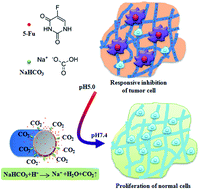
A smart and stable electrospun fibrous drug carrier was fabricated to release an incorporated drug in response to the local presence of acid and to provide a favourable micro-environment for tissue regeneration. To fabricate the electrospun fibrous scaffolds responsive to the local pH variation, sodium bicarbonate, a component of acid response, was facilely incorporated into drug-loaded electrospun fibers by co-electrospinning. The analysis of X-ray photoelectron spectroscopy, Fourier-transform infrared spectrometry, X-ray diffraction, Differential scanning calorimetry and water contact angles demonstrated that the sodium bicarbonate was successfully incorporated into the electrospun fibers. The in vitro drug release results showed that the release rate of an anti-cancer drug 5-fluorouracil in an acidic environment from the electrospun fibrous scaffolds was proportional to the concentration of sodium bicarbonate, and the structure of the electrospun fibrous scaffolds was well maintained before and after the drug release. The acid-responsive release of 5-fluorouracil for a short time was found to effectively inhibit osteosarcoma cell growth, and the electrospun fibrous scaffolds with long-term stability supported fibroblast adhesion and proliferation, indicating the potential for subsequent tissue regeneration.


Introduction

Malignant tumors are a leading cause of death worldwide because of their heterogeneous nature. The primary tumor treatment methods currently include surgical, chemotherapeutic, and radiation approaches. Surgical approaches are efficient for cases that are diagnosed early and for small tumors without remote metastases. For example, the growth of breast tumors,1 renal tumors,2 and bladder tumors3 can be inhibited by partial resection. However, partial resection is unable to remove all tumor cells; thus, local delivery of antitumor drugs is required to restrain the growth of remaining tumor cells and to induce their apoptosis.

Tumor tissue has different acidity compared to its vicinities.4 Tumor tissue typically has a more acidic environment caused by glycolysis, anaerobic respiration, and tumor phenotype.5 In resected tumor tissue, the remaining tumor cells may lead to an acidic environment in local tissue. Hence, various therapeutic strategies involving release of pH-responsive drug have been developed. One good example would be an amphiphilic copolymer containing pH-responsive sulfonamide functional groups and histidine; such copolymer undergoes a sharp transition between micellization and demicellization depending on ambient pH which results in rapid drug release at tumor tissues.6,7 Furthermore, various pH-responsive polymeric micelles with structural disruption according to tumoral acidic pH are developed to release the anti-cancer drug at the target acidic tissues.8 Overall, pH-responsive drug carriers have been shown to be promising and effective to restrain the growth of remaining tumor cells and induce their apoptosis. These stimulus-sensitive carriers are especially intelligent for their ability to adjust the drug release rates according to the pH of the surrounding acidic environment.

Importantly, the repair of damaged tissue after tumor resection is necessary in successful cancer treatment. Therefore, it would be desirable if an anti-tumor drug delivery vehicle could also support tissue repair. Most drug delivery vehicles to date are micro/nano carriers (e.g., microparticles micelles, microcapsules, hydrogels, liposomes, and microspheres), which are incapable of repairing tissue lesions despite their ability to cause tumor cell apoptosis.9–12 Currently, there are only few reports of drug delivery vehicle that can perform drug release and tissue repair simultaneously. Therefore, it is crucial to develop a drug delivery vehicle with both properties of pH-responsive drug release to induce tumor cell apoptosis and tissue regeneration after tumor resection to fully treat a tumor.

Electrospun fibers have a three-dimensional (3D) structure and a high surface to volume (S/V) ratio; such properties make them suitable for applications in tissue engineering and drug delivery. Acid-responsive electrospun fibers have gained much attention due to their site-specific delivery and controllable release profiles.13 In our previous study, poly (d, L-lactide)-poly(ethylene glycol) electrospun fibers were made acid-responsive by incorporating either orthoester or acetal functional groups into its biodegradable backbone. Unfortunately, the electrospun fibrous 3D scaffolds quickly degraded after drug release due to the acid-responsive nature of the functional groups in the polymer backbone.14 Overall, pH-responsive electrospun fibers that enable rapid drug release in an acidic environment as well as maintain their 3D structure to support the growth of normal tissue are needed.

In this study, pH-responsive poly-(L-lactide) (PLLA) fibers were electrospun along with anti-tumor drug 5-fluorouracil (5-Fu)15 and sodium bicarbonate (NaHCO3) simultaneously. This type of scaffold could maintain pH-sensitivity to adjust 5-Fu release and provide a slowly degrading scaffold for cell growth (Scheme 1). Biodegradable PLLA has proven capability as drug delivery carriers and has been approved by Food and Drug Administration (FDA).16 It was expected that in the presence of acid, NaHCO3 would react with acid to generate CO2 and the resultant CO2 would be released into air, without changing the chemical structure of the polymer.12,17 In addition, the reaction products of Na+, H2O and CO2 are safe in human body. Meanwhile, the 5-Fu could be released from the electrospun fibrous scaffolds through overflowing CO2 after the reaction of NaHCO3 with acid. The remaining electrospun PLLA fibrous scaffolds would work as a biotemplate to support cell attachment and proliferation.


image file: c4ra09483b-s1.tif
Scheme 1 Mechanism of drug (5-Fu) release from the pH-responsive electrospun fibers containing NaHCO3 for inhibition of tumor cells. After drug release, the 3D fibrous scaffolds will support normal cell growth and tissue regeneration.

Experimental methods

Materials

PLLA (Mw = 100 kDa, Mw/Mn = 1.6) was synthesized in our lab.14 Hexafluoroisopropanol (HFIP) and 5-Fu were of reagent grade and purchased from Aladdin Reagent Company (Shanghai, China). All other chemical reagents were of reagent grade or better and purchased from GuoYao Reagents Company (Shanghai, China), unless otherwise indicated.

Preparation of electrospun PLLA/NaHCO3 fibrous scaffolds

To prepare the electrospinning solution for each sample, 12.5 mg of 5-Fu was completely dissolved in 3.0 g of HFIP. Next, 500 mg of PLLA was added to the 5-Fu/HFIP solution at room temperature. Then, 0, 0.25, 0.30 or 0.40 ml of NaHCO3 saturated water solution (96 mg ml−1) was slowly added into each 5-Fu-PLLA/HFIP solution for fabricating three samples of PLLA, PLLA-1, PLLA-2 and PLLA-3 electrospun fibers. The solutions were then vigorously stirred with magnetic stirrers for 30 min.

The electrospinning solution was then put in a syringe pump. Electrospinning was performed with the following parameters: 15 kV jet voltage, 15 cm collecting distance and 0.04 ml min−1 flow rate. The electrospun fibrous scaffolds were collected on an aluminum foil-covered grounded plate.

Characterization

The morphology and fiber diameters of the electrospun scaffolds were determined using an environmental scanning electron microscope (ESEM, FEI, QUANTA250, Netherlands).14 The water contact angles (WCAs) of the surfaces of the fibrous scaffolds were measured by goniometry (Krüss GmbH DSA 100 Mk 2 goniometer, Hamburg, Germany) and averaged from at least ten separate trials per sample. Superficial chemical composition was determined using X-ray photoelectron spectroscopy (XPS, XSAM800, Kratos Ltd., Britain).18 UV-vis spectrophotometer (UV-vis; UNICAM, UV300) was used to calculate the amount of 5-Fu released. Chemical composition of the fibrous scaffolds was determined using Fourier-transform infrared (FT-IR) spectrometer (Nicolet 380, Thermo Scientific), acquired from 600 to 4000 cm−1. With 2Ɵ ranging from 5° to 70° and 0.35° min−1 scanning speed (k = 1.54060A), X-ray diffraction (XRD; Philips X'Pert PRO, the Netherlands) was used to investigate the crystalline structure of the electrospun fibrous membranes (20 × 20 mm2 for each sample). Thermal analysis was performed using a differential scanning calorimeter (DSC, NetzschSTA449C, Bavaria, Germany). For each sample, approximately 10 mg of the polymer fibers were examined in a perforated and covered aluminum pan under a nitrogen purge and heated from 25 °C to 100 °C at a rate of 10 °C min−1.

In vitro drug release study

For each sample of PLLA, PLLA-1, PLLA-2 and PLLA-3, 100 mg of the electrospun fibers were immersed in 25 ml of phosphate-buffered saline (PBS, pH = 7.4) or acetate buffer (pH = 5.0) followed by incubation in a shaker (100 rpm) at 37 °C for 48 h. At pre-determined time points, 1 ml of buffer was taken from each sample and was replaced with 1 ml of fresh PBS, and the collected buffer was subject to UV-vis spectroscopy at 266 nm. Then, with a calibration plot of 5-Fu standards with their concentrations ranging from 0 to 0.05 mg ml−1, the drug concentration in each of the collected buffer sample was determined by interpolation. The drug release percentage was then calculated according to the total amount of drug added into the electrospun fibrous scaffolds.

Cell adhesion and proliferation on fibrous scaffolds

The inhibition effects on tumor cells of the electrospun fibrous scaffolds before in vitro release were evaluated using human osteosarcoma cell line (MG-63) as a cell model. Cell adhesion and proliferation on fibrous scaffolds after 48 h of in vitro release in pH 5.0 acetate buffer were observed using multipotent C3H10T1/2 fibroblasts as a cell model. Cell culture medium was prepared from Dulbecco's modified Eagle's medium (DMEM) supplemented with 10% fetal bovine serum (FBS), 100 U ml−1 penicillin and 100 μg ml−1 streptomycin. Scaffold disks with 15 mm diameter were punched out and placed into 24-well plates and sterilized in 75% ethanol for 90 min. The scaffold disks were then washed twice with PBS prior to direct cell seeding at a density of 4 × 10⁴ cells per cm2 for adhesion study or 3 × 10³ cells per cm2 for proliferation study. The cells were then incubated in complete culture medium at 37 °C in 5% CO2 in a humidified incubator.

Cell adhesion on different surfaces was determined using a Live/Dead stain kit (Invitrogen; Eugene, OR). After 4 days of culture, cells were repeatedly washed in PBS before cell staining using the Live/Dead assay according to the manufacturer's instructions and observed under a fluorescence microscope (Leica DM 4000 B).

Cell proliferation on different specimens was tested by AlamarBlue® assay (AbD Serotec, UK). Cells were seeded into each well of a 24-well plate, mixed by shaking, and then AlamarBlue® reagent in an amount equal to 10% of the volume in the well was added under aseptic conditions. After incubation for 1, 2, or 4 days at 37 °C, 200 μl of the solution was transferred into a 96-well plate. Absorbance at 570 nm and 600 nm was determined using a spectrophotometer (Synergy 2, BioTek).

Statistical analysis

Data were presented as mean values with standard deviations (SD). All experiments were repeated three times. Using two-tailed Student's t-test, statistical difference between groups was declared when a p value was less than 0.05.

Results and discussions

Morphology of electrospun fibrous scaffolds

5-Fu and various concentrations of NaHCO3 were successfully co-electrospun into nanofibers. PLLA-1, PLLA-2, and PLLA-3 represented fibers with 5-Fu at a concentration of 2.5% (5-Fu/PLLA, w/w) with varying NaHCO3 concentrations of 5%, 6% and 8% (NaHCO3/PLLA, w/w), respectively. Fig. 1A demonstrated the uniform morphology of electrospun fibers. As concentration of NaHCO3 increased, the surface became smooth and the fiber diameter decreased. The fiber diameters of PLLA, PLLA-1, PLLA-2, and PLLA-3 with increasing concentrations of NaHCO3 were 936 ± 64, 417 ± 65, 258 ± 77 and 189 ± 58 nm, respectively. The net charge density of the electrospun solution could be increased because of addition of sodium salts, which might increase the force exerted on the jet during electrospinning, favoring the formation of small diameter fibers.19
image file: c4ra09483b-f1.tif
Fig. 1 (A) SEM photographs of electrospun PLLA (a), PLLA-1 (b), PLLA-2 (c), and PLLA-3 (d) fibers. (B) XPS spectra of electrospun PLLA fibers with different concentrations of NaHCO3.

Characterization of electrospun PLLA scaffolds

Various techniques were used to characterize the chemical and physical properties of the electrospun PLLA scaffolds. XPS (Fig. 1B) showed that the electrospun PLLA-1, PLLA-2, and PLLA-3 fibers all displayed the characteristic peaks of Na 1s (1071.2 eV) and Na KKL (500.0 eV),20 which were noticeably absent in the PLLA fibers with only two peaks (533.2 eV for O and 284.9 eV for C). This result corresponded with the WCA results (see below) and confirmed the presence of NaHCO3 on fiber surfaces, which might improve the wettability of fibers and accelerate the drug release.

Additional peaks of 1669 and 697 cm−1, corresponding to NaHCO3, were present in the FT-IR spectra of PLLA-1, PLLA-2, and PLLA-3 electrospun fibers (Fig. 2A), indicating successful incorporation of NaHCO3 into the fibrous scaffolds. XRD spectra found no crystallization changes in the electrospun PLLA fibers with NaHCO3 (PLLA-1, PLLA-2, and PLLA-3) compared to PLLA fibers (Fig. 2B), suggesting that crystallization of PLLA might not be affected by the addition of NaHCO3. DSC curves of PLLA, PLLA-1, PLLA-2, and PLLA-3 electrospun fibers indicated that the glass transition temperature (Tg) increased with the addition of NaHCO3 (Fig. 2C), possibly due to the NaHCO3 hybridization with the PLLA backbone, restricting the movement of the PLLA segments.


image file: c4ra09483b-f2.tif
Fig. 2 FTIR spectra (A), XRD patterns (B), DSC patterns (C) and WCAs (D) of the electrospun PLLA fibrous scaffolds with different concentrations of NaHCO3.

Surface wettability is crucial for electrospun fibrous drug carriers and tissue engineering scaffolds, because it determines the ability of water penetration throughout the polymer. Fiber hydrophilicity can enhance water intrusion, drug release, and polymer degradation. To determine surface wettability, WCAs were investigated (Fig. 2D) and were found to be approximately 138°, 106.4°, 54.1°, and 0° for PLLA, PLLA-1, PLLA-2, PLLA-3 fibers, respectively. This result indicates that as NaHCO3 concentrations increased, WCA reduced dramatically. The morphology of the electrospun fibers may influence its wetting property, and the different electrospun fiber diameter and pore size could change the WCAs.21 However, it could not transform the wettability from being hydrophobic to hydrophilic and the resultant WCAs would not be lower than 100°.21 Surface components of material are another key factor affecting material wettability.22 In this study, NaHCO3 acted as a hydrophilic component, generating a hydrophilic region and forming a hydrophilic surface. This process was also in accordance with Cassie impregnating wetting models.23–25 Therefore, the hydrophilicity of PLLA electrospun fibers as evident by the decrease of WCAs from 138° to 0°, was mainly resulted from the addition of NaHCO3. The change of hydrophilicity of the electrospun scaffolds may also indicate that NaHCO3 was distributed both inside and on the surface of the fibers.26

In vitro release and degradation behavior

To investigate the pH-responsive drug delivery of this system, the electrospun PLLA, PLLA-1, PLLA-2, and PLLA-3 fibrous scaffolds were immersed in buffer solutions with different pH values. The actual amount of loaded 5-Fu was determined to be close to what was expected. Fig. 3A showed the 5-Fu release profiles in pH 7.4 solutions, and the total drug release content was approximately 18.0% for PLLA, while higher release rates of 26.5%, 30.5%, and 32.8% were found for PLLA-1, PLLA-2, and PLLA-3, respectively. So we found that the NaHCO3 concentration was positively correlated to the rate of initial release and total amount of 5-Fu released. The relationship between the release profiles of electrospun fibers and various NaHCO3 concentrations in pH 5.0 solution was shown in Fig. 3B. Similar to the release behaviors in a physiological environment, the positive correlation also existed between the NaHCO3 concentration and the release rate (18.8%, 46.0%, 55.7%, and 84.6% for PLLA, PLLA-1, PLLA-2, and PLLA-3, respectively) at acidic environment. Comparing the release profiles in pH 7.4 and pH 5.0 buffers, the release rate of PLLA sample in the pH 5.0 environment was not significantly different compared to pH 7.4 environment. The electrospun PLLA-1 fibers had an initial burst release of 37.0% in the first 12 h and total release of 46.0% at pH 5.0 whereas 26.5% in total was released at pH 7.4. In addition, at pH 5.0, 43.5% of initial burst release in the first 12 h from PLLA-2 fibers was observed and 55.7% was released after 48 h; the latter was about twice as much as the drug release percentage at pH 7.4. At pH 5.0, with 63.7% initial burst release, 84.6% of 5-Fu leached out from the electrospun PLLA-3 fibers, which was more than twice as high as that released at pH 7.4. The above results have suggested that at neutral pH, fiber diameter as well as addition of NaHCO3 co-affected the drug release rates. Fibers with smaller diameter have increased drug release due to the larger surface area for water penetration.27 Fibers with higher NaHCO3 concentrations exhibited raised drug release rates due to the high solubility of NaHCO3, with more NaHCO3 attracting more water sorption for faster drug diffusion. The effect of NaHCO3 was profound particularly at acidic pH. This is because the NaHCO3 can react with hydrogen rapidly at acidic environment, resulting in carbon dioxide (gas) (NaHCO3 + H+ → Na+ + H2O + CO2(↑)). The resultant CO2 may create channels in the PLLA fibers during its release, allowing more ready water penetration and accelerating 5-Fu diffusion from the PLLA fibers.
image file: c4ra09483b-f3.tif
Fig. 3 In vitro release profiles of electrospun fibrous scaffolds with different concentrations of NaHCO3 in pH 7.4 (A) and pH 5.0 (B) buffer solutions. SEM images of electrospun PLLA fibrous scaffolds, with different concentrations of NaHCO3 after in vitro release in pH 7.4 (C) and pH 5.0 (D) buffer solutions for 48 h.

To summarize, a larger initial burst release as well as faster long-term release were achieved in a lower pH environment, which may be beneficial for inhibiting tumor cell growth. The differences of the release profiles between acidic and neutral buffers significantly increased as NaHCO3 content increased. The presence of NaHCO3 may contribute to acid-responsive release profiles and a high initial burst release; it is because that NaHCO3 reacted with the acid as it entered the polymer bulk, generating CO2, creating channels during its release and accelerating drug diffusion. The in vitro release profiles indicated that electrospun fibers incorporated with NaHCO3 accelerated the loaded drug release in an acidic environment and that the release rate could be controlled by fine-tuning the NaHCO3 content of the pH-responsive electrospun fibers.

The degradation behavior of the electrospun fibers, such as the disintegration of the polymer backbone and mass loss of fibrous scaffolds, may play an important role on the release profiles, including significant burst release and drug diffusion from the fiber matrix. However, the broken fibrous scaffolds due to degradation may make them unable to support normal cell growth. Intact 3D tissue engineering scaffolds are very important for promoting cell growth and proliferation. Fig. 3C and D showed the SEM morphology of the electrospun fibrous scaffolds after 48 hours of incubation at pH 7.4 and 5.0. After incubation for 48 hours, the 3D structure of the electrospun fibrous scaffolds maintained in all cases, although the appearance was slightly swollen compared to the original SEM morphology (Fig. 1A). The swelling may be due to matrix polymer chain relaxation during incubation. As concentration of NaHCO3 increased, the degree of degradation of fibers also increased, particularly at pH 5.0. PLLA-1 and PLLA-2 were found to be the most stable as tissue engineering scaffolds to facilitate normal cell proliferation after the treatment of tissue lesions.

In an acidic environment, the acid reacted with NaHCO3, generating CO2 that was quickly released from the fibers, which may accelerate the drug release from the fibers. By incorporating NaHCO3 into the polymer, a class of smart drug carriers was obtained that could precisely control the release of the encapsulated drugs by taking advantage of the differences in tumor tissue pH levels while maintaining the original structure of the polymer scaffold. Thus, from drug release profiles and degradation conditions, we concluded that by increasing NaHCO3 content, the release rate rose dramatically and the degradation was increased. In addition, after the composition analysis of these fibrous mats, PLLA-1 and PLLA-2 fibrous scaffolds appeared to be the most suitable because they maintained their original 3D structures and their hydrophobicity, while the drug can be rapidly released.

The mechanism of pH-responsive drug release

It was found that reducing pH could not significantly affect 5-Fu release from the naked PLLA fibers. However, 5-Fu release was significantly enhanced from the PLLA fibers with NaHCO3, particularly at acidic pH. This may be due to the high solubility of NaHCO3 and/or the reaction of NaHCO3 with hydrogen ions at acidic environment resulting in carbon dioxide (gas) (NaHCO3 + H+ → Na+ + H2O + CO2(↑)). The resultant CO2 may create channels during its release, allowing more ready water penetration and accelerating 5-Fu diffusion from the PLLA fibers. NaHCO3 is the key component contributing to the acid-responsive property of the PLLA fibers. After immersion in pH 5.0 buffer for 48 h, many tiny pores became present on the surface of the fibers (see example in Fig. 4). The development of these small pores may be due to the reaction of NaHCO3 with hydrogen ions at acidic environment, generating carbon dioxide (gas). The formation of these pores may allow more rapid drug diffusion.
image file: c4ra09483b-f4.tif
Fig. 4 Representative SEM images of PLLA-2 fibers showing the pore formation after immersion in pH 5.0 buffer for 48 h.

In vitro assay of tumor and fibroblasts culture

The abilities of electrospun fibrous scaffolds to inhibit tumor cells were investigated through experiments on osteosarcoma cells at an early stage. As shown in Fig. 5A, the surface of the PLLA fibers with no NaHCO3 had the highest number of cells adhered. However, the number of tumor cells decreased with increasing NaHCO3, as the tumor cells lowered the pH of the environment, and NaHCO3 reacted with the acid so that more 5-Fu was released in response, thereby inhibiting the growth of tumor cells. The result was further confirmed by measuring the proliferation of osteosarcoma cells (Fig. 5B). The electrospun PLLA fibrous scaffolds exhibited lower osteosarcoma cell proliferation after 4 days of culture, while superior inhibition effects were detected on fibers with NaHCO3 at early stage because more 5-Fu was released. Furthermore, the number of tumor cells clearly decreased with increasing NaHCO3.
image file: c4ra09483b-f5.tif
Fig. 5 Results from a live/dead assay (A and C) and percent reduction of AlamarBlue® (B and D) of osteosarcoma cells (A and B) and fibroblasts (C and D) on the surface of PLLA (a), PLLA-1 (b), PLLA-2 (c), and PLLA-3 (d) electrospun fibrous scaffolds after 4 days of culture. (All samples have been immersed for 48 h at pH 5.0 buffer solution prior to fibroblast seeding.)

To investigate the effect of the fibrous scaffold on normal cell proliferation after 5-Fu release at a later stage, experiments were carried out on normal cell samples after 48 h of drug release period in pH 5.0 buffer. Fibroblasts adhered to all surfaces (Fig. 5C), but a minimal number of cells adhered to the surface of PLLA fibers containing the maximal residual 5-Fu content after release for 48 h, while more cells adhered to PLLA fibers with NaHCO3. Furthermore, the fibroblasts exhibited an improved adherence with increasing NaHCO3 concentrations, that is, decreasing residual 5-Fu, which was confirmed by measuring cell proliferation (Fig. 5D). Cells were found to proliferate less on the electrospun PLLA and PLLA-1 fibrous scaffolds after 4 days of culture, while improved fibroblasts proliferation was found on other two scaffolds because of less residual 5-Fu in the fibers.

In addition, the surface morphology of electrospun fibers is very important for cell growth, and the fibers with micro- and nano-scale could regulate cell growth through their capacity to support and spatially distribute focal adhesion contacts. Analysis of adherent fibroblasts indicated that projected cell proliferation ratio increased systematically with increasing fiber diameter.28,29 However, in present study, the cell results showed that higher number of fibroblasts was found to adhere on the PLLA-2 and PLLA-3 fibers with smaller fiber diameter compared to PLLA and PLLA-1 (Fig. 5). It is thereby inferred that the behavior changes of the cells was mainly due to other factors, e.g., the presence of residual drug, instead of fiber morphology. In detail, the residual 5-Fu in fibers could significantly affect cell growth as evident that the cells grew better on the pH responsive fibers compared to the PLLA fibers after immersion in acids possibly due to less 5-Fu remaining in the pH-responsive fibers.

The inhibition of tumor cell growth and the promotion of normal cell proliferation on the surface of fibers are strongly influenced by NaHCO3 content. Smart electrospun fibrous scaffolds not only improved acid-responsiveness, speeding early stage drug release in acidic tumor tissues, but also maintained the 3D structure for normal cell growth at later stage. Compared to the electrospun PLLA fibrous scaffolds, the fibers with NaHCO3 were obviously superior in the inhibition of tumor cells (Fig. 5A and B) and the proliferation of normal cells (Fig. 5C and D).

Conclusions

In summary, we have successfully fabricated acid-responsive electrospun fibrous scaffolds containing NaHCO3, which provide a novel model for short-term acid-responsive and controlled anti-cancer drug release. These biomaterials also possess a stable 3D structure that is well-suited for normal cell growth after the treatment of tissue lesions. The release profiles indicated that the total release and burst release could be fine-tuned by varying the NaHCO3 concentrations, particularly in acidic environment. More importantly, the acid-responsive electrospun fibrous scaffolds maintained their 3D structure in both neutral and acidic buffer solutions after complete drug release. Moreover, there was an enhanced inhibition of osteosarcoma cells as well as an improved attachment and proliferation of fibroblasts after the in vitro 5-Fu release. Therefore, the electrospun PLLA fibrous scaffolds can find applications as acid-responsive and controllable short-term drug release systems and long-term tissue engineering scaffolds.

Acknowledgements

This work was supported by the National Natural Science Foundation of China (51373112 and 81271999).

References

  1. L. J. Solin, B. L. Fowble, D. J. Schultz and R. L. Goodman, Int. J. Radiat. Oncol., Biol., Phys., 1991, 21, 279–287 CrossRef CAS.
  2. K. Yoshimura, K. Okubo, K. Ichioka, N. Terada, Y. Matsuta and Y. Arai, J. Urol., 2001, 165, 1893–1896 CrossRef CAS.
  3. J. D. Figueroa, N. Malats, F. X. Real, D. Silverman, M. Kogevinas, S. Chanock, R. Welch, M. Dosemeci, A. Tardón and C. Serra, Hum. Genet., 2007, 121, 233–242 CrossRef CAS PubMed.
  4. K. Engin, D. Leeper, J. Cater, A. Thistlethwaite, L. Tupchong and J. McFarlane, Int. J. Hyperthermia, 1995, 11, 211–216 CrossRef CAS PubMed.
  5. I. F. Tannock and D. Rotin, Cancer Res., 1989, 49, 4373–4384 CAS.
  6. E. S. Lee, K. Na and Y. H. Bae, Nano Lett., 2005, 5, 325–329 CrossRef CAS PubMed.
  7. V. A. Sethuraman and Y. H. Bae, J. Controlled Release, 2007, 118, 216–224 CrossRef CAS PubMed.
  8. M. Oishi, K. Kataoka and Y. Nagasaki, Bioconjugate Chem., 2006, 17, 677–688 CrossRef CAS PubMed.
  9. J. H. Kimē, K. Emoto, M. Iijima, Y. Nagasaki, T. Aoyagi, T. Okano, Y. Sakurai and K. Kataoka, Polym. Adv. Technol., 1999, 10, 647–654 CrossRef.
  10. R. Jalil and J. Nixon, J. Microencapsulation, 1990, 7, 297–325 CrossRef CAS PubMed.
  11. S. C. Chen, Y. C. Wu, F. L. Mi, Y. H. Lin, L. C. Yu and H. W. Sung, J. Controlled Release, 2004, 96, 285–300 CrossRef CAS PubMed.
  12. C. J. Ke, T. Y. Su, H. L. Chen, H. L. Liu, W. L. Chiang, P. C. Chu, Y. Xia and H. W. Sung, Angew. Chem., 2011, 123, 8236–8239 CrossRef.
  13. C. Huang, S. J. Soenen, J. Rejman, B. Lucas, K. Braeckmans, J. Demeester and S. C. De Smedt, Chem. Soc. Rev., 2011, 40, 2417–2434 RSC.
  14. W. Cui, M. Qi, X. Li, S. Huang, S. Zhou and J. Weng, Int. J. Pharm., 2008, 361, 47–55 CrossRef CAS PubMed.
  15. L. Liu, P. Jin, M. Cheng, G. Zhang and F. Zhang, Chin. J. Chem. Eng., 2006, 14, 377–382 CrossRef CAS.
  16. L. Y. Cheng, X. M. Sun, C. M. Hu, R. Jin, B. S. Sun, Y. M. Shi, L. Zhang, W. G. Cui and Y. G. Zhang, Acta Biomater., 2013, 9, 9461–9473 CrossRef CAS PubMed.
  17. B. Choi, H. Park, S. Hwang and J. Park, Int. J. Pharm., 2002, 239, 81–91 CrossRef CAS.
  18. J. W. Zhao, S. Liu, B. Li, H. L. Yang, C. Y. Fan and W. G. Cui, Macromol. Biosci., 2013, 13, 885–892 CrossRef CAS PubMed.
  19. H. Fong, I. Chun and D. H. Reneker, Polymer, 1999, 40, 4585–4592 CrossRef CAS.
  20. H. Qi, P. Hu, J. Xu and A. Wang, Biomacromolecules, 2006, 7, 2327–2330 CrossRef CAS PubMed.
  21. W. G. Cui, X. H. Li, S. B. Zhou and J. Weng, Polym. Degrad. Stab., 2008, 93, 731–738 CrossRef CAS PubMed.
  22. W. G. Cui, L. Y. Cheng, H. Y. Li, Y. Zhou, Y. G. Zhang and J. Chang, Polymer, 2012, 5, 2298–2305 CrossRef PubMed.
  23. R. N. Wenzel, Ind. Eng. Chem., 1936, 28, 988–994 CrossRef CAS.
  24. B. Cassie-Baxter and S. Baxter, Trans. Faraday Soc., 1944, 40, 546–551 RSC.
  25. E. Bormashenko, R. Pogreb, T. Stein, G. Whyman, M. Erlich and A. Musin, Phys. Chem. Chem. Phys., 2008, 10, 40564–44061 RSC.
  26. S. Stankovich, R. D. Piner, X. Chen, N. Wu, S. T. Nguyen and R. S. Ruoff, J. Mater. Chem., 2006, 16, 155–158 RSC.
  27. W. G. Cui, X. H. Li, X. L. Zhu, G. Yu, S. B. Zhou and J. Weng, Biomacromolecules, 2006, 7, 1623–1629 CrossRef CAS PubMed.
  28. A. C. Bashur, L. A. Dahlgren and A. S. Goldstein, Biomaterials, 2006, 27, 5681–5688 CrossRef PubMed.
  29. S. Anand, M. R. Badami, M. Kreke, T. Shane, J. S. Riffle and A. S. Goldstein, Biomaterials, 2006, 27, 596–606 CrossRef PubMed.

Footnote

These authors contributed equally to this work.

This journal is © The Royal Society of Chemistry 2014
Click here to see how this site uses Cookies. View our privacy policy here.