HPTLC-densitometric determination and kinetic studies on antioxidant potential of monomeric phenolic acids (MPAs) from Bergenia species

Nishi Srivastavaab, Amit Srivastavaa, S. Srivastavaa, A. K. S. Rawat*a and A. R. Khan*b
aPharmacognosy and Ethnopharmacology Division, CSIR-National Botanical Research Institute, Lucknow-226001, India. E-mail: pharmacognosy1@rediffmail.com; Fax: +91-522-2207219; Tel: +91-522-2297816
bDepartment of Chemistry, Integral University, Lucknow-226001, India. E-mail: khanar70@yahoo.com; Tel: +91-522-2890730

Received 27th August 2014 , Accepted 2nd October 2014

First published on 2nd October 2014


Abstract

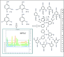
The aim of the present communication is the development of validated HPTLC method for simultaneous separation, detection, comparative quantification of monomeric phenolic acids (MPAs), such as vanillic acid (VA), syringic acid (SYA), gallic acid (GA), protocatechuic acid (PCA) in Bergenia species viz. Bergenia ciliata (BC) and Bergenia stracheyi (BS) (Paashanbheda; family Saxifragraceae) and Kinetics studies on antioxidant activity of focused metabolites. The analyses were performed on HPTLC pre-coated silica gel 60F254 plates with optimized solvent system toluene[thin space (1/6-em)]:[thin space (1/6-em)]ethyl acetate[thin space (1/6-em)]:[thin space (1/6-em)]formic acid (5[thin space (1/6-em)]:[thin space (1/6-em)]4[thin space (1/6-em)]:[thin space (1/6-em)]1 v/v/v) as mobile phase. Densitometric detection of MPAs was performed at 280 nm (λ max) wavelength. The contents of MPAs in both species were found (% in 10 mg ml−1) 0.007 ± 0.1–0.003 ± 0.4 (VA) (y = 3.326x − 1103, regression coefficient r = 0.998), 0.017 ± 0.4 − 0.002 ± 0.5 (SYA) (y = 3.410x − 1009, r = 0.998), 0.024 ± 0.2 − 0.012 ± 0.2 (GA) (y = 5.349x − 240.2, r = 0.999) and 0.027 ± 0.6 − 0.018 ± 0.2 (y = 3.6x − 461.5, r = 0.995). Quantitative variation was assumed as a result of samples collected from different altitudinal range. Two antioxidant assays DPPH and β-carotene were used kinetically in antioxidant potential assessment. Among both the species BC had higher DPPH antioxidant activity and antiradical kinetics than BS, MPAs and positive controls (TOCO), (BHT). Whilst in β-carotene assay highest antiradical activity was reported in PCA kinetically despite BHT than others. However, the deviation in CAA values of BC and BS extracts were very close to the PCA value. EC50 values, rate constant (k), rate of reaction (dx/dt), half-life and average life were also measured in both assays. On the basis of finding it can be concluded that the investigated MPAs were actively involved in antioxidant properties. The kinetic studies of MPAs revealed that H atom transfer from phenolic moieties to the ROS predicts the reactivity of antioxidants.


Introduction

The genus Bergenia (family Saxifragraceae) and its species viz. Bergenia ciliata (BC) and Bergenia stracheyi (BS), are an evergreen perennial herbs, generally distributed in Central and East Asia. It is also found in temperate Himalayas from Kashmir to Bhutan at high altitude 7000–10[thin space (1/6-em)]000 feet and in khasia hill at 400 feet.1 Previous studies on phytochemical analysis of B. ciliata have shown the isolation of bergenin (C-glycoside of 4-O-methyl gallic acid), gallic acid (3,4,5 trihydroxybenzoic acid), (+)catechin, leucocyanidin, (+)-catechin-3-gallate, (+) catechin-7-O-beta-D-glucopyranoside, paashaanolactone, β-sitosterol, β-sitosterol-D-glucoside, and (+)afzelechin.2 These phytochemicals have a range of biological activities such as antioxidant3,4 antidiarrheal, anti-inflammatory, menorrhagia, excessive hemorrhage5,6 antibacterial, antitussive7,8 and in the treatment of pulmonary infections.9

The pharmacological activity of extracts is dependent on the contents of active secondary metabolites in the plants. On varying the contents of secondary metabolites the activities also vary. Therefore, it is important to quantify the active secondary metabolites to find out the accurate pharmacological action of respective samples.

There are only few papers available on the analytical HPLC and HPTLC method development for the quantification of Bergenin and Gallic acid in different Bergenia species.10–12 No previous report is available on the simultaneous quantification of vanillic acid (VA), syringic acid (SYA), gallic acid (GA) and protocatechuic acid (PCA) in Bergenia species using high performance thin layer chromatography (HPTLC) (Fig. 1). Because of several advantages over other analytical methods, such as the rapidity, less amount of test sample and extremely limited solvent wastes, HPTLC has attracted massive interest as a most acceptable technique for the determination of pharmacologically interesting compounds in biological matrices such as plants and its different parts and even in formulations.13,14


image file: c4ra09330e-f1.tif
Fig. 1 Chemical structure of MPAs (VA = Vanillic acid, SYA = Syringic acid, GA = Gallic acid and PCA = Protocatechuic acid).

image file: c4ra09330e-f2.tif
Fig. 2 Images of TLC plate at wavelength λmax = 254 and λmax = 365.

Moreover, VA, SYA, GA and PCA were reported to possess various pharmacological effects, which may be closely correlated with its antioxidant activities.15,16 It has been well recognized that, several biochemical reactions involve the generation of ROS (reactive oxygen species) in human body. However, the balance between the generations of the diminution of ROS under normal conditions is controlled by antioxidant defense system. In case of certain pathological conditions, when ROS are not effectively eliminated by the antioxidant defense system, the dynamic balance between the generation and diminution of ROS is broken. The attack of excessive ROS and free radicals on carbohydrates, proteins, lipids, and DNA result into oxidative stress, which leads to various disorders and diseases.17 Antioxidants are compounds capable to either delay or inhibit the oxidation processes, which generate free radicals and reactive oxygen species. For the protection of bio-molecules against the attack of ROS, a no. of synthetic antioxidants such as 2- and 3-tert-butyl-4-methoxyphenol (i.e. butylated hydroxytoluene BHT, and tert-butylhydroquinone TBHQ) have been added to foodstuffs and also used for industrial processing in recent years, but because of their toxicity issues, their use is being questioned.18

To date, these synthetic antioxidants have been suspected of being harmful19 and cause severe side effects. Thus, in the recent years, search for natural antioxidants from plants is considerably focused. The plant derived antioxidants can be phenolic acid (flavonoids and tannins) nitrogen – containing compounds such as alkaloids, chlorophyll derivatives, amino acids, peptids), DL-α-Tocopherol acetate or ascorbic acid and its derivatives.20 Natural phenolic compounds are now proven as potent antioxidants, which quickly inhibit the generation of free ROS, compared to synthetic compounds. Therefore, plant extracts rich in polyphenolics are increasingly of interest to the food industry because they are capable to retard the oxidative degradation of lipids, and thereby improve the quality and nutritional value of the edible materials.

Consolidated comparative quantitative studies of MPAs using high performance thin layer chromatographic (HPTLC) and their antioxidant activity evaluation allows the analysts to determine the potency of each component within the total extract.21 Furthermore, it also allows the recovery of most active compounds and decides upon the best the development of technology for extraction, which enhances the quantity of potent compounds and to formulate products with these properties.22 In order to evaluate the antioxidant activity it is important to understand the mechanism of the reaction involved in scavenging for free radicals. According to DPPH assay the order of antioxidant activity (AA) is BC ∼ GA > PCA > SYA > BS > BHT ∼ TOCO ∼ VA of tested MPAs and extracts and in β-carotene the order of (AA) is BHT > PCA > TOCO > BC > GA > SYN > VA > BS. The results obtained from these two assays differ despite similar conditions used in the experiments. It seems important to notice that the compound, which is more active in DPPH assays, may not show the same potency in case of β-carotene assay. This contradiction can only be obvious, because the (AA) is not an inherent property of a particular compound, but depends on the nature of the free radical that is reacting with it. Free radical originated from hydrophilic reactions prefers polar compounds and those generated from lipophilic reactions like to be neutralized by non-polar antioxidants. Kinetic study is preferred to understand the order and mechanism of the reaction and it also helps in the estimation of different parameters required for the stability study of the compounds. Therefore, it was considered important to assess the comparative scavenging activity of each benzoic acid derivatives and extracts. For assessing the antioxidant activity DPPH and β-carotene assay were used.

Test

Chemicals. Vanillic acid (purity: 98% w/w), Syringic acid (purity: 99% w/w), Gallic acid (purity: 98% w/w) Protocatechuic acid (purity: 99% w/w), 1,1-diphenyl-2-picrylhydrazyl (DPPH˙) were procured from Sigma-Aldrich USA, DL-α-Tocopherol acetate (TOCO), Butylated hydroxy (BHT) from Laboratory Rasayan, linoleic acid, β-carotene from MP Biomedicals LLC and Tween 40 were procured from Merck. All the solvents used were of analytical grade from Rankem India.
Preparation of crude extracts. B. ciliata and B. stracheyi were collected from Lansdowne and Juda ka talab, Uttarakhand, India in the month of August and December 2012, deposited (voucher specimen no. 254021 and 262557) in repository of CSIR-National Botanical Research Institute, Lucknow (Uttar Pradesh) India (Table 1). After washing with tap water, rhizomes were chopped and dried under shade. The dried rhizomes (100 g) were crushed into powder and soaked in absolute methanol (4 × 250 ml) at room temperature (25 ± 2 °C) for 5 days. The suspension was filtered and evaporated to dryness by rotary evaporator (Buchi, USA). Methanolic extract of B. ciliata and B. stracheyi were further hydrolyzed in acidic medium as per reported by Srivastava et al., 2014.
Table 1 Details of collection of Bergenia species
Sample no. Plant State Region explored Collection stage GPS information Material
254021 B. ciliata Uttarakhand Lansdowne Pre-flowering 9400 feet, N 31°03.116′ E 78°11.096′ Whole plant
262557 B. stracheyi Uttarakhand Juda ka talab Pre-flowering 5400 feet, 29°50′N 78°41′E/29.83°N 78.68°E Whole plant


Acid-hydrolysis. Two grams of the extract was added into 10 ml of methanol containing 2 N HCl. These mixtures were refluxed in a thermostatically controlled water bath linked magnetic stirrer with continuous stirring at 80 °C for 30 min. The samples were cooled to room temperature and dried over vacuum to yield solid residue. Then, the extract was washed and dissolved in distilled water and eventually extracted three times with ethyl acetate.

HPTLC method

Apparatus. Camag Linomat V automated TLC applicator, Camag twin trough glass chamber, ascending. Camag TLC scanner model 3 equipped with Camag Wincats IV software were used during the study at temperature 27 ± 2 °C, relative humidity.
Chromatographic experiments. Sample solution and standards were applied on precoated silica gel 60F254 HPTLC plates with 6 mm band width using Camag 100 microlitre sample syringe (Hamilton, Switzerland) with a Linomat 5 applicator (Camag, Switzerland) under a flow of N2 gas. The Linear ascending development was carried out with Toluene/ethyl acetate/formic acid (5[thin space (1/6-em)]:[thin space (1/6-em)]4[thin space (1/6-em)]:[thin space (1/6-em)]1 v/v/v) as a mobile phase in a Camag glass twin trough chamber (20 × 10 cm). The saturation time of the TLC chamber in the mobile phase was optimized to 20 min for a good resolution of the tested markers and total run time was about 25 min at room temperature (27 ± 2°C), 50% ± 2% relative humidity. After the run, the plates were dried over hair drier and TLC image was obtained on wavelengths of λmax 254 and 365 nm (Fig. 2). Scanning of TLC plate was performed by Camag TLC Scanner 3 at λmax 280 nm under UV absorbance mode for all tracks, TLC plates were developed at a distance of approximately 80 mm from the point of application and the slit dimensions were 4 mm × 0.45 mm. The quantification evaluation of the plate was performed using peak area with linear regression amount of 1–6 μg/band (Table 2). Peak profiling was performed in ultra violet region at 280 nm (variable wavelength was used to get best absorbance range) to check the identity of the bands, the UV absorption spectrum of each standard was overlaid with the corresponding band in the samples track (Fig. 3). Standards solid chromatogram is illustrated in (Fig. 4).
Table 2 Statistical analysis of calibration curves in the HPTLC determination of MPAs (VA, SYA, GA & PCA)
Parameters VA SYA GA PCA
Accuracy 102.57 104.26 99.52 101.14
Rf value 0.47 ± 0.02 0.43 ± 0.01 0.23 ± 0.01 0.38 ± 0.01
Regression equation y = 3.326x − 1103 y = 3.410x − 1009 y = 5.349x − 240.2 y = 3.6x − 461.5
Slope 3.326 3.41 5.349 3.6
Intercept 1103 1009 240.2 461.5
Linearity range 1–6μg 1–6μg 1–6μg 1–6μg
95% Confidence limits of intercept −518.0196009 −94.16249112 267.4136872 1364.360849
Correlation coefficient (r) 0.998 0.998 0.999 0.995
LOD 510.7 778.06 275.23 602.83
LOQ 1547.58 2357.76 834.03 4457.3
SE of intercept 210.9533511 329.5067881 182.8382463 657.6350141
SD of intercept 514.7261768 803.9965629 446.1253209 1604.629434
P-value 0.006 0.03 0.0259173678 0.0521490957


image file: c4ra09330e-f3.tif
Fig. 3 Overlay spectra comparison of MPAs (VA, SYA, GA & PCA) with sample track (BC & BS).

image file: c4ra09330e-f4.tif
Fig. 4 All track chromatogram at wavelength λmax = 280 nm. Abbreviation-VA = Vanillic acid, SYA = Syringic acid, GA = Gallic acid and PCA = Protocatechuic acid, BC = Bergenia ciliata and BS = Bergenia stracheyi.

Assessment of antioxidant activity

Assays of total phenolic contents (TPC). Total phenolic content (TPC) was quantified as described by the method of Singh et al., 2010 (ref. 23) and expressed as mg gallic acid equivalents (GAE)/mg extract. The extract (1 mg ml−1), Folin–Ciocalteu's reagent (1 N) and 20% sodium carbonate were subsequently added. The test mixture was properly mixed on cyclomixer and maintained at room temperature for 30 min. Then, the volume of the mixture was maintained up to 25 ml with deionized water. The absorbance of the test mixture was recorded at A720 nm using ‘‘Thermo Scientific” Vis-UV spectrophotometer. TPC was determined using a standard curve with gallic acid (0–50 μg ml−1) as the standard.
Free radical scavenging activity (FRSA) assay. FRSA of the extracts was measured using DPPH˙ stable radical (Yen and Duh, 1994).24 In brief, each 0.1 ml extract was added to freshly prepared 2.9 ml DPPH˙ solution (6 × 10⁻⁵M) and vigorously mixed. The reduction of the DPPH˙ radical was continuously measured by monitoring the absorbance at A517 nm until stable values were obtained. The percentage of the remaining DPPH˙ (DPPHrem˙) was calculated as %DPPHrems˙ = DPPHt˙ = 60/DPPHt˙ = 0 and plotted against the sample concentration (Fig. 5). Results were expressed in terms of percent inhibition and efficiency concentration (EC50).
image file: c4ra09330e-f5.tif
Fig. 5 Comparison of DPPH % inhibition of each MPAs (VA, SYA, GA and PCA) at various concentrations.
β-carotene bleaching assay. Antioxidant activity of the extract was performed by autoxidation of β-carotene and linoleic acid coupled reaction method as reported by Singh et al., 2009.25 In brief, 2 mg of β-carotene was dissolved in 20 ml chloroform. Three milliliters of the β-carotene solution were added to 40 mg of linoleic acid and 400 mg of tween 40 emulsion followed by the addition of 100 ml distilled water. The solution was thoroughly mixed and 3 ml aliquot of this emulsion was mixed with the extract (1 mg ml−1) and incubated in a water bath at 50 °C for 60 min. Oxidation of this emulsified reaction mixtures was monitored by measuring the absorbance at A470 nm. The control contained solvent only in the place of the extract. AOA was expressed as percentage inhibition relative to the control.

Statistical analysis

The sample extracts were named into BC (hydrolyzed B. ciliata) and BS (hydrolyzed B. starcheyi) extract. The quantitative variation of MPAs in two species B. ciliata and B.stracheyi was performed. Data were analyzed by employing ANOVA at p < 0.05 significance level for statistically analyzing the results.

Result and discussion

Method validation

Specificity. The specificity of the methods was determined by analyzing the standards and samples bands. The bands for the MPAs (VA, SYA, GA, and PCA) in sample solution were confirmed by comparing the Rf and UV-spectra with the reference standards. A densitometer was used for providing whether the spot contains one compound or more by measuring its UV-spectrum at the up slope (peak start), apex (peak apex) and down slope (peak end). The value of correlation coefficient of up slope to apex (rsm) and apex to down slope (rme) are found (≈0.99), such that it can be concluded that the peak is pure (Table 3).
Table 3 Peak purity test for MPAs (VA, SYA, GA & PCA)
MPA Standard track r (s, m) Sample track r (s, m) BC Sample track r (s, m) BS Standard track r (e, m) Sample track r (e, m) BC Sample track r (e, m) BS
VA 0.999969 0.999263 0.998793 0.999942 0.997806 0.991686
SYA 0.998756 0.998426 0.999878 0.996367 0.996143 0.999623
GA 0.998256 0.998843 0.997241 0.996321 0.997944 0.987834
PCA 0.998779 0.999037 0.998944 0.9973 0.997562 0.99383


Calibration and quantification

The calibration curves for each standard MPAs (VA, SYA, GA, and PCA) were linear in the concentrations range of 1–6 μg/spot with correlation coefficient (r2) 0.998, 0.997, 0.999 and 0.991, respectively. The regression data obtained showed a good linear relationship (Table 2). Plate development and spot scanning, as well as quantification were performed as mentioned in Chromatographic experiments and calibration curve was constructed (Table 4).
Table 4 Quantification of MPAs (VA, SYA, GA and PCA) in Bergenia species
S.No. Sample Extract (MeOH) Applied Sample volume % Content of MPAs in extract (10 mg ml−1)
10 mg ml−1; 10 μl VA SYA GA PCA
1 B. ciliata Hydrolyzed extract 10 μl 0.007 ± 0.1 0.017 ± 0.4 0.024 ± 0.2 0.027 ± 0.6
2 B. stracheyi Hydrolyzed extract 10 μl 0.003 ± 0.4 0.002 ± 0.5 0.012 ± 0.2 0.018 ± 0.2


Accuracy

The accuracy of the methods was determined by analyzing the percentage recovery of the MPA in the samples. To obtain it, three sets were prepared from each species, i.e. B. ciliata and B. stracheyi. The samples were spiked with similar concentrations: 400 ng for each standard MPA (VA, SYA, GA, and PCA). The spiked samples were recovered in triplicate, and then analyzed by proposed HPTLC method. The average recoveries for each MPAs (VA, SYA, GA, and PCA) in B. ciliata were found to be 102.57%, 104.26%, 99.52%, 101.14%, whereas in B. stracheyi the average recoveries for each MPAs were found to be 101.92%, 100.82%, 97.49%, 99.16%, respectively, within the acceptable RSD% (Table 5).
Table 5 Recovery study to evaluate accuracy of method
MPA Amount present in BC in μg Amount present in BS in μg Amount added into sample Theoretical value in BC Theoretical value in BS Average amount found in mixture of BC Average amount found in mixture of BS Average recovery in BC Average recovery in BS
VA 740 290 400 1140 690 1169.3 710.5 102.5701754 102.9710145
SYA 1720 220 400 2120 620 2210.4 625.1 104.2641509 100.8225806
GA 2410 1170 400 2810 1570 2796.6 1530.6 99.52313167 97.49044586
PCA 2670 1820 400 3070 2220 3105 2201.4 101.1400651 99.16216216


Precision

Instrumental precision was checked by repeated scanning of the spot of standards MPAs (VA, SYA, GA, and PCA) five times each. The repeatability of the sample application and measurements of peak area was expressed in terms of percent relative standard deviation (% RSD). Intra-day precision study was achieved at different concentrations levels, 1–6 μg/spot of each standard MPAs (VA, SYA, GA, and PCA) were spotted three times within 24 h and expressed in terms of percent relative standard deviation %RSD (Table 6). For inter-day precision study, same concentrations levels of 1–6 μg/spot of each MPA were used over a period of 5 days and expressed as %RSD. The results showed no significant inter and intraday variation in the analysis of the MPAs (VA, SYA, GA, and PCA).
Table 6 Inter- and Intra-day precision of MPAs (VA, SYA, GA & PCA)
MPA Concentration (ng/spot) Intraday Interday
RSD% Mean RSD% RSD% Mean RSD%
VA 4000–6000 2.65 99.80 ± 2.64 2.29 100.22 ± 2.29
SYA 4000–6000 2.74 99.72 ± 2.73 2.75 100.7 ± 2.78
GA 4000–6000 2.01 99.27 ± 1.99 1.37 100.28 ± 1.37
PCA 4000–6000 4.71 99.82 ± 4.67 2.13 102.19 ± 1.37


Limit of detection (LOD) and quantification (LOQ)

In order to estimate the limit of detection (LOD) and limit of quantification (LOQ), the signal to noise ratio was determined. LOD was considered as 3[thin space (1/6-em)]:[thin space (1/6-em)]1 and LOQ as 10[thin space (1/6-em)]:[thin space (1/6-em)]1. In the present study, LOD for MPAs (VA, SYA, GA, and PCA) estimation in samples was found to be 510.70, 778.06, 275.23, and 602.83 ng/band, respectively, whereas LOQ for MPAs (VA, SYA, GA, and PCA) estimation in samples was found to be 1547.58, 2357.76, 834.03 and 4457.30 ng/band (Table 2).

Robustness

Robustness is a measure of the method to remain unaltered by small, but deliberate variations in the method conditions, and it indicates the reliability of the method. For robustness study different mobile phase compositions, developing TLC distance and different TLC plate lots were assessed (Table 7).
Table 7 Robustness testing of the HPTLC methoda
Parameters RSD% of peak area
(VA) (SYA) (GA) (PCA)
a RSD = Relative standard deviation.
Time interval difference between spotting and plate development 0.32 0.36 0.43 0.56
Mobile phase composition 0.39 0.7 0.73 0.76
Time interval between drying and scanning 0.37 0.5 0.67 0.78


Kinetic studies

Previously many reports had shown strong antioxidant activity of plant polyphenols in various model systems. We were quite interested to investigate if polyphenols rich hydrolyzed extracts of Bergenia species could show antioxidant potential in in vitro system, and the results showed the higher quantity of TPC in extracts of BC, the greater the antioxidant activity is compared to the BS extract. In our previous report, it was documented that acid hydrolysis is important practical approach to recover optimum quantity of phenolic compounds.22 On the basis of previous finding we prepared the acid hydrolyzed samples and analyzed total phenolic contents.

The TPC varies from 24.2 to 179.1 μg GAE/mg extract (Fig. 6). The order of TPC descended in following order: BC > BS. Incidentally, BC with highest poly-phenolic contents also had higher amount of targeted compounds (i.e. VA, SYA, GA and PCA) as evident from HPTLC analysis.


image file: c4ra09330e-f6.tif
Fig. 6 Comparative total phenolic contents in hydrolyzed and un-hydrolyzed BC and BS extract.

The DPPH˙ radical has been widely used to estimate the free radical scavenging capacity of various antioxidants. The free radicals are scavenged by antioxidants that provide stability to the free radicals by electron or hydrogen donation. The un-reacted or remaining level of DPPH˙ in the reaction medium was calculated by using the following relation.

% of remaining DPPH˙ = 100 × [As517 nm (t = 30)/Ac517 nm] (eqn (1)), where As represents absorbance of sample at 517 nm (λ max) measured at (t = 30 min) and Ac represents absorbance of control at 517 nm (λ max) measured at (t = 0). It was observed that the % of the remaining DPPH· level linearly decreased with increasing B. ciliata and B. stracheyi concentrations to a certain level, and then leveled off. Along with extracts each identified MPAs (VA, SYA, GA and PCA) concentration effect on % of remaining DPPH˙ was also assessed. The effectiveness of the extracts of both the species of Bergenia in scavenging the free radicals was separately estimated as the half maximal effective concentration (EC50) (which refer to the concentration of drug, which induce a response half way between the baseline and maximum after a specified exposure time) of both the extracts in the reaction mixture that caused the decrease in the initial concentrations of DPPH˙ by 50%, denoted as EC50. EC50 values for the extracts of both the species of Bergenia and MPAs (VA, SYA and PCA) along with TOCO and BHT (used as positive control) are presented in (Table 7). The results of the investigated extracts, MPAs, TOCO and BHT, showed stronger free radical scavenging activity of BC extract over BS extracts, MPAs, TOCO and BHT. The effect of the extracts, MPAs (VA, SYA and PCA), positive control TOCO and BHT on the kinetics of free radical scavenging capacity for the investigated antioxidants is compared in (Fig. 7). In Fig. 8 the values of As 517 nm (t = x)-Ac517 nm are presented as the function of time and a concentrations of antioxidants, MPAs and positive control TOCO and BHT in the reaction mixture of amounting 0.1 mg mL−1. In (Fig. 7) Y-axis value As 517 nm (t = x)-Ac 517 nm refers to the concentrations of DPPH˙ scavenged at variable time interval (t = x). From Fig. 5, it is clear that in the presence of extracts of BC extract rapid initial decrease of DPPH˙ concentrations are followed by slow gradual disappearance of DPPH˙. Antioxidants quench the free radicals by two major mechanisms: by hydrogen atom transfer or via electron transfer that may also occur in parallel.26 However, the end result is the same regardless of the mechanism, but the kinetics differ.27 The contribution of the particular mechanism depends upon the compound involved (Fig. 9). DPPH˙ quenching is considered to be mainly based on electron transfer mechanism whilst hydrogen atom transfer mechanism is marginal reaction pathway.28 The reaction was initiated with transfer of either electron or hydrogen atom from antioxidants to the free radicals. Because it is clear from Fig. 7 that there are significant variations between the slopes after the completion of the initial fast step that do not rank in the manner as the EC50 values do. These variations are related to the role of slow secondary reactions, which may be due to the dimerization or disproportionation of initially formed phenol-derived radicals. To analyze the first rapid step of DPPH˙ quenching, different kinetic models have been proposed.29,30 To study the dynamic behavior of the system being analyzed, some mathematical models proposed by Saguy and Karel, 1980 (ref. 31) were used. It has already been established that antioxidant acidity follows the first order kinetics.32


image file: c4ra09330e-f7.tif
Fig. 7 The dependence of Ac 517 nm–As 517 nm (t = x) on time of incubation at a BC and BS extract and MPAs concentrations in the reaction mixture of 0.1 mg ml−1. Symbols represent experimental values; curves are plotted according to the parameters from eqn (1).

image file: c4ra09330e-f8.tif
Fig. 8 The dependence of Ac 470 nm–As 470 nm (t = x) on time of incubation at a BC and BS extract and MPAs concentrations in the reaction mixture of 1 mg ml−1. Symbols represent experimental values, curves are plotted according to the parameters from eqn (1).

image file: c4ra09330e-f9.tif
Fig. 9 Availability of hydrogen free radicals from MPAs to DPPH free radicals.

To evaluate the mechanism and time dose-response of antioxidants in this investigation, a general reaction rate equation for first order kinetics can be written as follow −dx/dt = kf (x)m (eqn (2)), where x representing the concentration of reactant at time t, k represents rate of reaction of order m. In the above equation m = 1 and rate constant k are calculated at different time intervals depicted in (Table 8) Half-life of each were also calculated using first order half-life equation t1/2 = 0.693/k. Half-life of any compound represents the time at which half of concentration remains. Similarly, average life of MPAs were also calculated using the equation τ = 1/k.

Table 8 Antioxidant activity evaluation using first order kinetic for DPPH and β-carotene assay, rate constant, rate of reaction, ED50, CAA, half-life, average life
  Rate constantD K (mean ± SD) (1 × 10⁻² (min−1) Rate of reactionD (average) EC50D (t = 0.25,10,20,30 min) Half LifeD t1/2 (mean) Average lifeD (τ) (mean) Rate constantβ K (mean ± SD) (1 × 10⁻³ (min−1) Rate of reactionβ (average) CAAβ Half lifeβ t1/2 (mean) Average lifeβ (τ) (mean)
BC 88.27 ± 0.05 0.144788 54.08277t=0.2 0.768055 1.108305 30.129218 ± 0.07 0.01697 0.856718 24.82295 35.81955
54.90861t=10
55.06106t=20
52.24576t=30
BS 88.82 ± 0.03 0.021078 53.84259t=0.25 0.76422 1.102771 22.473847 ± 0.04 0.014521 0.764849 31.09774 44.87409
66.86047t=10
72.03846t=20
5.586592t=30
VA 76.30 ± 0.05 0.027918 223t=0.25 0.863595 1.246168 35.6869547 ± 0.05 0.186336 0.822632 20.88828 30.14182
87.75t=10
41.18519t=20
151.6875t=30
SYA 83.96 ± 0.06 0.190908 7.87879t=0.25 0.799937 1.154311 34.978968 ± 0.06 0.017452 0.829644 23.03764 33.24335
8.83268t=10
47.70115t=20
64.01786t=30
GA 102.11 ± 0.05 0.057927 57.5t=0.25 0.681032 0.98273 34.964833 ± 0.05 0.016605 0.849656 23.41162 33.78299
194.7475t=10
41.79063t=0
43.31719t=30
PCA 91.51 ± 0.07 0.110352 56.63452t=0.25 0.745764 1.076139 36.06857 ± 0.06 0.014859 0.913298 24.06178 34.72118
54.69366t=10
54.13303t=20
55.33425t=30
BHT 108.73 ± 0.03 0.036225 83.33333t=0.25 0.645982 0.932153 25.405815 ± 0.05 0.009022 0.981896 28.90811 41.71444
53.97154t=10
56.96815t=20
58.56125t=30
TOCO 103.51 ± 0.06 0.019808 89.57143t=0.25 0.673302 0.971576 29.313845 ± 0.04 0.017703 0.861469 25.81279 37.24789
80.30435t=10
109.2903t=20
87.16667t=30


The rate of reaction (Rs) was calculated at different time intervals t = 0–0.25 min (initial rate), t = 0.25–5 min (reaction propagation), t = 5–10 min (after the completion of initial step) and at t = 15, 20, 25 min and t = 30 min (at the end of the observation when the reactions are presumably completed)and are depicted in (Table 7). To find the accurate EC50, a graph (Fig. 5) was plotted in between % inhibition and concentrations at different intervals and positive correlation coefficient of linear equation showed the value (r2 => 0.9). EC50 values were calculated by taking mean of minimum base to maximum range on Y-axis to the X-axis.

To understand the Kinetics of antioxidant activity we plotted a graph between % inhibition and rate of reaction, which showed positive correlation in the case of DPPH˙ antioxidant activity. Higher the value of rate of reaction, more will be the activity. To date this relationship has not been reported in previous studies. In first order kinetics the rate of reaction is directly proportional to the concentration of reactants at time t. Similarly, proportional relations were observed in the % inhibition and concentration. This relation led into the correlation of % inhibition, which is proportional to the rate of reaction (Fig. 10). In DPPH˙ scavenging activity, the availability of proton is responsible for attaining the stability of free radicals. The decreasing absorbance value indicates the stability of the free radical achieved by proton donated by targeted samples. In other words, the de-colorization of sample solution shows the positive antioxidant activity. The higher the ability of the samples to decolorize the DPPH˙ solution the more the potency of the samples will be. The overall results of the kinetic study are summarized in (Table 7). The order of DPPH˙ antioxidant activity is BC ∼ GA > PCA > SYA > BS > BHT ∼ TOCO ∼ VA. Results of (Fig. 5) concentrations Verses time also support the above statements.


image file: c4ra09330e-f10.tif
Fig. 10 Correlation of rate of reaction of DPPH free radical scavenging activity with % inhibition.

Similarly, kinetic approach was also used for assessing antioxidant activity of extracts, MPAs (VA, SYA, GA, PCA), positive control TOCO and BHT in β-carotene antioxidant assay. In plant and living system multiple phases in which lipids and water coexist with some emulsifier, therefore, it becomes important to study the antioxidant assay using a heterogeneous system or emulsion is also required. The antioxidant activity using emulsions are defined as β-carotene antioxidant assay. The emulsion system of linoleic acid was used to estimate the antioxidant activity of the extracts, MPAs and positive controls. The temperature of the reaction was maintained under 50 °C to avoid or minimize the formation of side products. In the reaction mixture free radicals (peroxy radicals-ROO)were formed from the oxidation of linoleic acid that attack on β-carotene (target molecule) and result in rapid de-colorization of the reaction medium. The mechanism of de-colorization of β-carotene can be slowed down by the subsequent addition of antioxidants, which donates hydrogen atom to quench the free peroxy radicals by converting them into lipid derivatives RCOOH via the following mechanism.

 
ROO˙ + β-carotene → bleaching (1)
 
ROO˙+ AH → RCOOH + A (2)

The Kinetic profile of auto-oxidation of polyunsaturated fatty acid was evaluated using the observed data from β-carotene-assay. β-carotene was exposed to free peroxy radicals (eqn (1) and (2)) formed from the emulsion of linoleic acid in the presence of antioxidants i.e. extracts, MPAs and positive control TOCO and BHT. The Kinetic of β-carotene assay was assessed in the same way as in the case of DPPH˙ quenching using the same expression [−dx/dt = kf(x)m] m = 1 (eqn (2)). The value Ac 470 nm-As 470 nm (t = x) refer to the change in the concentrations of β-carotene, which was obtained by the measurement of the absorbance of the sample, As 470 nm (t = x) at t = 20, 40, 60, 80, 100 and 120 min. The curve was plotted between value Ac 470 nm-As 470 nm (t = x) as a function of time in (Fig. 8). The extracts concentrations used in emulsion were 1 mg ml−1. Similarly, DPPH˙ free radical scavenging kinetics, the mathematical model that most satisfactorily describes the time dependence of Ac 470 nm-As 470 nm (t = x) for extracts, MPAs, positive controls TOCO and BHT is the as a function of time.

The antioxidant activity coefficient (CAA) was calculated according to following (eqn (3))

 
CAA = 1−[AS470nm(t=0) − As470nm(t=120)/Ac470nm(t=0) − Ac470nm(t=120)]. (3)

In (eqn (3)) AS470nm (t=0) denotes the initial absorbance of the sample along with antioxidants at time = 0 and As470nm(t=120) denotes the absorbance of sample at t = 120 min. Similarly, Ac470nm(t=0) shows the absorbance of control at t = 0 and Ac470nm(t=120) shows the absorbance of control at t = 120 min. The results obtained from both assays were almost similar and extract BC was found to be more active over BS, MPAs, TOCO despite BHT. The following order of activity based on CAA was achieved BHT > PCA > TOCO > BC > GA > SYN > VA > BS. The higher the value of CAA, the higher the β-carotene bleaching activity will be. In contrast to the DPPH˙ antioxidant activity, the relation between inhibition and rate was not found to be positive as in β-carotene. The correlation coefficient was obtained from the graph of % inhibition and the rate showed the R2 = 0.87. It has already been noted in the DPPH˙ antioxidant activity that greater the capacity of de-colorization of the sample solution, the higher will be the activity whilst in the case of β-carotene the inverse of the DPPH˙ observation was found, the lower the capacity of de-colorization or the higher the color retention of the sample solution, the higher will be the antioxidant activity (Fig. 11).


image file: c4ra09330e-f11.tif
Fig. 11 Correlation of rate of reaction of β-carotene free radical scavenging activity with % inhibition.

The need for the kinetic study was to resolve the problem of present time, use of single-time dose response of one commercial antioxidants as calibration curve to compute the equivalently antioxidant activity of sample was considered as common and incorrect practice because of the availability of computational applications that provide the adequate tools to work with different variables in non-linear models also. The study of dose-response at one single time and expecting to find linear relation often lead to unreliable values hiding the real aspects of the actual responses. Multiple times dependent dose response can be used to find linear regression curve and that can be used to describe the whole kinetic profile.

Conclusion

Simple, precise and reproducible HPTLC method for simultaneous separation and quantification of biologically active phenolics acids (VA, SYA, GA and PCA), was developed and validated for the first time in Bergenia species. High contents of targeted MPAs were observed in B. ciliata compared to B. stracheyi and were also validated through measurement of TPC contents. On the basis of findings the higher antioxidant activity was reported in the same that was having high contents of TPC. In conclusion, PCA was proved to be more effective than other tested compounds in both lipid and aqueous mediums. Its contents varied from BC to BS and proved that PCA play a major role in the antioxidant activity of BC. Antioxidant activities were evaluated using kinetic approach to establish the whether quenching ability is directly associated with concentrations of active metabolites or not. The correlation between rate of reaction and % inhibition is established for first time in the present communication.

Conflict of interest

The authors declare that there are no conflicts of interest.

Abbreviations

BCBergenia ciliate
BSBergenia stracheyi
MPAsMonomeric phenolic acids
VAVanillic acid
SYASyringic acid
GAGallic acid
PCAProtocatechuic acid
ROSReactive oxygen species
RNSReactive nitrogen species
BHTButylated hydroxytoluene
TOCOα-Tocopherol acetate

Acknowledgements

The authors are thankful to the Director CSIR-NBRI, Lucknow, India for providing research facilities at Central Instrumental facility (CIF). Nishi Srivastava is also thankful to CSIR, New Delhi for the award of Senior Research Fellowship (SRF).

References

  1. Anonymous, The Wealth of India. Raw materials, Council of Scientific and Industrial Research (CSIR), New Delhi, 1948, vol. 1, p. 179 Search PubMed.
  2. A. P. Tucci, M. F. Delle, B. Marini and B. Giovanni, Annals 1st super sanita, 1969, vol. 5, pp. 555–556 Search PubMed.
  3. M. S. Blois, Nature, 1958, 181, 1199–2000 CrossRef CAS.
  4. S. M. Klein, G. Cohen and A. I. Cederbaum, Biochemistry, 1981, 20, 6006–6012 CrossRef CAS.
  5. K. R. Kirtikar and B. D. Basu, Indian Medicinal Plants, ed. B. Singh and S. M. Pal, Dehradun, India, 1975, vol. 2, pp. 993–994 Search PubMed.
  6. K. M. Nadkarni, Indian Materia Medica, Popular Prakashan, Bombay, India, 1976, vol. 1, p. 1113 Search PubMed.
  7. S. Sinha, T. Murugesan, K. Maiti, J. R. Gayen, B. Pal, M. Pal and B. P. Saha, Fitoterapia, 2001, 72(5), 550–552 CrossRef CAS.
  8. S. Sinha, T. Murugesan, M. Pal and B. P. Saha, Phytomedicine, 2001, 8(4), 298–301 CrossRef CAS PubMed.
  9. N. K. Gehlot, V. N. Sharma and D. S. Vyas, Indian J. Pharmacol., 1976, 8, 92 Search PubMed.
  10. D. P. Singh, S. K. Srivastava, R. Govindrajan and A. K. S. Rawat, Acta Chromatogr., 2007, 19, 246–252 CAS.
  11. N. Srivastava, A. Srivastava, S. Srivastava, A. R. Khan and A. K. S. Rawat, Res. J. Phytochem., 2013, 114, 238–244 Search PubMed.
  12. N. Srivastava, S. Srivastava, S. Verma and A. K. S. Rawat, J. Biomed. Res., 2014, 28(4), 328–331 CrossRef PubMed.
  13. T. Watanabe and S. Terabe, J. Chromatogr. A, 2000, 880, 311–322 CrossRef CAS.
  14. P. Bhandari, N. Kumar, A. P. Gupta, B. Singh and V. K. Kaul, Chromatographia, 2006, 64, 599–602 CAS.
  15. G. F. Shi, L. J. An, B. Jiang, S. Guan and Y. M. Bao, Neurosci. Lett., 2006 Search PubMed.
  16. T. H. Chou, H. Y. Ding, W. J. Hung and C. H. Liang, Experimental Dermatology, John Wiley & Sons A/S, 2010, vol. 19, pp. 742–750 Search PubMed.
  17. X. Y. Zhao, H. D. A. J. Sun Hou, Q. S. Zhao, T. T. Wei and W. J. Xin, Biochim. Biophys. Acta, Gen. Subj., 2005, 725, 103–110 CrossRef PubMed.
  18. P. Valentao, E. Fernandes, F. Carvalho, P. B. Andrade, R. M. Seabra and M. L. Bastos, J. Agric. Food Chem., 2002, 50, 4989–4993 CrossRef CAS PubMed.
  19. H. C. Grice, Food Chem. Toxicol., 1988, 26(8), 717–723 CrossRef CAS.
  20. Y. S. Velioglu, G. Mazza, L. Gao and B. D. Oomah, J. Agric. Food Chem., 1998, 46, 4113–4117 CrossRef CAS.
  21. A. N. Shikov, O. N. Pozharitskaya, S. A. Ivanova and E. A. Poltanov, et al., in Proceedings of the 9th International congress Phytopharm 2005, ed. V. G. Makarov, V. A. Severtsev and G. P. Yakovlev, St- Petersburg, 2005, pp. 450–457 Search PubMed.
  22. N. Srivastava, S. Verma, S. Pragyadeep, S. Srivastava and A. K. S. Rawat, J. Planar Chromatogr.--Mod. TLC, 2014, 27, 69–71 CrossRef CAS.
  23. H. B. Singh, B. N. Singh, S. P. Singh and C. S. Nautiyal, Bioresour. Technol., 2010, 10, 6444–6453 CrossRef PubMed.
  24. G. C. Yen and P. D. Duh, J. Agric. Food Chem., 1994, 42, 629–632 CrossRef CAS.
  25. B. N. Singh, B. R. Singh, R. L. Singh, D. Prakash, R. Dhakarey, G. Upadhyay and H. B. Singh, Food Chem. Toxicol., 2009, 47, 1109–1116 CrossRef CAS PubMed.
  26. D. Huang, B. Ou and R. L. Prior, J. Agric. Food Chem., 2005, 53, 1841–1856 CrossRef CAS PubMed.
  27. R. L. Prior, X. Wu and K. Schaich, J. Agric. Food Chem., 2005, 53, 4290–4303 CrossRef CAS PubMed.
  28. M. C. Foti, C. Daquino and C. Geraci, J. Org. Chem., 2004, 69(7), 2309–2314 CrossRef CAS PubMed.
  29. P. Goupy, C. Dufour, M. Loonis and O. Dangles, J. Agric. Food Chem., 2003, 51(3), 615–622 CrossRef CAS PubMed.
  30. M. A. Murado and J. A. Vázquez, J. Agric. Food Chem., 2010, 58(3), 1622–1629 CrossRef CAS PubMed.
  31. I. Saguy and M. Karel, Food Technol., 1980, 34(2), 78–85 CAS.
  32. M. S. Al-Saikhan, L. R. Howard and J. C. Miller, J Food Sci., 1995, 60, 341–343 CrossRef CAS PubMed.

This journal is © The Royal Society of Chemistry 2014
Click here to see how this site uses Cookies. View our privacy policy here.