DOI:
10.1039/C4RA09215E
(Paper)
RSC Adv., 2014,
4, 54684-54693
Rheological and fluorescent properties of riboflavin–poly(N-isopropylacrylamide) hybrid hydrogel with a potentiality of forming Ag nanoparticle†
Received
25th August 2014
, Accepted 29th September 2014
First published on 1st October 2014
Abstract
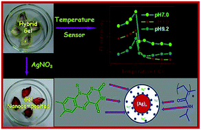
The riboflavin (R) and poly(N-isopropylacrylamide) (PNIPAAM) hybrid hydrogels have been prepared using the free radical polymerization of N-isopropylacrylamide in the presence of N,N′-methylene bisacrylamide as a cross linker for 1, 2 and 3 mM concentrations of R. The invariance of storage (G′) and loss (G′′) moduli over a wide range of angular frequency, where G′ > G′′ for R–PNIPAAM system characterize it to be behaving as a gel in the hybrid state. Both G′ and G′′ decrease with the increase of R concentration, but the decrease is four times higher in the former than in the latter. R–PNIPAAM gel has a higher critical strain value than PNIPAAM gels and it gradually increases with the increase in R concentration indicating that R is acting as a supramolecular cross-linker. The fluorescent intensity of R–PNIPAAM gels increases with the increase in R concentration and its variation with temperature at different pH shows an increase in the intensity value with temperature, showing the maximum at ∼30 °C because of the coil-to-globule transition of PNIPAAM chains, suggesting that the R–PNIPAAM gel can be used to be a probe for temperature detection. The sensitivity index of the fluorescent temperature sensing of R–PNIPAAM gel is moderate and it is highest at pH 7. R becomes gradually released from the R–PNIPAAM gel when dipped into water (pH 7) at 20 °C, initially at a slower rate, then at a higher rate, and finally it shows a leveling for the release of 19% of embedded R at 24 h of aging. Silver nanoparticles that are grown in the R–PNIPAAM gels by immersing the slashed gel pieces in AgNO3 solution stay at both the surface and the interior of the R–PNIPAAM fibres, maintaining the gel structure intact with a decrease in critical strain and causing a quenching of the fluorescence intensity of R–PNIPAAM gels. The average size and size distribution of AgNPs linearly increase with R concentration at a constant AgNO3 concentration.
Introduction
Polymeric hydrogels are obtained from the physical/chemical crosslinking of water soluble polymers forming a three dimensional network, which can immobilize a large amount of water molecules. These hydrogels are endowed with a myriad of applications in everyday life.1–3 Amongst all the polymeric hydrogels poly(N-isopropylacrylamide) (PNIPAAM) has attracted considerable attention due to its interesting thermal properties. The linear hydrophilic chains of PNIPAAM undergo a reversible coil-to-globule transition to form hydrophobic aggregates above its lower critical solution temperature (LCST, ∼32 °C).4,5 Utilizing this property, PNIPAAM has been extensively used for the fabrication of optoelectronic devices6–8 and bio-medical appliances.9–16 Because of this coil-to-globule transition, PNIPAAM hydrogel also serves to be an effective candidate for designing temperature sensors.17–20
Yamashita and co-workers have reported an interpenetrating gel network of PNIPAAM and poly(sodium acrylate), which has been successfully used as an adsorbent of heavy metal ions.21 Haraguchi and co-workers have demonstrated the effects of cross-linker contents on various physical properties of two different types of poly(N-isopropylacrylamide) (PNIPAAM) hydrogels, e.g. (i) nanocomposite type PNIPAAM hydrogels (NC gel) produced by using clay nanoparticles and (ii) the conventional chemically cross-linked PNIPAAM hydrogels (OR gel) produced using organic cross-linker N,N′-methylene bisacrylamide (BIS).22 The NC gels have exhibited extraordinary mechanical toughness, tensile moduli and tensile strengths, whereas the OR gels always exhibit weak and brittle natures, even on altering the concentration of BIS. The temperature-dependent adsorption of proteins on PNIPAAM hydrogel microspheres prepared by the precipitation polymerization of N-isopropylacrylamide (NIPAM) in water at 70 °C has been reported by Kawaguchi and co-workers.23 The physical interactions in the egg phosphatidylcholine liposome embedded into a PNIPAAM hydrogel matrix were studied along with the release mechanism of the encapsulated contents in the liposome at different temperatures.24 Nanocomposite hydrogels based on PNIPAAM and graphene-oxide (GO) were recently reported.25 GO was functionalized with glycidyl methacrylate (GMA) and it was incorporated into the PNIPAAM hydrogels, exhibiting a large volumetric change in response to infrared (IR) light illumination because of the highly efficient photothermal conversion of GO–GMA. In a recent report carbon nanotube-coated macroporous PNIPAAM hydrogel was fabricated and the composite hydrogel displayed a thermally induced shrinkage when electrically heated.26
There are also some reports on fluorescent hydrogels based on PNIPAAM.17–20,27,28 In some cases, the gels are rendered fluorescent by adopting rigorous synthetic methods by covalently attaching pyrene and fullerene (C60),17 3-hydroxyflavones,18 and 4-N-(2-acryloyloxyethyl)-N-methylamino-7-N,N-dimethylaminosulfonyl-2,1,3-benzoxadiazole (DBD-AE)20 to PNIPAAM chains. In some other cases fluorescent Au nano dots27 and CdTe quantum dots28 are embedded into the PNIPAAM gel matrix. Here, we have used a biologically important and highly luminescent small molecule, riboflavin, which has a lot of biological function. (−) Riboflavin (R) ([α]21 = −114.9°) is a very important bio-molecule (Vitamin B2) whose action is intimately related to the function of flavin nucleotides in biochemical reactions,29,30 and it also acts as a photoreceptor in the phototropism of plants.31 R has three different redox states: fully oxidized, one-electron reduced, and fully reduced. Each of these redox states exist in a cationic, neutral, and anionic form depending on the pH of the medium. Tuning the redox states, R can act as a very useful candidate for the synthesis of different metal nanoparticles. Recently, hybrid gels have also attracted intense research interest due to their excellent physical and mechanical properties.32,33 Here, we have prepared a PNIPAAM based fluorescent hydrogel by a very simplistic approach. We have used the highly luminescent biomolecule riboflavin (R) to prepare R–PNIPAAM hybrid hydrogel by dissolving the monomer NIPAAM in the R solutions of different concentrations followed by subsequent polymerization, in which R also acts as a supramolecular cross linker.
Covalent crosslinking between linear polymer chains creates a three dimensional network, but it reduces the structural flexibility of the system to certain extent because of the rigid nature of the covalent bonds. Introduction of a supramolecular synthon into the polymeric scaffolds can produce a cross linked three dimensional network constructed by noncovalent bonds. This type of supramolecular crosslinking generates a new class of smart polymeric materials with enhanced flexibility. However, there are only few reports on supramolecular cross linking.34–37 In an elegant work, Kretschmann and co-workers have reported a hydrogel based on supramolecular crosslinking between adamantyl-containing copolymers and cyclodextrin dimers.38 Here, we have incorporated a small molecule R, acting as a supramolecular cross linker, between the PNIPAAM chains. We have investigated the rheological properties of the R–PNIPAAM hybrid hydrogels to determine the changes in the mechanical properties of the gel compared to that of the pure PNIPAAM gels. R–PNIPAAM gel has a higher critical strain value than that of PNIPAAM gels and it gradually increases with increasing R concentration, indicating that R is acting as a supramolecular cross-linker.
Therefore, the major aim of this work is to study the influence of supra-molecular crosslinking using a fluorescent biomolecule, riboflavin (R), by preparing a fluorescent hydrogel based on PNIPAAM without using rigorous synthetic methods. The structures of R and PNIPAAM are depicted in Scheme 1. From the structures presented in Scheme 1, there is a possibility of supramolecular crosslinking through the >NH, >C
O and –OH groups of R with the >C
O and >NH groups of PNIPAAM. Here, we want to report, in addition to the covalent cross-linking present in the PNIPAAM gel, the effect of supramolecular cross-linking on the mechanical and fluorescent properties of the hydrogel by varying the concentration of the supramolecular cross-linker at a constant covalent cross-linking density of the PNIPAAM gel. Moreover, it is important to know how the properties (e.g. fluorescent, reducing, and nanoparticle stabilization) of the supramolecular cross-linker vary in the gel matrix. With this aim, we have prepared a fluorescent hydrogel of PNIPAAM by doping R into the PNIPAAM gel matrix. R acts to be a supramolecular cross-linker, and it enhances the mechanical as well as the fluorescence properties of the hydrogel. The fluorescence activity of the R–PNIPAAM gel is monitored with temperature at three different pH values. We have elegantly combined the temperature sensitivity of PNIPAAM with the pH dependent fluorescence property of R to develop a probe for temperature sensing, and an index of sensitivity has been obtained. We have investigated the reducing property of the supramolecular crosslinked R to produce AgNPs in the R–PNIPAAM gel and also tuned the size and distribution of AgNPs by varying the concentration of R. Because of the supramolecular nature of the linkages there is a possibility of leaching out of R from the PNIPAAM fibres in the gel, and an insight into the mechanism of the leaching process for aging in aqueous medium at room temperature (30 °C) is also delineated here.
 |
| Scheme 1 Chemical structures of (a) PNIPAAM and (b) riboflavin. | |
Experimental procedure
Materials
(−) Riboflavin (R), N-isopropylacrylamide (NIPAAM), N,N′-methylene bisacrylamide and ammonium persulfate were purchased from Aldrich Chemical Co., USA. Silver nitrate was purchased from SRL, Mumbai, India.
N,N,N′,N′-Tetramethyl ethylene diamine was purchased from Loba Chemie, Mumbai, India. They were used as received and water was doubly distilled before use.
Preparation of Riboflavin doped PNIPAAM hydrogels
1, 2 and 3 mM solutions of R were prepared in buffer solutions of three different pH, 4, 7, and 9.2. Required quantities of R were dissolved in the buffer solutions by heating them in screw capped vials to prepare the R–PNIPAAM gels. The R–PNIPAAM gels were prepared using free radical polymerization technique39 in aqueous medium. 300 mg of NIPAAM was dissolved in 6 ml R solution (pH 7), and to this solution 12 mg N,N′-methylene bisacrylamide, 24 μl N,N,N′,N′-tetramethyl ethylene diamine, 4 mg ammonium persulfate were added, and it was kept undisturbed at 20 °C. Gel formation was observed within 2–3 hours. The R–PNIPAAM gel appeared as a bright yellow semi-solid mass. For temperature sensing purposes, the gels were prepared in a quartz cell of 1 cm path length. Three different R–PNIPAAM gels were prepared by varying the R concentrations as 1, 2 and 3 mM, and they are designated as R–PNIPAAM1, R–PNIPAAM2 and R–PNIPAAM3 gels.
Rheology
To understand the mechanical properties of the gels, rheological experiments were performed with an advanced rheometer (AR 2000, TA Instrument, USA) using cone plate geometry on a Peltier plate. The diameter of the plate was 40 mm and the cone angle was 4° with a plate gap of 121 μm. The R concentration in the R–PNIPAAM system was varied from 1 mM to 3 mM concentration.
Spectroscopy
For fluorescence studies the hydrogel samples were prepared in sealed cuvettes and the fluorescence studies were carried out in a Horiba Jobin Yvon Fluoromax 3 instrument. Each gel sample in a quartz cell of 1 cm path length was excited at 373 nm wavelength and the emission scans were recorded from 400 to 700 nm using an excitation slit width of 3 nm and emission slit width of 5 nm with a 1 nm wavelength increment having an integration time of 0.1 s. The FT-IR spectra of pure components and the xerogels were recorded using KBr pellets in a Perkin Elmer FT-IR instrument (FT-IR-8400S).
Dynamic light scattering (DLS)
The DLS experiments of the Ag nanoparticle embedded R–PNIPAAM1, R–PNIPAAM2, and R–PNIPAAM3 gel samples were carried out in a Malvern instrument. The laser source was a He–Ne laser, placed at an angle of 173° and equipped with a non-invasive back scatter detector using the method of cumulants. The AgNP embedded gels were diluted with water and sonicated for half an hour before the measurement.
Release of R from the R–PNIPAAM gel
To study the release of R from the R–PNIPAAM1 gels, 3 ml of the R–PNIPAAM1 gel was prepared and immersed in 10 ml of water (one time) and kept at 20 °C. The supernatant solution was taken in a quartz cell of 1 mm path length and UV-vis spectra were obtained at different time intervals.
Synthesis of Ag nanoparticles in R–PNIPAAM gels
The R–PNIPAAM1, R–PNIPAAM2 and R–PNIPAAM3 gels were washed carefully with water to remove unreacted reagents. Then, the gels were slashed into pieces of almost equal dimension. The slashed pieces were dipped into 10 ml of 1 mM AgNO3 solution. Nanoparticle (NP) formation was activated by the diffusion of AgNO3 inside the gel matrix, which was detected within few minutes by observing a color change of the gels from bright yellow to bright red. Nanoparticle formation was allowed to proceed for 30 minutes. Then, the Ag nanoparticle embedded R–PNIPAAM1, R–PNIPAAM2 and R–PNIPAAM3 gel pieces (R–PNIPAAM–Ag) were taken out of the AgNO3 solution and carefully washed with water. The UV-vis spectra of the R–PNIPAAM1–Ag gel were recorded with a Hewlett-Packard UV-vis spectrophotometer (model 8453). The slashed gel pieces were taken in microscopic cover slips and the absorbance spectra were recorded.
Microscopy
The morphology of the R–PNIPAAM1–Ag gel was investigated by transmission electron microscopy (TEM). A small portion of the R–PNIPAAM1–Ag gel was diluted and drop casted on a carbon coated copper grid (300 mesh) and the samples were dried in open air at 30 °C overnight before the experiment was performed.
Results and discussion
In the gelation process, the riboflavin doped chemically cross linked PNIPAAM gel is produced through the cross linker N,N′-methylene bisacrylamide (BIS). It has a bright yellow colour with self-sustainable morphology and it can be slashed into pieces which are stable for weeks.
Mechanical properties
The gel formation was characterized by the rheological experiments. Gels are viscoelastic material endowed with the unique properties of storage and dissipation of energy. The storage modulus (G′) indicates how the energy is stored in the system, whereas the loss modulus (G′′) indicates how the energy is dissipated from the system. There are only few reports about the mechanical properties of PNIPAAM hydrogels and this is the first report of a hybrid hydrogel of PNIPAAM with a small molecule. Here, we have used a fluorescent biomolecule, riboflavin (R), which also behaves as a supramolecular cross linker, and have successfully tuned the mechanical properties of the hydrogel by varying the amount of the supramolecular cross linker R. Frequency sweep experiments were carried out at room temperature (25 °C) under a 1% strain and G′ and G′′ values were plotted against angular frequency (ω) (Fig. 1a and b). The frequency sweep plots exhibit an invariance of G′ and G′′ values over a wide range of angular frequency values for both PNIPAAM and R–PNIPAAM1 gels. Moreover, the values of G′ are higher than G′′, which characterize both the systems to behave as gels. It is evident from the figure that PNIPAAM gel has 37% higher storage modulus than that of R–PNIPAAM1 gel. With further increase in R concentration, the storage modulus significantly decreases, as shown in Fig. 2a and S1.† Thus, for a 3 mM concentration of riboflavin there is a 22 times decrease in storage modulus and 6 times decrease in loss modulus than those of pure PNIPAAM gel.
 |
| Fig. 1 (a) and (b) Oscillation frequency dependency of the modulus values G′ and G′′ of PNIPAAM and R–PNIPAAM1 gels, respectively. (c) G′ and G′′ vs. % strain plot of PNIPAAM gel and (d) G′ and G′′ vs. % strain plot of R–PNIPAAM1 gel at a constant frequency of 1 Hz. | |
 |
| Fig. 2 (a) Plot of modulus (G′, G′′) values of the R–PNIPAAM gels with varying concentration of R (b) plot of critical % strain values of the R–PNIPAAM gels with the varying concentration of R. | |
Here, it is necessary to compare the above rheological data with those of other systems reported in the literature. Zhang and co-workers have reported PVA/PNIPAAM semi-IPN hydrogels prepared by radical polymerization using N,N′-methylenebisacrylamide (BIS) as the crosslinker.40 The amount of crosslinking was maintained constant for all the cases and with increasing the NIPAAM concentration, the G′ values were found to increase. However, two sets of IPN hydrogels containing PVA with different molecular weight, but same PVA content, showed similar G′ values. Chetty and co-workers have reported hydrogels of poly(N-isopropylacrylamide-co-N,N′′-methylene-bisacrylamide) by redox free radical polymerization in aqueous medium.41 A 1.0 M (11% (w/v)) NIPAAM and 0.1 M (1.5% (w/v)) solution of BIS was used for the polymerization, and the molar ratio of BIS to NIPAAM was varied from 0.01 to 0.1. They observed that the G′ values of the hydrogels increased (1917 Pa to 7143 Pa) with increasing cross-linker concentration. However, in our work, we have maintained the concentration of the monomer NIPAAM (5% (w/v)) and the concentration of the chemical crosslinker N,N′-methylene bisacrylamide (0.2% (w/v)) constant, but varied the concentration of the supramolecular crosslinker R, and a decrease of G′ value from 315 Pa to 15 Pa with increasing R content is observed (Fig. 2a). This is considerably a contrasting behavior than that of the chemically cross linked gels and a probable reason from the view point of the supramolecular cross-linking of riboflavin is discussed here.
In PNIPAAM gel, the individual polymer chains are supramolecularly linked with each other via interchain H-bonding interactions in addition to the chemical cross-linking through the cross-linker N,N′-methylene bisacrylamide (cf. FTIR spectra). Thus, the energy is efficiently stored in the PNIPAAM gel by transferring through both the chemical crosslinks and also through the supramolecular links between different PNIPAAM chains. However, when riboflavin enters between the PNIPAAM chains the interchain separation increases, causing a decrease in the propensity of supramolecular H-bond formation between the chains, though there occurs fewer new supramolecular crosslinking via riboflavin. This lowering of H-bonding density between the PNIPAAM chains causes a decrease in the storage of energy through the PNIPAAM chains, though some contribution to energy storage may come from the supramolecularly anchored small riboflavin molecules. However, the latter contribution is considerably less compared to that through PNIPAAM chains, which cannot compensate for the loss due to the larger separation of the PNIPAAM chains. The exact reason for the lesser degree of decrease in loss modulus compared to that of storage modulus on the addition of R to the PNIPAAM gel is not known and it may be explained in the following way. In the PNIPAAM gel the energy is dissipated to the solvent molecules from the fibril surface, where the PNIPAAM chains are also supramolecularly connected through the H-bonds. Upon the addition of R, these bonds decrease in number, as discussed above, and the decrease in loss modulus is expected to be comparable to that in storage modulus. However, here we observe about four times smaller decrease than that of the former. A possible reason is that the energy is lost through the surface of the fibres to the solvent molecules and it is not directly linked to the supramolecular H-bonds between the chains.
The strain sweep experiments performed at a constant frequency of 1 rad s−1 (Fig. 1c and d) indicate that G′ crosses G′′ at a 322% strain indicating the critical strain (minimum strain for gel breaking) of R–PNIPAAM1 gel (Fig. 1d), and it is higher than that of the PNIPAAM gel (203%, Fig. 1c). With further increase in R concentration, the critical strain shows almost a linear increase (Fig. 2b and S2†). A probable reason may be that riboflavin is acting as a supramolecular cross linker between the PNIPAAM chains and the crosslinking density increases with increase in R concentration. The supramolecular crosslinks afford an additional stability to the R–PNIPAAM1 gels, thus a larger strain is required to rupture the gel. In R–PNIPAAM2 and R–PNIPAAM3 gels the supramolecular crosslinking density increases, causing almost a linear increase in the critical strain value with increase in R concentration. Chetty and co-workers observed that the critical strain increases from 1.6% to 7.9% with increasing cross-linker ([BIS]/[NIPAAM]) concentration from 0.01 to 0.1.41 However, in the present system the critical strain increases from 203% to 584% with the addition of 3 mM R solution. This significant increase of critical strain is an interesting observation particularly for the case of supramolecular crosslinking.
FTIR spectra
The supramolecular interaction between the components is evident from the FTIR spectra (Fig. 3). The peak at 1638 cm−1 in the PNIPAAM gel, which may be ascribed to the secondary amide C
O stretching of PNIPAAM,42 is shifted to 1643 cm−1 in the R–PNIPAAM gel indicating H bonding between the >C
O group with the –N–H and –O–H groups of riboflavin. The R has small peaks at 3100–3600 cm−1 for the –N–H and –O–H vibrations and these are not observed in the R–PNIPAAM1 gel probably due to the H-bonding interactions. This type of supramolecular interaction helps the R–PNIPAAM1 gel to endure a considerably higher strain than the PNIPAAM gel before breaking. It is also important to note that in the PNIPAAM gel, the –N–H vibration peak at 3435 cm−1 is very broad compared to those of R, which is an indication of the presence of interchain H-bonding interactions between the PNIPAAM chains through its >C
O groups (Scheme 1).
 |
| Fig. 3 FTIR spectra of Riboflavin and PNIPAAM and R–PNIPAAM1 xerogels. | |
Fluorescence properties
The photoluminescence properties of riboflavin have been extensively studied,29,43 and its emission efficiency is dependent upon the polarity of the solvent. Riboflavin shows bright green fluorescence when it is excited at 373 nm, having a quantum yield of 0.27 at pH 7.44 The hydrogen bond forming solvents mitigate the photoluminescence efficiency due to the quenching of excitons through these bonds.29 The PNIPAAM gel is not fluorescent active but the R–PNIPAAM gel becomes fluorescent active due to the presence of riboflavin (Fig. 4a). In addition, as the concentration of riboflavin increases in the gel, the fluorescence intensity increases. A small red shift in the emission peak is also observed in the R–PNIPAAM gel with increase in R concentration. The increase in fluorescence intensity may be attributed to the increase in fluorophore concentration and the red shift may arise due to the better stabilization of excitons at the aggregated state of the fluorophore at its higher concentration. It is to be noted here that the emission intensity of riboflavin solutions at identical concentration are considerably less than that at the gel state (Fig. 4a). Thus, the fluorescence intensity of R is highly increased when embedded in the PNIPAAM gel. For R–PNIPAAM gels, with the addition of 1, 2 and 3 mM R solution to the PNIPAAM gel, the fluorescence intensity increases by approximately 4, 24 and 41 times respectively, compared to the solution state. This abrupt increase in the fluorescence intensity of R from the solution to embedded gel state is due to the hydrophobic environment inside the gel fibril, prohibiting the quenching of R by the water molecules. It is also interesting to note that due to supramolecular crosslinking of R in the PNIPAAM gel, there is a blue shift of the emission peak by 6, 3 and 3 nm for R–PNIPAAM1, R–PNIPAAM2 and R–PNIPAAM3 gels, respectively.
 |
| Fig. 4 (a). Fluorescence spectra of R solutions and the corresponding R–PNIPAAM gels (b) temperature dependent fluorescence spectra of R–PNIPAAM1 gel at pH 7, (c) variation of fluorescent intensity of R–PNIPAAM1 gels with temperature at different pH. | |
Temperature sensing
As PNIPAAM is a thermosensitive polymer, we studied the temperature dependent fluorescence properties of R–PNIPAAM1 gel at different pH. Fig. 4b shows the temperature dependent fluorescence spectra of R–PNIPAAM1 gel at pH 7 (Fig. S3 and S4† at pH 4 and pH 9.2) and the variation of fluorescent intensity of R–PNIPAAM1 gels with temperature at different pH is shown in Fig. 4c. In each case, the fluorescence intensity at first increases with increase in temperature and reaches maximum at ∼30 °C showing an inflection point. Subsequently, it undergoes a sudden fall in the intensity value, and then becomes almost constant. The initial increase in fluorescence intensity with temperature may be ascribed to the commencement of the coil to globule transition of PNIPAAM chains leading to the formation of hydrophobic aggregates, thus decreasing the quenching of R with water. The R molecules trapped inside the hydrophobic cores experience less H bonding with the solvent (water) molecules, thus experiencing a lesser degree of quenching of excitons by solvent molecules29,30,45 and this causes an increase in the fluorescence intensity. After 30 °C, because of the phase transition (lower critical solution temperature, LCST) of PNIPAAM, the R–PNIPAAM1 gel becomes turbid and the fluorescence intensity suddenly decreases due to scattering.46 Therefore, the inflection point can be regarded to be the LCST phase transition temperature of PNIPAAM in the presence of 1 mM R. It is evident from Fig. 4c that the fluorescence intensities at pH 9.2 are lower than those at pH 4 and pH 7, particularly at and below 35 °C. This is because at neutral pH (pH = 7) the imido H atom of R remains unaffected but at higher pH (9.2) the labile imido H atom is abstracted, and this disturbs the conjugation of the isoalloxazine ring,47 consequently the quenching of the fluorescence intensity occurs. Moreover, at pH 4 the protonation of ring nitrogens and carbonyl oxygen occurs, which hinders the conjugation of the isoalloxazine ring to certain extent, and hence lower fluorescence intensity is exhibited than at pH 7.
In both Fig. 4b and c a sharp transition within a very narrow temperature range (30–32 °C) is observed. This behavior strongly suggests that the R–PNIPAAM1 gel can be used as a probe for the detection of a specific temperature. Two major advantages of this system are: (i) it can be designed in a very simple way via polymerization of the monomer, NIPAAM, by incorporating fluorescent molecule R, and (ii) R and NIPAAM both are biocompatible and might be useful in biological thermometry.19
To investigate whether our hydrogel can function as a temperature sensor, we calculated the sensitivities of the hydrogel based sensors at different pH. The lowest sensitivity limit (Tlow) and the highest sensitivity limit (Thigh) of temperature were obtained from the criterion shown by Uchiyama and co-workers.48 The temperature sensitivity of the sensors can be defined by the parameters ΔT = Tlow − Thigh, fluorescent enhancement (FE) factor (the ratio of the fluorescence intensities of Tlow and Thigh) and the FE/ΔT values. All these values are presented in Table 1. The FE/ΔT values are considered to be an index of sensitivity and their values are found to be between 0.32 and 1.1. The values indicate that this hydrogel based temperature sensor is most sensitive at pH 7 and it is more sensitive than the temperature sensors developed using other principles like exciplexes,49 excimers,50 twisted intramolecular charge-transfer complexes,51,52 and spin crossover complexes53 (cf. maximum FE/ΔT = 0.063). However, the present system has smaller values than those obtained by Iwai and co-workers, where they developed molecular thermometers based on PNIPAAM labeled with the fluorescent molecule benzofurazan (FE/ΔT ∼ 0.6 to 2.7).20
Table 1 Index of sensitivity (FE/ΔT) of fluorescent temperature sensing of the R–PNIPAAM1 gel at different pH
pH |
Thigh |
Tlow |
ΔT |
FE |
FE/ΔT |
7 |
32 |
30 |
2 |
2.2 |
1.1 |
4 |
45 |
30 |
15 |
4.8 |
0.32 |
9.2 |
35 |
30 |
5 |
2.6 |
0.52 |
Release of R from the R–PNIPAAM gel
Because of the porous nature of PNIPAAM hydrogel,54 there is a possibility that embedded R molecules in the R–PNIPAAM gels may gradually leach out as R is supramolecularly linked with the PNIPAAM chains in the gel fibre. To study the time scale of riboflavin release from the R–PNIPAAM1 gel, 3 ml of gel were submerged in 10 ml water at 20 °C at pH 7 and UV-vis spectra of the supernatant solution were obtained at certain time intervals (Fig. 5a). The gel was found to be stable in aqueous environment even after immersion for more than two days, but it is evident from the UV-vis spectra (Fig. 5) that R is gradually released from the R–PNIPAAM1 gel as the absorbance values of the two peaks of R at 354 and 448 nm (π–π* transitions of R43) gradually increase with time. The plots of absorbance values of the two peaks with time (Fig. 5b) indicate that initially the release of R is slow, then it rapidly increases and finally it gets retarded, showing saturation. It is observed that ∼19% of R is released from the R–PNIPAAM1 system after 24 hours, and after that no increase in absorbance intensity is observed. The initial slow release of R may be attributed to the release of R captured near the inner periphery of the fibre surface where the R molecules experience an inner drag due to the surface tensional forces of the R–PNIPAAM1 nanofibres. This slow release creates some new channels, through which the embedded R present at the interior of the fibre leaches out at a faster rate. This observation certainly indicates that R prefers to stay at the interior of the fibre due to a higher degree of supramolecular interaction with the PNIPAAM chains from different sides. However, due to the strong H-bonding ability of water with R, it gradually diffuses out of the fibre surface till the chemical potential of R at the fibre interior becomes equal to that in the water. In the R–PNIPAAM1, gel equilibrium is almost attained after 24 h of aging at 20 °C, releasing 19% (w/w) of embedded R.
 |
| Fig. 5 (a) UV-vis spectra of the supernatant solution for the study of R release at different time intervals from R–PNIPAAM1 gel (b) plot of absorbance of the peaks at 354 and 448 nm with time (inset: Enlarged plot of absorbance values with time for release up to 150 min). | |
Synthesis of R–PNIPAAM–Ag gel nanocomposite
Riboflavin (vitamin B2, R) is the most frequently encountered organic co-factor in nature, which possesses three different redox states: fully oxidized, one-electron reduced, and fully reduced55 and each of them exists in corresponding anionic, neutral and cationic form depending on the pH of the solution. The reduction potential of R (−0.3 versus SCE)30 allows it to reduce Ag+ to Ag, (0.80 V versus SCE).56 R can also act as a stabilizing agent of silver nanoparticles57 and PNIPAAM is also known to stabilize silver nanoparticles.58 Here, we would like to observe whether riboflavin, which is present in the gel matrix as a supramolecular cross-linker, can act as an effective reducing agent to produce Ag nanoparticles (NPs), and if so whether the gel remains stable after the generation of the AgNPs. Here, we synthesized AgNPs in the R–PNIPAAM1 gel in a very simple approach (cf. experimental section). The R–PNIPAAM1 gel possesses high mechanical stability, thus it can be slashed into pieces (Fig. 6a). The slashed pieces of the gel are dipped into AgNO3 solutions. As soon as the Ag+ ions diffuse into the gel matrix they are reduced to Ag, and more Ag+ ions then diffuse, get reduced to Ag(0) in the gel matrix and the nanoparticles begins to grow. The Ag nanoparticles are then stabilized inside the R–PNIPAAM1 gel by complexation with R and PNIPAAM chains. R stabilizes the Ag nanoparticles by the complexation of absorbed Ag+ ions with the flavin moiety, and it is also stabilized through the complexation with the PNIPAAM chain (Scheme 2). This causes a rapid color change in the gel pieces from bright yellow to red indicating the formation of Ag nanoparticles (Fig. 6a and b). However, after the formation of Ag nanoparticles, a quenching of the fluorescence intensity of the hydrogel system occurs, which is clearly evident from the images of R–PNIPAAM1 and R–PNIPAAM–Ag gel pieces under UV light of wavelength 350 nm (Fig. 6c and d). Quantitative data for fluorescence quenching after the Ag NP formation is evident from Fig. 6e, where a significant quenching of fluorescence intensity (∼135 times) compared to that of R–PNIPAAM1 gel has occurred. This is because AgNPs are well known fluorescence quenchers, and because R stabilizes the Ag nanoparticles, it efficiently quenches the fluorescence of R by providing alternate routes for the decay of the excitons.57
 |
| Fig. 6 Images of (a) the R–PNIPAAM1 hydrogel pieces (b) Ag nanoparticle embedded bulk R–PNIPAAM1 gel pieces after half an hour of the addition of AgNO3. (c) and (d) R–PNIPAAM1 and R–PNIPAAM1–Ag gels, respectively, under UV light of wavelength 350 nm. (e) Fluorescence spectra of R–PNIPAAM1 and R–PNIPAAM1–Ag gels. | |
 |
| Scheme 2 Schematic representation of the stabilization of Ag nanoparticles by R–PNIPAAM gel. | |
The formation and stabilization of Ag nanoparticles in the R–PNIPAAM1 gel is further confirmed by transmission electron microscopy (TEM) (Fig. 7). It is evident from the careful investigation of Fig. 7a that the Ag nanoparticles are grown both at the interior (blurred black spots) of the fibres and also on the surface (bright black spots) of the fibres of the gel. Thus, the AgNP formation keeps the gel network intact, in particular at the concentration studied here. The Ag nanoparticle formation is also evidenced from the UV-vis spectra of Ag nanoparticle embedded R–PNIPAAM1 gels (Fig. S5†). The peaks at 380 and 477 nm correspond to the π–π* transition peak of R in the R–PNIPAAM1 gels and the 535 nm peak corresponds to the plasmon band of AgNPs.57
 |
| Fig. 7 TEM images of Ag nanoparticle embedded in R–PNIPAAM1 hydrogel. | |
To get an insight on Ag-nanoparticle formation in the gel fibres, we conducted a dynamic light scattering (DLS) study on the AgNP embedded R–PNIPAAM gels made at different concentrations of R, after rigorous sonication. It is important to note from the DLS results (Fig. 8) that average sizes of AgNPs are 48 ± 9.1, 72 ± 15.1 and 87 ± 17.1 nm, produced from R–PNIPAAM1, R–PNIPAAM2 and R–PNIPAAM3 gels, respectively. It indicates a linear increase of average particle sizes of AgNPs with increasing AgNO3 concentration. A probable way of explaining the results is that Ag+ ion first diffuses into the gel matrix, and as soon as it approaches the embedded R in the gel fibres, it gets reduced to Ag(0). Thus, a greater number of Ag+ ions gradually diffuse into the gel matrix and get reduced to Ag (0) causing the nucleation of AgNPs. These AgNPs then grow through the force of attraction (cohesive force) between the Ag (0) particles, which easily moves through the nanopores present in the gel fibres.59,60 Thus, a large number AgNPs grow within the gel matrix and as the R concentration increases, the diameter of the AgNPs increases due to formation of higher number of Ag (0) particles causing a linear variation in size of AgNPs. The half height width of the Gaussian fitted curves of the DLS data (Fig. 8) are 22.6, 38.6 and 43.5 nm for R–PNIPAAM1, R–PNIPAAM2 and R–PNIPAAM3 gels, respectively, indicating the wider distribution of particle size with increasing R concentration. A possible reason for wider particle size distribution is that increasing R-concentration in the gel causes an increase in the number of the nucleating sites of AgNPs within the gel fibres, and the particle size at these sites are therefore not very uniform due to the competition of growth processes at different nuclei causing the wider distribution of particle size of AgNPs.
 |
| Fig. 8 DLS data of AgNPs produced from (a) R–PNIPAAM1, (b) R–PNIPAAM2 and (c) R–PNIPAAM3 gels, respectively (d) plot of AgNP diameter and distribution of particle sizes of AgNPs with increase of R concentration in the R–PNIPAAM gel. | |
Rheological experiments were also performed to investigate the mechanical properties of the Ag nanoparticle embedded R–PNIPAAM1 (R–PNIPAAM1–Ag) hydrogels. The value of storage modulus (G′) of the Ag nanoparticle embedded R–PNIPAAM1 hydrogels are almost the same as that of R–PNIPAAM1 gel, but they are lower than that of the pure PNIPAAM gels (Fig. S6†), indicating that the formation of Ag nanoparticles does not affect the strength of the R–PNIPAAM1 gel. The strain sweep experiments (Fig. S7†) shows that G′ crosses G′′ at a 254% strain indicating that the critical strain of the Ag nanoparticle embedded R–PNIPAAM1 gel is lower than that of R–PNIPAAM1 gel by 68%. One probable reason is the lesser degree of H-bonding between R and PNIPAAM because some sites of R and PNIPAAM are actively used to stabilize the Ag nanoparticles.
Conclusion
In the R–PNIPAAM hybrid hydrogels prepared by the free radical polymerization of NIPAAM in the presence of N,N′-methylene bisacrylamide as cross linker, R acts as a supramolecular cross-linker. The invariance of storage (G′) and loss (G′′) moduli over a wide range of angular frequency, where G′ > G′′ characterize the R–PNIPAAM hybrid system to be a gel. However, with increase in the concentration in R both G′ and G′′ decrease. The decrease has been attributed to the decrease in supramolecular H-bonds between the PNIPAAM chains due to the incorporation of R. The critical strain value of R–PNIPAAM gel gradually increases with increase in R concentration confirming the supramolecular cross-linking property of R, in addition to the presence of chemical cross-linking with N,N′-methylene bisacrylamide in the system. PNIPAAM gel is not fluorescent but the R–PNIPAAM gels are fluorescent and the intensity increases with increase in R concentration. Its variation with temperature at different pH shows an increase in the intensity value with temperature, showing the maximum at ∼30 °C, due to the coil to globule transition of PNIPAAM chains, suggesting that the R–PNIPAAM gel can be used as a probe for temperature detection. The index of sensitivity of the fluorescent temperature sensing of R–PNIPAAM gel is moderate and it is highest at pH 7. R is gradually released from the R–PNIPAAM gel, initially at a slower rate, then at a higher rate and finally it shows a leveling off, with a release of 19% of embedded R after 24 h AgNPs grown in the R–PNIPAAM gels stay at both the surface and interior of the R–PNIPAAM fibres, keeping the gel structure intact and causing a quenching of the fluorescence intensity of R–PNIPAAM gels. With the increase of R concentration, the average size and AgNPs size distribution linearly increase for a constant AgNO3 concentration.
Acknowledgements
P.C and P.B acknowledge CSIR, New Delhi for providing the fellowship. We also acknowledge unit of Nanoscience project at IACS for financial support.
References
- S. Vinogradov, E. Batrakova and A. Kabanov, Colloids Surf., B, 1999, 6, 291–304 CrossRef.
- R. Esfand and D. A. Tomalia, Drug Discovery Today, 2001, 6, 427–436 CrossRef CAS.
- S. Stolnik, L. Illum and S. S Davis, Adv. Drug Delivery Rev., 1995, 16, 195–214 CrossRef CAS.
- M. Heskins and J. E. Guillet, J. Macromol. Sci., Part A: Pure Appl. Chem., 1968, 2, 1441–1455 CrossRef CAS.
- H. G. Schild, Prog. Polym. Sci., 1992, 17, 163–249 CrossRef CAS.
- J. M. Weissman, H. B. Sunkara, A. S. Tse and S. A. Asher, Science, 1996, 274, 959–960 CrossRef CAS.
- L. Dong, A. K. Agarwal, D. J. Beebe and H. Jiang, Nature, 2006, 442, 551–554 CrossRef CAS PubMed.
- A. Sidorenko, T. Krupenkin, A. Taylor, P. Fratzl and J. Aizenberg, Science, 2007, 315, 487–490 CrossRef CAS PubMed.
- Z. Cheng, S. Liu, P. W. Beines, N. Ding, P. Jakubowicz and W. Knoll, Chem. Mater., 2008, 20, 7215–7219 CrossRef CAS.
- L. E. Bromberg and E. S. Ron, Adv. Drug Delivery Rev., 1998, 31, 197–221 CrossRef CAS.
- P. F. Kiser, G. Wilson and D. Needham, Nature, 1998, 394, 459–462 CrossRef CAS PubMed.
- N. Murthy, M. C. Xu, S. Schuck, J. Kunisawa, N. Shastri and J. M. Fréchet, Proc. Natl. Acad. Sci. U.S.A., 2003, 100, 4995–5000 CrossRef CAS PubMed.
- J. Kim, S. Nayak and L. A. Lyon, J. Am. Chem. Soc., 2005, 127, 9588–9592 CrossRef CAS PubMed.
- Y. Zhang, S. Furyk, D. E. Bergbreiter and P. S. Cremer, J. Am. Chem. Soc., 2005, 127, 14505–14510 CrossRef CAS PubMed.
- Y. Li, Y. Tang, R. Narain, A. L. Lewis and S. P. Armes, Langmuir, 2005, 21, 9946–9954 CrossRef CAS PubMed.
- M. Das, S. Mardyani, W. C. W. Chan and E. Kumacheva, Adv. Mater., 2006, 18, 80–83 CrossRef CAS.
- S. W. Hong, D. Y. Kim, J. U. Lee and W. H. Jo, Macromolecules, 2009, 42, 2756–2761 CrossRef CAS.
- C. Y. Chen and C. T. Chen, Chem. Commun., 2011, 47, 994–996 RSC.
- C. Gota, K. Okabe, T. Funatsu, Y. Harada and S. Uchiyama, J. Am. Chem. Soc., 2009, 131, 2766–2767 CrossRef CAS PubMed.
- K. Iwai, Y. Matsumura, S. Uchiyama and A. P. de Silva, J. Mater. Chem., 2005, 15, 2796–2800 RSC.
- K. Yamashita, T. Nishimura and M. Nango, Polym. Adv. Technol., 2003, 14, 189–194 CrossRef CAS.
- K. Haraguchi, T. Takehisa and S. Fan, Macromolecules, 2002, 35, 10162–10171 CrossRef CAS.
- H. Kawaguchi, K. Fujimoto and Y. Mizuhara, Colloid Polym. Sci., 1992, 270, 53–57 CAS.
- Y. Liu, Z. Li and D. Liang, Soft Matter, 2012, 8, 4517–4523 RSC.
- C.-W. Lo, D. Zhu and H. Jiang, Soft Matter, 2011, 7, 5604–5609 RSC.
- J. Wu, Y. Ren, J. Sun and L. Feng, ACS Appl. Mater. Interfaces, 2013, 5, 3519–3523 CAS.
- L. Y. Chen, C. M. Ou, W. Y. Chen, C. C. Huang and H. T. Chang, ACS Appl. Mater. Interfaces, 2013, 5, 4383–4388 CAS.
- J. Li, X. Hong, Y. Liu, D. Li, Y. W. Wang, J. H. Li, Y. B. Bai and T. J. Li, Adv. Mater, 2005, 17, 163–166 CrossRef CAS.
- G. R. Penzer and G. K. Radda, Q. Rev., Chem. Soc., 1967, 21, 43–65 RSC.
- V. Massey, Biochem. Soc. Trans., 2000, 28, 283–296 CrossRef CAS.
- K. V. Thimann and G. M. Curry, in Comparative Biochemistry, ed. M.Florkin and H. S.Masson, Academic Press, New York, 1960, p. 281 Search PubMed.
- V. R. R. Kumar, V. Sajini, T. S. Sreeprasad, V. K. Praveen, A. Ajayaghosh and T. Pradeep, Chem. Asian J., 2009, 4, 840–848 CrossRef CAS PubMed.
- D. Dasgupta, S. Srinivasan, C. Rochas, A. Ajayaghosh and J. M. Guenet, Langmuir, 2009, 25, 8593–8598 CrossRef CAS PubMed.
- T. Haino, E. Hirai, Y. Fujiwara and K. Kashihara, Angew. Chem., Int. Ed., 2010, 49, 7899–7903 CrossRef CAS PubMed.
- F. Wang, J. Zhang, X. Ding, S. Dong, M. Liu, B. Zheng, S. Li, L. Wu, Y. Yu, H. W. Gibson and F. Huang, Angew. Chem., 2010, 122, 1108–1112 CrossRef.
- E. A. Appel, F. Biedermann, U. Rauwald, S. T. Jones, J. M. Zayed and O. A. Scherman, J. Am. Chem. Soc., 2010, 132, 14251–14260 CrossRef CAS PubMed.
- X. Ji, Y. Yao, J. Li, X. Yan and F. Huang, J. Am. Chem. Soc., 2013, 135, 74–77 CrossRef CAS PubMed.
- O. Kretschmann, S. W. Choi, M. Miyauchi, I. Tomatsu, A. Harada and H. Ritter, Angew. Chem., Int. Ed., 2006, 45, 4361–4365 CrossRef PubMed.
- L. Chen, M. Liu, L. Lin, T. Zhang, J. Ma, Y. Songa and L. Jiang, Soft Matter, 2010, 6, 2708–2712 RSC.
- J.-T. Zhang, R. Bhat and K. D. Jandt, Acta Biomater., 2009, 5, 488–497 CrossRef CAS PubMed.
- A. Chetty, J. Kovács, Zs. Sulyok, Á. Mészáros, J. Fekete, A. Domján, A. Szilágyi and V. Vargha, eXPRESS Polym. Lett., 2013, 7, 95–105 CrossRef CAS.
- Y. Cui, C. Tao, Y. Tian, Q. He and J. Li, Langmuir, 2006, 22, 8205–8208 CrossRef CAS PubMed.
- P. F. Heelis, Chem. Soc. Rev., 1982, 11, 15–39 RSC.
- P. Bairi, B. Roy and A. K. Nandi, Chem. Commun., 2012, 48, 10850–10852 RSC.
- S. Manna, A. Saha and A. K. Nandi, Chem. Commun., 2006, 4285–4287 RSC.
- A. C. Kumar, H. Erothu, H. B. Bohidar and A. K. Mishra, J. Phys. Chem., B, 2011, 115, 433–439 CrossRef CAS PubMed.
- A. Saha, S. Manna and A. K. Nandi, Soft Matter, 2009, 5, 3992–3996 RSC.
- S. Uchiyama, Y. Matsumura, A. P. de Silva and K. Iwai, Anal. Chem., 2003, 75, 5926–5935 CrossRef CAS PubMed.
- N. Chandrasekharan and L. A. Kelly, J. Am. Chem. Soc., 2001, 123, 9898–9899 CrossRef CAS.
- J. Lou, T. A. Hatton and P. E. Laibinis, Anal. Chem., 1997, 69, 1262–1264 CrossRef CAS.
- C. F. Chapman, Y. Liu, G. J. Sonek and B. J. Tromberg, Photochem. Photobiol., 1995, 62, 416–425 CrossRef CAS PubMed.
- I. D. Figueroa, M. El Baraka, E. Quinones, O. Rosario and M. Deumie, Anal. Chem., 1998, 70, 3974–3977 CrossRef CAS.
- M. Engeser, L. Fabbrizzi, M. Licchelli and D. Sacchi, Chem. Commun., 1999, 1191–1192 RSC.
- C. S. Biswas, V. K. Patel, N. K. Vishwakarma, A. K. Mishra, S. Saha and B. Ray, Langmuir, 2010, 26, 6775–6782 CrossRef CAS PubMed.
- P. V. Jaiswal, V. S. Ijeri and A. K. Srivastava, Anal. Sci., 2001, 17, i741–i744 Search PubMed.
- M. N. Nadagouda and R. S. Varma, J. Nanomater., 2008, 782358–782366 Search PubMed.
- B. Roy, P. Bairi and A. K. Nandi, Analyst, 2011, 136, 3605–3607 RSC.
- F. Tang, N. Ma, X. Wang, F. He and L. Li, J. Mater. Chem., 2011, 21, 16943–16948 RSC.
- D. Dasgupta and A. K. Nandi, Macromolecules, 2005, 38, 6504–6512 CrossRef CAS.
- D. Dasgupta and A. K. Nandi, Macromolecules, 2007, 40, 2008–2018 CrossRef CAS.
Footnote |
† Electronic supplementary information (ESI) available: Rheological data, temperature dependent fluorescence spectra, UV-vis spectra etc. See DOI: 10.1039/c4ra09215e |
|
This journal is © The Royal Society of Chemistry 2014 |
Click here to see how this site uses Cookies. View our privacy policy here.