Enhanced supercapacitor performance by incorporating nickel in manganese oxide

Preety Ahuja, Sanjeev Kumar Ujjain, Raj Kishore Sharma* and Gurmeet Singh
Department of Chemistry, University of Delhi, Delhi 110 007, India. E-mail: drrajksharma@yahoo.co.in; Fax: +91-11-27666646 ext. 156

Received 21st August 2014 , Accepted 23rd October 2014

First published on 23rd October 2014


Abstract

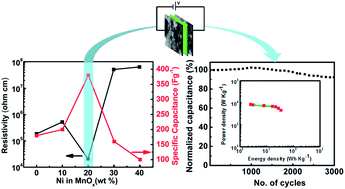
Nickel manganese mixed oxides (NiyMn1−yOx; 0 ≤ y ≤ 0.4) have been synthesized by in situ inclusion of nickel during the growth of manganese oxide (MnOx). The effect of nickel concentration in MnOx is investigated by cyclic voltammetry, current–voltage characteristics, scanning electron microscopy and N2 adsorption–desorption analysis. Variations in electronic conductivity and specific capacitance suggest that nickel concentration in the MnOx matrix significantly affects the supercapacitor electrode performance. At Ni/Mn ∼0.25, i.e. Ni0.2Mn0.8Ox, the material crystallizes into spinel NiMn2O4 as a prominent phase and exhibits a specific surface area (118 m2 g−1) with a granular morphology. Furthermore Ni0.2Mn0.8Ox exhibited low resistivity (2.07 × 10⁴ Ohm cm) and consequently high specific capacitance ∼380 F g−1, endowing additional merits. The fabricated supercapacitor device (Ni0.2Mn0.8Ox//Ni0.2Mn0.8Ox) delivers 35 W h kg−1 energy density and 3.74 kW kg−1 power density with remarkably high capacitive retention ∼92% after 3000 galvanostatic charge–discharge cycles. These encouraging results show great potential in developing energy storage devices from manganese oxide based electrodes incorporating nickel in the lattice.


1. Introduction

Electrochemical supercapacitors have attracted much attention as energy storage devices due to their promising applications in portable electronics and hybrid electrical vehicles.1,2 Supercapacitors store charge via (a) charge accumulation at the electrode/electrolyte interface in the form of an electrical double layer (EDL) and (b) by the pseudocapacitance that arises from faradic redox transitions that go deep inside the bulk material.

Carbon materials store charge via EDL whereas transition metal oxides and conducting polymers follow redox process for charge storage.3,4 EDL is surface dependent and therefore the resulting capacitance values are low however the charge–discharge rates are high. Due to the bulk contribution, puseudocapacitors give high specific capacitance (CSP) whereas the response time limits their application. In order to exploit high CSP and energy density, metal oxides have been extensively studied and the response time or charge–discharge time is modified by improving the charge collection through a support material, preferably carbon. Researchers recently focussed on mixed metal oxides as potential supercapacitor electrode materials because pristine transition metal oxides are poor conductors resulting in high equivalent series resistance with limited capacity and power density.5 Besides good electronic conduction, mixed transition metal oxides could offer richer redox reactions beneficial for electrochemical applications.6

Recently, many nanocomposites like Co3O4@MnO2, Co3O4–Ni(OH)2, MnO2–NiO are attracting great interest for supercapacitors owing to their improved traits over pristine counterparts.7–12 Various nickel and cobalt composites result in high electronic conductivity consequently enhancing the electrochemical performance.11 Liu et al. synthesized high surface area spinel nickel manganese oxide for supercapacitor with high CSP 243 F g−1 at 5 mV s−1. Fan et al. reported MnO2–NiO nanoflake-assembled tubular array on stainless steel substrate by template method where ZnO is employed as in situ sacrificial template.9 Although high capacitance values are achieved using nano architectures, the bulk material performance and the electrode cyclability could be further improved.

In this work, we investigated the in situ inclusion of nickel in manganese oxide (NiyMn1−yOx; 0 ≤ y ≤ 0.4) lattice and tested the composite material as supercapacitor electrodes. NiMn2O4 crystallizes as a prominent phase when 20 wt% NiCl2 is added to 80 wt% aqueous MnCl2 solution for oxidative growth of manganese oxide. Ni0.2Mn0.8Ox exhibits highest electrical conductivity and surface area among other compositions. Proposed supercapacitor, Ni0.2Mn0.8Ox//Ni0.2Mn0.8Ox provides high energy density 35 W h kg−1 with ∼92% retention of initial CSP after 3000 galvanostatic charge–discharge cycles.

2. Experimental

2.1 Materials

Nickel chloride (NiCl2·6H2O), manganese chloride (MnCl2·4H2O), sodium hydroxide (NaOH), sodium sulphate (Na2SO4), isopropyl alcohol (IPA) and isooctane were purchased from Merck. Perfluorinated ion exchange resin (Nafion) was procured by Sigma Aldrich. Sodium sulphosuccinate (AOT) was used as surfactant (supplied by Alfa Aesar).

2.2 Synthesis of nickel manganese mixed oxide

Three aliquots of reverse microemulsions of water/AOT/isooctane13 were prepared having 0.8 M MnCl2, 0.2 M NiCl2 and 2 M NaOH aqueous solutions respectively. These were thoroughly mixed in a reaction vessel and stirred for 2 h. Excessive volume of IPA was added to break the miceller arrangement after completion of the reaction. The resulting precipitate was collected after washing several times with IPA and deionized water. Solid product obtained after washing was dried overnight at 80 °C. Different compositions of NiyMn1−yOx; 0.1 ≤ y ≤ 0.4 were prepared as given in Table 1 (y is molar concentration of nickel in MnOx). MnOx and Ni(OH)2 were also synthesized by the same method under similar conditions.14
Table 1 Percentage of nickel in manganese oxide
Sample name NiCl2·6H2O (g) MnCl2·4H2O (g) NaOH (g) Ni/Mn (wt%)
Ni0.1Mn0.9Ox 0.48 3.56 1.60 10
Ni0.2Mn0.8Ox 0.95 3.16 1.60 25
Ni0.3Mn0.7Ox 1.43 2.77 1.60 43
Ni0.4Mn0.6Ox 1.90 2.37 1.60 65


2.3 Preparation of electrode

10 mg of composite material (NiyMn1−y Ox; 0.1 ≤ y ≤ 0.4) was ultrasonically mixed with 5 wt% Nafion in IPA. The mixture after 30 min sonication was spray deposited on 1 cm2 area of the polished graphite sheets (2 × 1 cm) with the help of N2 gas. After spray deposition, the film was dried at 80 °C in air and weighed to estimate the loading of the composite material. Loading was found to be approximately 0.3 mg cm−2.

2.4 Characterization

Conductivity measurements were carried out on pellet (dia. 10 mm) using Keithley 2400 source meter. Perkin Elmer model 1257 X-ray photoelectron spectroscope (XPS) was employed to estimate the percentages of different metal oxide phases present in the specimen. Powder X-Ray Diffraction (XRD) measurements were performed with Bruker D8 Advance X-ray diffractometer. Microstructural investigations were carried out using Zeiss Ultra 55 Field emission scanning electron microscope (FESEM). Surface area of the samples were analyzed by N2 adsorption–desorption isotherms using Micromeritics ASAP-2020. High resolution transmission electron microscopy (HRTEM) and Selected Area Electron Diffraction (SAED) was conducted on Phillips Technai T-300 microscope. Electrochemical tests were performed with CHI 604D electrochemical analyzer. A piece of platinum gauze and Ag/AgCl were assembled as the counter and reference electrode respectively. Polished graphite with metal oxide/mixed metal oxide coating served as the working electrode. Cyclic voltammograms (CV) were recorded by polarizing the working electrode between 0 to 0.9 V vs. Ag/AgCl in 0.5 M aqueous Na2SO4 electrolyte. The charge–discharge characteristics of the supercapacitor cell were evaluated at different current density (two electrode assembly) using an Arbin instrument (model: BT2000, USA). CSP was calculated from galvanostatic charge–discharge curve according to following equation:3,15
image file: c4ra09027f-t1.tif
where I is the current, ΔVt is the change in potential with time and m is mass loading on the electrode.

The energy density (E in W h kg−1) and power density (P in W kg−1) were expressed as

image file: c4ra09027f-t2.tif

image file: c4ra09027f-t3.tif
where C is measured device capacitance, m is total mass loading on the electrodes, V is the operating potential range and R is the equivalent series resistance calculated from IR drop in galvanostatic discharge curve.

3. Results and discussion

The effect of Ni incorporation in MnOx matrix was studied using cyclic voltammetry (CV) and current (I)–voltage (V) characteristics for NiyMn1−y Ox; 0.1 ≤ y ≤ 0.4. Fig. 1 shows the variation of CSP and electrical resistivity with different nickel percentage in MnOx. Ni0.1Mn0.9Ox did not show much change in CSP but the resistivity is slightly increased. Ni0.2Mn0.8Ox, compared to MnOx shows more than twice increase in CSP with drastic decrement in resistivity. Upon 20 wt% inclusion of Ni, CSP of MnOx (180 F g−1) remarkably increases to 380 F g−1 with one order decrease in resistivity (1.85 × 10⁵ Ohm cm of MnOx to 2.07 × 10⁴ Ohm cm of Ni0.2Mn0.8Ox). With further increase in nickel content, Ni0.4Mn0.6Ox shows detrimental effect as CSP decreases and resistivity increases. More importantly, drastic increment in CSP of Ni0.2Mn0.8Ox is investigated by its structural and electrochemical analysis.
image file: c4ra09027f-f1.tif
Fig. 1 Effect of Ni (wt%) on resistivity and CSP of NiyMn1−yOx; 0 ≤ y ≤ 0.4.

Fig. 2 shows scanning electron micrographs and the surface area analysis using BET. The surface microstructure of MnOx, Ni0.2Mn0.8Ox and Ni0.4Mn0.6Ox exhibit granular morphology. MnOx in Fig. 2a, shows aggregated spherical grains of dia. ∼ 10 nm. Ni0.2Mn0.8Ox exhibited an increase in grain size (dia. ∼ 15 nm) (Fig. 2b). Upon increasing Ni concentration to 40 wt% (Ni0.4Mn0.6Ox), the composite exhibited dense microstructural features with very high aggregation (Fig. 2c). It appears that the increased concentration of Ni increases the aggregation.


image file: c4ra09027f-f2.tif
Fig. 2 SEM micrographs (a–c) and N2 adsorption–desorption isotherms (d–f) of MnOx, Ni0.2Mn0.8Ox and Ni0.4Mn0.6Ox.

N2 adsorption–desorption isotherm analysis of MnOx show high specific surface area (80.5 m2 g−1) with Type IV isotherm with H1 type hysteresis loop (Fig. 2d). Upon inclusion of 20 wt% nickel (Ni0.2Mn0.8Ox), surface area significantly increases to 118 m2 g−1. Nearly 50% enhancement in the specific surface area is attributed due to the complex pore network as in case of H2 hysteresis loop (Fig. 2e). Increasing the Ni more than 20 wt%, the surface area shows a decrease (Fig. 2f) and is attributed to the dense morphology of the composite having high Ni concentration. Pore size distribution of MnOx (ESI S1) demonstrate the presence of mesopores (5–20 nm) whereas in Ni0.2Mn0.8Ox, majority of pores fall in the optimal sizes of 2–5 nm for supercapacitors16 and thus supports the high surface area of Ni0.2Mn0.8Ox, consequently resulting in enhanced capacitance.

XPS analysis of MnOx show two peaks of MnO2 and Mn3O4 in Mn 2p at 641.8 eV and 642.43 eV respectively.17 Areas covered under MnO2 and Mn3O4 peak are estimated to be 85% and 15% respectively (Fig. 3a) ascertaining the simultaneous presence of MnO2 and Mn3O4 in MnOx. As shown in Fig. 3b, the survey scan of Ni0.2Mn0.8Ox confirm the presence of Mn, Ni and O in the composite. Inset shows Ni 2p XPS which is assigned to Ni 2p3/2 and Ni 2p1/2 peaks with two satellites (sat.). The spin energy separation of Ni 2p3/2 and Ni 2p1/2 is 17.5 eV which is characteristic of Ni(OH)2 phase.18 Mn 2p XPS spectrum of Ni0.2Mn0.8Ox shows a single peak at 642.4 eV (Fig. 3c), analogous to that of MnOx ascertaining the absence of Mn3O4 in Ni0.2Mn0.8Ox.


image file: c4ra09027f-f3.tif
Fig. 3 (a) Mn 2p X-ray photoelectron spectrum (XPS) of MnOx, (b) XPS survey scan with Ni 2p core level spectra in inset (c) deconvoluted Mn 2p spectrum of Ni0.2Mn0.8Ox, (d) X-ray diffraction patterns of MnOx and Ni0.2Mn0.8Ox, (e) XRD patterns of Ni0.2Mn0.8Ox powder annealed at 250 °C, 450 °C and 650 °C and (f) thermogravimetric analysis of Ni0.2Mn0.8Ox.

XRD analysis of MnOx and Ni0.2Mn0.8Ox further supported XPS results. Fig. 3d shows XRD patterns of Manganese oxide (MnOx) and Ni0.2Mn0.8Ox. Diffraction pattern of MnOx endorses the presence of different phases i.e. MnO2 and Mn3O4.19 XRD pattern of Ni0.2Mn0.8Ox exhibits three major diffraction peaks at 18°, 35.6° and 63.5° indexed to (111), (311) and (440) planes of spinel NiMn2O4 respectively.20,21 The diffraction pattern showed relatively amorphous character with indication of the presence of some secondary phases like MnO2, Ni(OH)2 and NiMnO3, interestingly there is no sign of Mn3O4. An important observation of the work is that in situ incorporation of 20 wt% Ni in MnOx results in NiMn2O4 as prominent phase formation with complete suppression of manganese higher oxide (Mn3O4) phases. The suppression of Mn3O4 is attributed to its distorted spinel structure having Mn+2 ions in tetrahedral sites and Mn+3 ions in octahedral sites.22 These Mn+3 ions causes Jahn–Teller distortion which is the result of departure from ideal interactions among bonding orbital and gives rise to lattice instability.23 Ni+2 ions can replace Mn+3 ions from octahedral site and such a substitution might require an equivalent amount of remaining Mn+3 ions to lose electrons and become Mn+4 in order to maintain the charge balance consequently resulting in NiMn2O4. Such a cation distribution offers lesser lattice alterations during electrochemical cycling resulting in enhanced electrochemical stability of NiMn2O4.8

Annealing carried out at different temperatures helped understanding the growth of NiMn2O4 by intermixing of secondary phases. Fig. 3e shows diffraction patterns of Ni0.2Mn0.8Ox powder after 2 h annealing at different temperatures. XRD pattern of Ni0.2Mn0.8Ox without any annealing treatment show three major diffraction peaks at 2θ value of 18°, 35.6°, 63.5° confirming the presence of NiMn2O424 with some other less intense peaks of MnO2, Ni(OH)2 and NiMnO3. Annealing results in grain growth and intermixing of the secondary phases. The Ni0.2Mn0.8Ox sample after 250 °C annealing shows the presence of prominent NiMn2O4 with some secondary phases of MnO2 and NiO and a shoulder NiMnO3 at 36.7°. Here, Ni(OH)2 converts to NiO which shows a peak at 61.2° corresponding to (220) plane. Further elevation in annealing temperature (450 °C) shows the crystalline XRD pattern of NiMn2O4 with presence of NiMnO3. Quantification of the above XRD pattern by Xpert High Score software via Rietveld method (ESI S2) suggest the percentage of prominent phase NiMn2O4 (81.3%) and NiMnO3 (18.7%). It is assumed that the secondary phases of MnO2 and NiO intermix and form NiMn2O4. The formation of NiMnO3 could be due to dissociation of NiMn2O4 phase as discussed in later section. The XRD pattern of Ni0.2Mn0.8Ox annealed at 650 °C made this aspect clear and showed higher (44.5%) percentage of NiMnO3.25 The increase in intensity of the peaks corresponds to gradual evolution to crystalline phases.

Thermo-gravimetric analysis of Ni0.2Mn0.8Ox supports the phase transformation as observed in the X-ray diffraction. Due to loss of the adsorbed and/or structurally bonded water, TGA thermogram of Ni0.2Mn0.8Ox (Fig. 3f) shows ∼18% weight loss below 120 °C. Further weight loss ∼7% between 120–250 °C corresponds to the decomposition of Ni(OH)2 to NiO and H2O according to the following probable reaction:

Ni(OH)2 → NiO + H2O

Gradual 8% weight loss between 350–450 °C is attributed to the formation of NiMn2O4 and evolution of oxygen according to the reaction:26

2MnO2 + NiO → NiMn2O4 + 1/2O2

Major 36% weight loss between 450–700 °C is attributed to the evaporation of MnO from NiMn2O4 according to the following reaction which is in agreement with the XRD results of Ni0.2Mn0.8Ox oxide annealed at 650 °C:

NiMn2O4 → NiMnO3 + MnO(g)

As MnO is evaporated, characteristic peaks in the XRD pattern corresponds to NiMn2O4 and NiMnO3 which shows the simultaneous presence of both.

TEM micrographs in Fig. 4a shows spherical particles with dia. ∼ 20 nm. Continuous lattice fringes (Fig. 4b) reveals d spacing 0.25 nm (311) of NiMn2O4. Three different planes (111), (311) and (440) of NiMn2O4 are observed in SAED pattern which are in agreement with XRD results (Fig. 4c). SEM of Ni0.2Mn0.8Ox (Fig. 4d) shows interconnected spherical nanoparticles forming porous channels. EDX results of spherical particles show Ni/Mn ratio ∼0.25 which is in agreement with experimental value (Fig. 4e).


image file: c4ra09027f-f4.tif
Fig. 4 (a) TEM of Ni0.2Mn0.8Ox and (b) HRTEM shows d spacing ∼0.25 nm, (c) SAED pattern, (d) SEM and (e) EDX of Ni0.2Mn0.8Ox with atomic percentage of Ni[thin space (1/6-em)]:[thin space (1/6-em)]Mn ∼0.25.

The electrochemical performance of Ni0.2Mn0.8Ox is investigated by cyclic voltammetry (CV) and Electrochemical impedance spectroscopy (EIS). CV of Ni0.2Mn0.8Ox show much larger area indicating higher charge storage (∼380 F g−1) than MnOx (∼180 F g−1) and Ni(OH)2 (∼95 F g−1), (Fig. 5a). This CSP is higher than previous MnO2-based nanocomposites electrodes, such as the graphene/MnO2-based textile (315 F g−1) and MnO2 nanosheets/graphene (263 F g−1).12,27,28 Moreover, absence of any peak indicates pseudoconstant charging and discharging over whole voltammetric cycle.29 The possible charge storage mechanism in Ni0.2Mn0.8Ox (NiMn2O4 prominent phase) may be explained as:

NiMn2O4 + Na+ + e → NaNiMn2O4


image file: c4ra09027f-f5.tif
Fig. 5 (a) Cyclic voltammograms (CV) of MnOx, Ni(OH)2 and Ni0.2Mn0.8Ox at 5 mV s−1, (b) Nyquist plot of Ni0.2Mn0.8Ox in high frequency region. Inset shows Nyquist plot of MnOx and Ni0.2Mn0.8Ox, (c) effect of annealing temperature on CV response of Ni0.2Mn0.8Ox electrode. Inset shows variation of specific capacitance vs. annealed temperatures, (d) cycling life test of MnOx and Ni0.2Mn0.8Ox at 50 mV s−1 and (e and f) Mn 2p XPS spectra of MnOx and Ni0.2Mn0.8Ox after 10[thin space (1/6-em)]000 CV cycles.

Complex plane plot of Ni0.2Mn0.8Ox reveals resistive and capacitive response with change in frequency (Fig. 5b). The equivalent circuit for the best fit conditions is shown in the inset. High frequency region of the Nyquist plot, represented by suppressed semicircle shows internal resistance RS 5.3 Ω and the high to mid frequency region represents RC circuit (RCT, charge transfer resistance and Cdl, double layer capacitance). RCT (1 Ω), lower than earlier reported values of MnO2–NiO composite indicates intimate contact formation between the current collector and electrolyte contributing to facile charge transfer.9 A 45° sloped line in the mid frequency range corresponds to Warburg impedance which is related to ion diffusion resistance. Inset shows Nyquist plot of MnOx and Ni0.2Mn0.8Ox exhibiting similar behaviour in high and low frequency region. Besides, the impedance line of MnOx possess an arc in mid frequency region restricting the accessibility of ions causing low capacitance of MnOx. More importantly, capacitive character of Ni0.2Mn0.8Ox attains earlier than that of MnOx as evident from higher knee frequency below which maximum energy is easily accessible.

Effect of annealing on CSP of Ni0.2Mn0.8Ox is shown by Fig. 5c. Inset shows variation of CSP vs. annealing temperature. Ni0.2Mn0.8Ox has NiMn2O4 as prominent phase which shows high CSP 380 F g−1. It is important to mention that the samples were dried at 80 °C for 1 h. Extending this temperature to 250 °C for 2 h results in CSP decrement to 290 F g−1 which may be due to improved crystallanity of the material.30 At 450 °C, single phase XRD pattern (NiMn2O4) shows CSP 220 F g−1 which is in close proximity to the reported values.12 Above 450 °C, crystalline evolution takes place and the crystal lattice is rigid and not easily expanded (or contracted). This consequently retards the protonation (or deprotonation) reaction of the oxide leading to drastic decrement of CSP at 650 °C.

Cycling life stability of MnOx and Ni0.2Mn0.8Ox is explored by relatively long time cycling at 50 mV s−1 (Fig. 5d). Result shows that CSP of Ni0.2Mn0.8Ox shows an increment in initial cycles followed by ∼99% retention up to 10[thin space (1/6-em)]000 cycles. Initial increment is probably attributed to activation process involving the increment in the number of available active sites allowing the trapped ions to gradually diffuse out.31 CSP decay in MnOx is found to be 50% after 10[thin space (1/6-em)]000 cycles. This remarkable difference in cycling life stability with and without nickel in MnOx electrode is investigated by XPS analysis before and after electrochemical cycling.

Core level Mn 2p XPS of MnOx after redox cycling (unlike before cycling) shows intense peak of Mn3O4 with relatively less intense MnO2 peak (Fig. 5e). Ni0.2Mn0.8Ox after electrochemical cycling shows a single peak of MnO2 with no Mn3O4 (Fig. 5f). This demonstrate considerable conversion of MnO2 to less electroactive Mn3O4 phase during cycling. This implicates that the decrement in charge storing capacity of MnOx was due to the gradual formation of Mn3O4. It is noteworthy that in situ availability of nickel (20 wt%) in MnOx inhibits Mn3O4 formation not only during growth but also during redox cycling, thereby enhancing the electrochemical performance of Ni0.2Mn0.8Ox.

As a demonstration of application potential of Ni0.2Mn0.8Ox, a symmetric supercapacitor, Ni0.2Mn0.8Ox//Ni0.2Mn0.8Ox is fabricated and examined via GCD measurements. Fig. 6a shows GCD curves of Ni0.2Mn0.8Ox//Ni0.2Mn0.8Ox at different current densities. Deviation from the linear variation of voltage with time is attributed to pseudocapacitive nature of the material. Small IR drop associated with the discharge curve ascertains low equivalent series resistance (esr) of the cell which can be calculated from the slope of linear correlation of IR drop with current density.32,33


image file: c4ra09027f-f6.tif
Fig. 6 (a) Galvanostatic charge–discharge (GCD) curves at different current densities, (b) variation of IR drop and specific capacitance vs. current density, (c) Ragone plot and (d) GCD cycling performance of Ni0.2Mn0.8Ox//Ni0.2Mn0.8Ox supercapacitor cell. Inset shows overview of last cycles.

Variation of CSP and IR drop at different current density in Fig. 6b display lesser esr ∼0.03 Ω favouring high power delivery in practical applications making it a potential material for supercapacitor. Moreover, CSP decreases with increase of current density as lesser surface area is accessible at higher current densities. It delivers energy density 35 W h kg−1 with power density 3.74 kW kg−1 at 0.5 A g−1 (Fig. 6c). It still maintains an energy density 5.10 W h kg−1 and power density 7.44 kW kg−1 even at high current density 12.5 A g−1.

Long term cycling performance of Ni0.2Mn0.8Ox//Ni0.2Mn0.8Ox was investigated by GCD cycling at 2.5 A g−1 (Fig. 6d). Inset shows overview of charge–discharge cycles. Slight increment in CSP during initial 1000 cycles as observed in various reports,31 followed by 8% decrement in CSP in further cycles. Such a high retainability of initial CSP after 3000 cycles endows a long term stable material for charge storage in supercapacitor.

4. Conclusions

In situ inclusion of nickel during manganese oxide (NiyMn1−yOx; 0 ≤ y ≤ 0.4) growth and its effect on structural, electrical and electrochemical properties is investigated. Upon 20 wt% addition of nickel in MnOx (Ni0.2Mn0.8Ox) the composite exhibited spinel NiMn2O4 as prominent phase with highest surface area (118 m2 g−1), Consequently the superior performance in terms of highest conductivity and CSP (∼380 F g−1). Galvanostatic charge–discharge measurements of fabricated supercapacitor, Ni0.2Mn0.8Ox//Ni0.2Mn0.8Ox demonstrate high energy and power density (35 W h kg−1, 3.74 kW kg−1). Furthermore, it also retains 92% initial capacitance after 3000 cycles ascribing excellent cycling stability.

Acknowledgements

Authors gratefully acknowledge the financial support from DST India through project SR/S1/PC-31/2010 and Delhi University for Docotoral Research Grant. Preety Ahuja thankfully acknowledges the research fellowship from UGC India. SK Ujjain thank the INSPIRE fellowship from DST. Authors thank Dr R. Nagarajan, Department of Chemistry, University of Delhi for electrical characterisation instrumentation and Inorganic Materials and Catalysis group for the support through analysis of surface area using N2 Sorption facility under DST(SR/S1/PC-11/2011).

References

  1. Z. Li, J. Wang, L. Niu, J. Sun, P. Gong, W. Hong, L. Ma and S. Yang, J. Power Sources, 2014, 245, 224–231 CrossRef CAS.
  2. G. Wang, L. Zhang and J. Zhang, Chem. Soc. Rev., 2012, 41, 797–828 RSC.
  3. S. K. Ujjain, K. Deori, R. K. Sharma and S. Deka, ACS Appl. Mater. Interfaces, 2013, 5, 10665–10672 Search PubMed.
  4. P. Simon and Y. Gogotsi, Nat. Mater., 2008, 7, 845–854 CrossRef CAS PubMed.
  5. D. L. Fang, B. C. Wu, Y. Yan, A. Q. Mao and C. H. Zheng, J. Solid State Electrochem., 2012, 16, 135–142 CrossRef CAS.
  6. J. M. Luo, B. Gao and X. G. Zhang, Mater. Res. Bull., 2008, 43, 1119–1125 CrossRef CAS.
  7. J. P. Liu, J. Jiang, C. W. Cheng, H. X. Li, J. X. Zhang, H. Gong and H. J. Fan, Adv. Mater., 2011, 22, 2076–2081 Search PubMed.
  8. C. H. Tang, X. Yin and H. Gong, ACS Appl. Mater. Interfaces, 2013, 5, 10574–10582 CAS.
  9. J. Lu, J. Jiang, M. Bosman and H. J. Fan, J. Mater. Chem., 2012, 22, 2419–2426 RSC.
  10. B. Liu, B. Liu, Q. Wang, X. Wang, Q. Xiang, D. Chen and G. Chen, ACS Appl. Mater. Interfaces, 2013, 5, 10011–10017 CAS.
  11. J. H. Zhong, A. L. Wang, G. R. Li, J. W. Wang, Y. N. Ou and Y. X. Tong, J. Mater. Chem., 2012, 22, 5656–5665 RSC.
  12. M. Zhang, S. Guo, L. Zheng, G. Zhang, Z. Hao, L. Kang and Z. H. Liu, Electrochim. Acta, 2013, 87, 546–553 CrossRef CAS.
  13. R. K. Sharma, H. S. Oh, Y. G. Shul and H. Kim, J. Power Sources, 2007, 173, 1024–1028 CrossRef CAS.
  14. D. Liu, B. B. Garcia, Q. Zhang, Q. Guo, Y. Zhang, S. Sepehri and G. Cao, Adv. Funct. Mater., 2009, 19, 1015–1023 CrossRef CAS.
  15. P. Ahuja, V. Sahu, S. K. Ujjain, R. K. Sharma and G. Singh, Electrochim. Acta, 2014, 146, 429–436 CrossRef CAS.
  16. R. Ding, L. Qi and H. Wang, J. Solid State Electrochem., 2012, 16, 3621–3633 CrossRef CAS.
  17. M. Chigane, M. Ishikawa and M. Izaki, J. Electrochem. Soc., 2001, 148, D96–D101 CrossRef CAS.
  18. I. C. Chang, T. T. Chen, M. H. Yang, H. T. Chiu and C. Y. Lee, J. Mater. Chem. A, 2014, 2, 10370–10374 CAS.
  19. Y. Z. Zheng, H. Y. Ding and M. L. Zhang, Mater. Res. Bull., 2009, 44, 403–407 CrossRef CAS.
  20. Y. Torii, A. Tsuzuki, K. Kato, Y. Uwamino, B. H. Choi and M. J. Lee, J. Mater. Sci., 1996, 31, 2603–2607 CrossRef CAS.
  21. Menaka, N. Garg, S. Kumar, D. Kumar, K. V. Ramanujachary, S. E. Lofland and A. K. Ganguli, J. Mater. Chem., 2012, 22, 18447–18453 RSC.
  22. J. D. Hem, C. J. Lind and C. E. Roberson, Geochim. Cosmochim. Acta, 1989, 53, 2811–2822 CrossRef CAS.
  23. T. N. Ramesh and P. V. Kamath, J. Power Sources, 2008, 175, 625–629 CrossRef CAS.
  24. Y. Torii, A. Tsuzuki, K. Kato, Y. Uwamino, B. H. Choi and M. J. Lee, J. Mater. Sci., 1996, 31, 2603–2607 CrossRef CAS.
  25. S. H. Kim, S. J. Kim and J. Kim, J. Power Sources, 2006, 163, 294–299 CrossRef CAS.
  26. J. D. Hem and C. J. Lind, Geochim. Cosmochim. Acta, 1991, 55, 2435–2451 CrossRef CAS.
  27. G. H. Yu, L. B. Hu, M. Vosgueritchian, H. L. Wang, X. Xie, J. R. McDonough, X. Cui, Y. Cui and Z. N. Bao, Nano Lett., 2011, 11, 2905–2911 CrossRef CAS PubMed.
  28. Z. P. Li, J. Q. Wang, X. H. Liu, S. Liu, J. F. Ou and S. R. Yang, J. Mater. Chem., 2011, 21, 3397–3403 RSC.
  29. X. Lang, A. Hirata, T. Fujita and M. Chen, Nat. Nanotechnol., 2011, 6, 232–236 CrossRef CAS PubMed.
  30. J. W. Lang, L. B. Kong, W. J. Wu, M. Liu, Y. C. Luo and L. Kang, J. Solid State Electrochem., 2009, 13, 333–340 CrossRef CAS.
  31. T. Liu, S. Xu, L. Wang, J. Chu, Q. Wang, X. Zhu, N. Bing and P. K. Chu, J. Mater. Chem., 2011, 21, 19093–19100 RSC.
  32. L. F. Chen, Z. H. Huang, H. W. Liang, Q. F. Guan and S. H. Yu, Adv. Mater., 2013, 25, 4746–4752 CrossRef CAS PubMed.
  33. H. Jiang, C. Li, T. Sun and J. Ma, Chem. Commun., 2012, 48, 2606–2608 RSC.

Footnote

Electronic supplementary information (ESI) available. See DOI: 10.1039/c4ra09027f

This journal is © The Royal Society of Chemistry 2014
Click here to see how this site uses Cookies. View our privacy policy here.