DOI:
10.1039/C4RA08987A
(Communication)
RSC Adv., 2014,
4, 54140-54143
Engineered nanoporous gold microspheres for stochastic sensing
Received
20th August 2014
, Accepted 10th October 2014
First published on 10th October 2014
Abstract
Engineered nanoporous gold microspheres were designed and used as new materials for stochastic microsensors. Carcinoembryonic antigen was used as model analyte to prove the stochastic sensing capabilities of the new material. A new sensor based on gold particles was used for the assay of the carcinoembryonic antigen in biological fluids. Stochastic sensing was used to determine the antigen in the samples. The working concentration range of the stochastic microsensor (1.6 × 10⁻⁸ to 1.6 × 10⁻⁵ mg mL−1) as well as its sensitivity (3.0 × 10³ s mg mL−1) and limit of quantification (16 ng mL−1) made possible its reliable utilization for screening tests of whole blood samples for the carcinoembryonic antigen.
Introduction
Bayley and Cremer demonstrated in 2001 (ref. 1) the basic principles of stochastic sensing through a protein pore; they were also stating the importance of stochastic sensing for qualitative and quantitative analysis and the principles associated (1/ton = A + B × Concanalyte; toff – used for qualitative analysis). Stochastic sensing (introduced for the first time in bioanalysis and biomedical analysis by Stefan–van Staden) is a powerful technique in biomedical analysis.2–5 Pattern recognition of different antigens and neurotransmitters was possible by determining the signatures of each of them in the diagrams obtained using stochastic sensors and microsensors.2–5
Of high importance were both qualitative and quantitative analysis of the analytes, obtained in the two stage process of the stochastic sensing when a potential of a certain value was applied. The mechanism of current development consists on two stages: stage 1 on which the analyte extracted from the solution into the membrane–solution interface blocks the channel, the intensity of the current recorded being zero for a certain period of time, named signature of the analyte which is measured using toff (the channel is losing the conductivity, and the intensity of current became 0); and stage 2 when the analyte (A) is interacting with the wall, the following equilibrium equation is taking place:
where Ch is the channel, and
i is the interface.
Due to the fact that toff value depends on: the size of the analyte, of its capacity of unfolding, geometry of the analyte, and velocity to pass through the channel, it is difficult to find two or more analytes that will give the same value for toff; all these should correlate with size and geometry of the channel/pore. At this stage it is difficult to predict which geometry or size will be the best for a certain analyte, especially for proteins like CEA, because the proteins are unfolding before gong through the pores.1 Accordingly, this method is selective, and it is not necessary to add antibodies, or other analytes in order to improve the selectivity. The principle of the recognition (based on blocking of a channel for a certain time) made from this method the only reliable electrochemical method for recognition (qualitative analysis) of a biomolecule in biological samples. Furthermore, the analytes are getting into the channel in a certain order, accordingly with their size and geometry, from the smallest to the biggest; these processes are giving the form of the signal.
In this paper, stochastic sensors based on engineered nanoporous gold microspheres were designed and used for chemical analysis. Carcinoembryonic antigen (CEA) was chosen as model analyte to prove the stochastic sensing capabilities of the new designed microsensor. Carcinoembryonic antigen is a glycoprotein discovered by Gold and Freedman,6,7 often associated with certain cancers. It is hardly present in the serum of healthy adults; but it appeared in the serum of patients with cancer.8–15 To date, CEA (a protein biomarker used in cancer diagnosis and assessment of treatment) was detected using different techniques, including chemiluminescence,16 radioimmunoassay,17 ELISA (applied as standard method in clinical laboratories),18 and electrochemical techniques.19–22 Circulating tumor cells are also analysed reliable using electrochemical and fluorescence based methods.23–27
Experimental
Materials and reagents
CEA (carcinoembryonic antigen), monosodium phosphate, disodium phosphate were purchased from Aldrich (Milwaukee, USA); paraffin oil and NaN3 were purchased from Fluka (Buchs, Switzerland). Monosodium phosphate and disodium phosphate were used for preparation of phosphate buffer 0.1 mol L−1, pH = 7.4. Deionized water obtained from a Millipore Direct-Q 3 System (Molsheim, France) was used for the preparation of all solutions. All standard solutions were prepared in buffer solution pH = 7.4, with NaN3 0.1%.
Instrumentation
All voltammograms were recorded using a PGSTAT 12 potentiostat/galvanostat connected to a three-electrode cell, and connected to a computer via an Eco Chemie (Utretch, The Netherlands) software version 4.9. A Pt electrode and an Ag/AgCl electrode served as the counter and reference electrodes for the cell. The working electrode was the microsensor based on engineered nanoporous gold microspheres. The pH measurements were performed using a CyberScan PCD 6500 Multiparameter.
Design of the stochastic microsensor
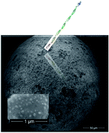
Engineered nanoporous 14 K gold microspheres were obtained from NILPRP. In order to obtain the nanopores on its surface, the particle was chemically corroded in an acid solution consisting of HCl:HNO3 in a 3:1 ratio, for 15 minutes and then rinsed with deionized water. As different sizes of gold microsphere were obtained, micrometers were used to select the microspheres with a certain diameter – of 300 μm. A typical SEM image of the microsphere surface is presented in Fig. 1a. A zoom in an area with a pore (able to show better the surface morphology) is shown in Fig. 1b. The stochastic microsensor was prepared as following: a 300 μm diameter gold nanoporous microparticle was tightly fixed inside a plastic tube together; a silver wire (diameter 150 μm) served as electrical connection to the external circuit (Fig. 2). The active surface of the sensor was rinsed in between measurements with distilled water, and gently dried with absorbent paper.
|
| Fig. 1 SEM image of (a) 24 K gold microparticle; (b) engineered pore in the 24 K gold microparticle. | |
|
| Fig. 2 Schematic diagram of the stochastic sensor. | |
Stochastic sensing
For the stochastic sensing, a chronoamperometric technique was selected for the measurements of ton and toff at a constant potential of 125 mV. The applied potential is the one driving the analyte through the channel/pore; accordingly, one should chose a value for which the signature value (toff) can be read reliable – usually it is preferred to do not be lower than 1 ms. The electrodes were dipped into a cell containing solutions of antigen of different concentrations. Values of ton were read on the diagram, and the equation of calibration 1/ton = f(Conc.) for CEA was determined using statistics. The unknown concentrations of the CEA in whole blood samples were determined from the calibration equations.
Results and discussions
Response characteristics of the sensor based on gold particles used for screening of CEA
The toff of 2.9 s serving as signature of CEA in biological fluids was determined while ton values were used for assessing the equation of calibration (1/ton = A + B × Concentration). Statistic calculations used for the equation of calibrations were extended to determine the working concentration range, sensitivity, and the limit of quantification of the stochastic microsensor. The stochastic microsensor proposed for the assay of CEA covered a linear concentration range between 16 and 1.6 × 10⁴ ng mL−1 with a sensitivity of 3.00 × 10³ s mg mL−1. The lowest concentration of CEA that could be reliably determined was 16 ng mL−1. The microsensor was stable over a period of six months, when used daily for measurements, its sensitivity varying with a RSD less than 0.1%.
Analytical applications
Whole blood samples were obtained from eight confirmed breast cancer patients from the Universitary Hospital in Bucharest (ethics committee approval no. 11/2013). These samples were used for screening tests using the proposed stochastic microsensor without any pretreatment. Pattern recognition of CEA based on its signature was performed using typical diagrams recorded using the stochastic microsensor (Fig. 3). The signature (toff value) of CEA was identified in the patterns recorded (Fig. 3) for whole blood samples – these results being in agreement with the standard method (ELISA) performed in the clinical laboratories of the hospital; after identifying the signature of the analyte, the ton was measured and inserted in the calibration graph for determining the concentration of CEA (arrows were used in Fig. 3 to show on the pattern selected where ton and toff values were measured). Table 1 shown the quantities of CEA found in the eight whole blood samples using both: the stochastic sensing as well as ELISA (the standard method). Blood samples were collected from ten healthy people (negative control) and analysed with the proposed method as well with ELISA; the signature of CEA (value of toff) was not found in any diagram recorded for these patients, this result being confirmed by ELISA method.
|
| Fig. 3 Specific diagram obtained for the pattern recognition of CEA in whole blood sample using the stochastic microsensor. | |
Table 1 Analysis of CEA from whole blood samples using the stochastic microsensor based on nanoporous gold microsphere and ELISAa
Sample no. |
CEA, ng mL−1 |
t-test |
Sensor based on gold particles |
ELISA |
All values are the average of ten determinations. |
1 |
2.06 ± 0.12 |
1.98 ± 0.32 |
1.89 |
2 |
2.10 ± 0.10 |
2.00 ± 0.40 |
1.90 |
3 |
1.40 ± 0.13 |
1.20 ± 0.38 |
1.95 |
4 |
1.09 ± 0.09 |
0.89 ± 0.29 |
2.10 |
5 |
1.64 ± 0.12 |
1.32 ± 0.30 |
2.25 |
6 |
1.32 ± 0.15 |
1.15 ± 0.29 |
1.76 |
7 |
1.37 ± 0.11 |
1.28 ± 0.42 |
1.70 |
8 |
0.93 ± 0.09 |
0.74 ± 0.28 |
2.90 |
Statistical analysis based on pair t-test was performed for a confidence level of 99.00%. The results have shown that there is no significant difference between the values obtained using the stochastic microsensor and ELISA (standard method) at 99.00% confidence level (tteoretical = 4.032). The comparison refers to paired data.
Conclusions
The new material based on engineered nanoporous gold microsphere is an excellent material for the design of stochastic microsensors with features for biomedical analysis. For carcinoembryonic antigen – selected as model analyte, high sensitivity and low limit of determination was recorded. The microsensor was validated using whole blood samples obtained from confirmed patients. The stochastic microsensor was stable for a 6 month period when used daily.
Acknowledgements
This work was supported by PNII Program Capacity, 2012–2014, Contract no. 3ERC-like/2012, PNII Ideas project number PN-II-ID-PCE-2011-3-0953 and by the Sectorial Operational Programme Human Resources Development (SOP HRD), financed from the European Social Fund and the Romanian Government under the contract number POSDRU/159/1.5/S/137390/.
References
- H. Bayley and P. S. Cremer, Nature, 2001, 413, 226–230 CrossRef CAS PubMed.
- R. I. Stefan-van Staden, I. Moldoveanu, D. F. Sava, C. Kapnissi-Christodoulou and J. F. van Staden, Chirality, 2013, 25, 114–118 CrossRef CAS PubMed.
- R. I. Stefan-van Staden and J. F. van Staden, J. Mod. Med. Chem., 2013, 1, 86–91 Search PubMed.
- R. I. Stefan-van Staden and I. Moldoveanu, J. Electrochem. Soc., 2014, 161, B45–B48 CrossRef CAS.
- R. I. Stefan-van Staden, I. Moldoveanu and J. F. van Staden, J. Neurosci. Methods, 2014, 229, 1–7 CrossRef CAS PubMed.
- S. Oikawa, C. Inuzuka, M. Kuroki, Y. Matsuoka, G. Kosaki and H. Nakazato, Biochem. Biophys. Res. Commun., 1989, 164, 39–45 CrossRef CAS PubMed.
- P. Gold and S. O. Freedman, J. Exp. Med., 1965, 121, 439–462 CrossRef CAS PubMed.
- J. Jiang, S. Zhao, Y. Huang, G. Qin and F. Ye, J. Chromatogr. A, 2013, 1282, 161–166 CrossRef CAS PubMed.
- S. Benchimol, A. Fuks, S. Jothy, N. Beauchemin, K. Shirota and C. P. Stanners, Cell, 1989, 57, 327–334 CrossRef CAS PubMed.
- D. M. Goldenberg, R. M. Sharkey and F. J. Primus, JNCI, J. Natl. Cancer Inst., 1976, 57, 11–22 CAS.
- Y. T. Shi, R. Yuan, Y. Q. Chai and X. L. He, Electrochim. Acta, 2007, 52, 3518–3524 CrossRef CAS.
- F. Y. Kong, X. Zhu, M. T. Xu, J. J. Xu and H. Y. Chen, Electrochim. Acta, 2011, 56, 9386–9390 CrossRef CAS.
- L. Hernández, A. Espasa, C. Fernández, A. Candela, C. Martín and S. Romero, Lung Cancer, 2002, 36, 83–89 CrossRef.
- M. J. Duffy, A. van Dalen, C. Haglund, L. Hansson, R. Klapdor, R. Lamerz, O. Nilsson, C. Sturgeon and O. Topolcan, European J. Cancer, 2003, 39, 718–727 CrossRef CAS.
- M. J. A. Engelen, H. W. A. de Bruijn, H. Hollema, K. A. ten Hoor, P. H. B. Willemse, J. G. Aalders and A. G. J. van der Zee, Gynecol. Oncol., 2000, 78, 16–20 CrossRef CAS PubMed.
- W. Dungchai, W. Siangproh, J. M. Lin, O. Chailapakul, S. Lin and X. Ying, Anal. Bioanal. Chem., 2007, 387, 1965–1971 CrossRef CAS PubMed.
- M. Herlyn, M. Blaszczyk, H. F. Sears, H. Verrill, J. Lindgren, D. Colcher, Z. Steplewski, J. Schlom and H. Koprowski, Hybridoma, 1983, 2, 329–339 CrossRef CAS PubMed.
- Y. Kumada, Y. Ohigashi, Y. Emori, K. Imamura, Y. Omura and M. Kishimoto, J. Immunol. Methods, 2012, 385, 15–22 CrossRef CAS PubMed.
- X. Cao, N. Wang, S. Jia, L. Guo and K. Li, Biosens. Bioelectron., 2013, 39, 226–230 CrossRef CAS PubMed.
- T. Shi, R. Yuan, Y. Q. Chai and X. L. He, Electrochim. Acta, 2007, 52, 3518–3524 CrossRef.
- Y. F. Y. Kong, X. Zhu, M. T. Xu, J. J. Xu and H. Y. Chen, Electrochim. Acta, 2011, 56, 9386–9390 CrossRef.
- X. Sun and Z. Ma, Anal. Chim. Acta, 2013, 780, 95–100 CrossRef CAS PubMed.
- M. Maltez-da Costa, A. de la Escosura-Muñiz, C. Nogués, L. Barrios, E. Ibáñez and A. Merkoçi, Nano Lett., 2012, 12, 4164–4171 CrossRef CAS PubMed.
- M. Maltez-da Costa, A. de la Escosura-Muñiz, C. Nogués, L. Barrios, E. Ibáñez and A. Merkoçi, Small, 2012, 8, 3605–3612 CrossRef CAS PubMed.
- I. Ivanov, J. Stojcic, A. Stanimirovic, E. Sargent, R. K. Nam and S. O. Kelley, Anal. Chem., 2013, 85, 398–403 CrossRef CAS PubMed.
- J. Liu, Y. Qin, D. Li, T. Wang, Y. Liu, J. Wang and E. Wang, Biosens. Bioelectron., 2013, 41, 436–441 CrossRef CAS PubMed.
- M. Moscovici, A. Bhimji and S. O. Kelley, Lab Chip, 2013, 13, 940–946 RSC.
|
This journal is © The Royal Society of Chemistry 2014 |
Click here to see how this site uses Cookies. View our privacy policy here.