Systematic investigation of quinoxaline derivatization of sialic acids and their quantitation applicability using high performance liquid chromatography

Libo Wanga, Dan Wangb, Xiang Zhoub, Lijun Wu*a and Xue-Long Sun*b
aCollege of Pharmacy, Harbin Medical University, Baojian Road 157, Nangang District, Harbin 150081, P. R. China. E-mail: wulijun_111@hotmail.com; Tel: +86 451 86686092
bDepartment of Chemistry, Chemical and Biomedical Engineering and Center for Gene Regulation in Health and Disease (GRHD), Cleveland State University, 2121 Euclid Avenue, Cleveland, Ohio 44115, USA. E-mail: x.sun55@csuohio.edu; Fax: +1 216 687 9298; Tel: +1 216 687 3919

Received 20th August 2014 , Accepted 11th September 2014

First published on 12th September 2014


Abstract

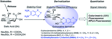
Quinoxaline derivatization has been the most commonly used approach for sialic acid quantitation of biological samples and glycoproteins by either HPLC or LC-MS/MS. However, the reported methods still suffer from many disadvantages, such as instable/expensive reagents, instable derivatives, and/or multiple products, which prevent their practical applications for SA-focused clinical screening and biopharmaceutical analysis. In this study, we investigated the quinoxaline derivatization of SAs with different commercially available phenyldiamines, and found a stable and inexpensive 4,5-dimethylbenzene-1,2-diamine (DMBA), that selectively reacted with SA to form a stable quinoxaline derivative efficiently and with strong UV and fluorescence absorption. The optimized DMBA derivatization conditions were successfully applied to quantitation of free and total SAs (N-acetyl-D-neuraminic acid (Neu5Ac) and N-glycolyl-D-neuraminic acid (Neu5Gc)) in fetal bovine serum (FBS) and glycoprotein fetuin using HPLC with fluorescence detection. The limit of detection (LOD) for Neu5Ac and Neu5Gc were 6.00 and 8.80 pg, and the limits of quantitation (LOQ) for Neu5Ac and Neu5Gc were 18.0 and 29.0 pg on column, respectively. This method was also applied to determine total SA in human plasma samples. This DMBA derivatization method provides a robust, easy-handling, sensitive and cost effective approach for quantitation of SAs in glycoconjugates and biomatrices.


Introduction

Sialic acids (SAs), also known as neuraminic acids, often terminate cell surface glycoconjugates and are involved in many physiological and pathological processes, such as immunological processes, hormonal response, signal transmission, and tumor progression.1,2 For example, cancers and cancer stages are associated with a significant over representation of SAs on the cancer cell surface.3,4 In addition, SAs exist in both free and conjugated forms such as glycoproteins and glycolipids in tissues and fluids such as serum, urine, and saliva. It is known that the amounts of free SAs and conjugated SAs are elevated in plasma of cancer patients in comparison to healthy individuals.5–8 Furthermore, the elevation of total SAs in serum of cardiovascular diseases is a strong predictor of cardiovascular mortality.9 Therefore, SAs have been considered as a biomarker for certain types of cancers, cardiovascular diseases, and some other diseases. Development of efficient analytical methods for structural and functional studies of SAs and sialylglycans are very important and highly demanded.10 In addition, the ability to detect and quantify the changes in both free SA and conjugated SA is an important aid for clarifying the great significance of SAs in both physiological and pathological conditions and in the diagnosis of diseases. On the other hand, glycoprotein sialylation and the identity of the sialic acids are very critical to therapeutic glycoprotein's efficacy, pharmacokinetics, and potential immunogenicity.11 Therefore, identification and quantitation of SAs are very important for therapeutic glycoprotein production and applications.

N-acetyl-D-neuraminic acid (Neu5Ac) and N-glycolyl-D-neuraminic acid (Neu5Gc) are common SAs present in glycoconjugates. So far, several methods, including colorimetric12,13 and fluorometric14,15 assays, liquid chromatography coupled with different detection techniques, such as pulsed amperometric detection (PAD),16,17 UV,18 fluorescence,19–25 and mass spectrometry (MS),26–38 have been developed for quantitation of SAs. Chromatographic methods have the advantage of quantitatively differentiating various forms of SAs. For example, quinoxaline derivatization followed by HPLC with fluorescence detection has been widely used for SA quantitation.20,21,23–25,27,36–38 1,2-Diamino-4,5-methylenedioxybenzene (DMB) has been most commonly used for SA derivatization and quantitation since it gives strong fluorescence intensity for HPLC detection.20,21,23–25 Recently, DMB derivatization has been expanded to LC-MS/MS SAs quantitation since it also affords good MS response.27,36,38 However, both DMB reagent and its SA derivative are very instable due to their light and oxygen sensitivity. Therefore, DMB reagent and its SA derivative need to be stabilized by reducing agents such as sodium hydrosulfite and 2-mercaptoethanol during derivatization reaction. In our studies, we found that DMB changed color even during a short weighing process, and the chromatographic peak area of the SA-DMB varied day to day.37,39 Therefore, fresh DMB solution has to be prepared for each assay, which is very time consuming and often causes analytical variations. In addition, the high cost of DMB is another problem considering that its reagent solution needs to be prepared freshly each time. In this study, we evaluated four commercially available compounds having the same phenyldiamine structure as DMB for SA derivatization and their HPLC analysis applicabilities (Fig. 1). We found that inexpensive 4,5-dimethylbenzene-1,2-diamine (DMBA) was a very stable reagent, and it reacted with Neu5Ac to form a single, stable quinoxaline derivative, which had very strong UV and fluorescence response too. Further, the DMBA derivatization was successfully applied to the HPLC analysis of SAs in fetal bovine serum (FBS), glycoprotein fetuin and human plasma.


image file: c4ra08930h-f1.tif
Fig. 1 Quinoxaline derivatization of sialic acids (SAs) for its HPLC quantitation application.

Results and discussion

UV spectroscopy monitoring of stability of penyldiamines and their sialic acid quinoxaline derivatives

DMB has been widely used for SA quantitation as it reacts selectively with α-keto carboxylic acid of SA to form the quinoxalinone that has strong fluorescent absorption. However, DMB is instable due to its light and oxygen sensitivity and very expensive as well, all of which prevent its practical use in SA quantitation applications. In this study, we systematically examined four commercially available phenyldiamines having the similar 1,2-diamino-benzene structure as DMB for SA derivatization and HPLC analysis (Fig. 1). In general, the quinoxlinone derivatives of SA have UV absorption at 340–360 nm. First, we used UV spectroscopy to monitor and compare the stabilities of phenyldiamine DMB, OPD, DAT, DMBA and their Neu5Ac derivatives with or without reducing agent sodium hydrosulfite (Na2S2O4) (Fig. 2). Various UV absorptions were observed for both DMB and DMB-Neu5Ac derivative with and without Na2S2O4, which indicated the instability of both DMB and its derivative during the reaction. The same conclusion could be obtained from the UV absorptions of DMB-Neu5Ac derivative even though two reducing agents sodium hydrosulfite and 2-mercaptoethanol were added during derivatization reaction. Meanwhile, OPD and DAT as well as their derivatives showed very simple UV absorptions in the presence of Na2S2O4, which mean that they were quite stable in the presence of reducing agent. However, in the absence of Na2S2O4, their UV spectra showed additional absorption due to their structural changes. In contrast, for DMBA-Neu5Ac derivatization with or without Na2S2O4, there was no additional absorption observed except the absorption from the derivative that increased steadily with reaction time increased. These results clearly indicated that both DMBA and its SA derivative are very stable and could serve as a better derivation reagent for quantitative analysis of SA. In addition, DMBA is thousand times cheaper than DMB as the current commercially availability.
image file: c4ra08930h-f2.tif
Fig. 2 UV spectra of phenydiamines (DMB, DAT, OPD and DMBA) and their Neu5Ac (SA) derivatizations at 60 °C from 0 to 6 h with and without Na2S2O4. (the blue lines are the absorption of DMB, DAT, OPD, DMBA and the red lines are the absorption of derivatives in each of the spectra, and the absorption intensity of each reaction solution increases along with the extension of reaction time).

1H NMR spectroscopy monitoring of the reaction kinetics of sialic acid quinoxaline derivatization

The formation of SA quinoxalinone derivatives and their reaction kinetics were also monitored by 1H NMR spectroscopy, in which the chemical shifts of protons at C-3 of SA and aromatic protons of phenyldiamines were found shift to the lower field along with the quinoxalinone derivatives forming (Fig. 3). Apparently, the formation of SA quinoxaline derivatives completed in 2 h according to the proton signals (δ 2.53, 2.13) at C-3 of SA shifting to low field completely. Interestingly, from the 1H-NMR spectrum of DAT-Neu5Ac derivative, two methyl proton signals of δ 2.67, 2.69 and two ABX system protons of aromatic groups at δ 7.43–7.94 were observed (Fig. 3A), which indicated that there were two quinoxalinone derivatives formed. This is due to the asymmetrical methyl substitution of DAT, which has two addition pathways upon reacting with α-keto carboxylic acid of SAs. In contrast, only one quinoxalinone derivative was observed for DMBA (Fig. 3B), OPD, and DMB (ESI Fig. S1A and B), all of which have symmetric substitution on the phenyl ring. Furthermore, in the spectra of Neu5Ac-DMB derivative (ESI Fig. S1B), the signals of δ 7.22 and some small peaks not belong to the Neu5Ac-DMB derivative were observed too, which verified that DMB is not good reagent for Neu5Ac derivatization. Finally, the SA quinoxalinone derivative of DMBA has been purified from the reaction solution as a white powder and fully characterized with ESI-MS (m/z 414.0043, [M-H2O + Na]+) and 1D and 2D NMR data (ESI Fig. S3–S6 and Table S1). These further proved that DMBA could serve as an excellent SA derivatization reagent.
image file: c4ra08930h-f3.tif
Fig. 3 1H-NMR spectra of Neu5Ac (SA) derivatizations with DAT (A) and DMBA (B) at 60 °C from 0 to 6 h (in D2O).

Optimization of SAs derivatization with DMBA for HPLC analysis

Our preliminary experiments showed that the DMBA derivatization product of Neu5Ac had strong excitation at 379 nm and emission at 432 nm (Fig. 2S in ESI), which were employed as the fluorescent channels for HPLC analysis. In order to find out the optimized derivatization condition, the standard Neu5Ac was reacted with DMBA at 25, 50, 60, 70, 80, and 90 °C for 30–180 min, respectively. As shown in Fig. 4, a maximum intensity of SA was obtained following DMBA derivatization at 60 °C for 1 h. Therefore, we chose this derivatization condition for SAs HPLC quantitation of biological samples below.
image file: c4ra08930h-f4.tif
Fig. 4 HPLC peak areas of Neu5Ac with DMBA derivatization at 25, 50, 60, 70, 80, 90 °C for 30 to 180 min at each temperature.

Quantitation of total and free silaic acids in FBS and glycoprotein fetuin based on quinoxaline derivatization and HPLC-fluorescent detection

Optimization of acidic release of free SAs from FBS. The most commonly used method to liberate Neu5Ac and Neu5Gc from biological samples is acidic hydrolysis. In order to determine the total SAs, a complete hydrolysis of glycosidic linkage must be achieved while maintaining the stability of Neu5Ac and Neu5Gc in the hydrolysis medium. Hydrochloric acid was widely used as a hydrolysis catalyst in the literatures.28,32 In the present study, we investigated the free SAs release from FBS with hydrochloric acid at different concentrations (10, 25, 50, and 100 mM) for 30–240 min. As a result, 100 mM hydrochloric acid at 80 °C for 60 min afforded the maximum content of Neu5Ac (Fig. 5), therefore, this condition was employed to release free SAs from FBS and fetuin as well prior to DMBA derivatization and HPLC analysis.
image file: c4ra08930h-f5.tif
Fig. 5 Measurements of total Neu5Ac in FBS after hydrolysis with different concentrations of HCl (10, 25, 50, and 100 mM) at 80 °C for 30 to 240 min.
Quantitation of total and free Neu5Ac and Neu5Gc in FBS, fetuin, and human plasma. The SA-released samples from FBS and fetuin were derivatized with DMBA under the optimized conditions above followed by HPLC analysis under fluorescent detection. As shown in Fig. 6, DMBA-derivatives of Neu5Ac and Neu5Gc were well separated in all samples using our HPLC conditions. As a result, the total Neu5Ac and Neu5Gc were 1.13 and 0.06 μg μL−1 in FBS, and the total Neu5Ac and Neu5Gc were 6.50% (w/w) and 0.20% (w/w) in fetuin, respectively (Table 2). In addition, the free Neu5Ac and Neu5Gc were measured as 6.90 × 10⁻³ and 6.00 × 10⁻³ μg μL−1 in FBS by DMBA derivatization and HPLC analysis after protein precipitation. Our measurements were much close to the reported values.24 Upon quintuplicate experiments, the coefficient of variations (CV) for quantitation of free and total SAs in FBS and fetuin were in the range of 3–9%, indicating our assay were very reproducible. In addition, using our DMBA derivatization combined with HPLC-fluorescence assay (2 μL injection volume), the LOD and LOQ for Neu5Ac were 6.00 pg and 18.0 pg, respectively, while, the LOD and LOQ for Neu5Gc were 8.80 pg and 29.0 pg, respectively. Finally, the SA-released samples from human plasma sample was derivatized with DMBA under the optimized conditions above followed by HPLC analysis (Table 1). The total SA concentration of 0.52 μg μL−1 was obtained. To the best of our knowledge, this is the first study to quantify SA in human plasma using HPLC method.
image file: c4ra08930h-f6.tif
Fig. 6 Representative chromatogram showing separation of Neu5Ac and Neu5Gc derivitized with DMBA at 60 °C for 1 h: (A) standard Neu5Gc and (B) Neu5Ac, (C) fetal bovine serum and (D) fetuin. Peaks: 1 from DMBA, 2 from Neu5Gc-DMBA, 3 from Neu5Ac-DMBA.
Table 1 Quantitation of sialic acids in fetal bovine serum, fetal fetuin, and human plasma by DMBA derivatization and HPLC analysis
  Neu5Ac Neu5Gc
Total Free Total Free
a Average ± SD, CV: coefficient of variations, nd: not detectable.
Fetal bovine seruma (μg μL−1) (CV) 1.130 ± 0.06 (5.3%) (6.90 ± 0.30) × 10−3 (4.3%) 0.060 ± 0.002 (3.3%) (6.00 ± 0.50) × 10−3 (4.3%)
Fetal fetuina (%, w/w) (CV) 6.500 ± 0.200 (3.3%) nd 0.200 ± 0.007 (3.5%) nd
Human plasmaa (μg μL−1) (CV) 0.520 ± 0.020 (3.8%) nd nd nd


Table 2 Gradient time program for HPLC determination
Time (min) Methanol concentration (%)
0 5
5 30
10 35
20 35
25 60
30 98
35 98
35.01 5
40 5


Recoveries of Neu5Ac and Neu5Gc standards spiked into FBS. In order to further evaluate the suitability of this assay for quantitation of SAs in FBS, we added Neu5Ac and Neu5Gc standards into aliquots FBS samples after 100 mM hydrochloric acid hydrolysis at 80 °C for 60 min, and then measured the Neu5Ac and Neu5Gc contents in the samples using the DMBA derivatization followed by HPLC-fluorescent detection. The increased contents in the samples with the addition of Neu5Ac and Neu5Gc were 101% (CV = 1%) and 99.5% (CV = 2%) of the added SAs recovered, respectively. These results suggested that there were excellent recoveries of Neu5Ac and Neu5Gc.

Conclusions

We have thoroughly investigated quinoxaline derivatization of SAs with commercially available phenyldiamines and their HPLC quantitation applicability. DMBA, a stable and inexpensive phenyldiamine, selectively reacted with SA to form a single stable derivative efficiently with strong UV and fluorescent absorptions. Further, the SA-DMBA derivatization provided a convenient and cost-effective HPLC analysis of SAs as demonstrated by quantitation of Neu5Ac and Neu5Gc in both FBS and glycoprotein fetuin. Finally, this method was successfully applied to determine the total SA in human plasma. Considering such advantages as great stability of both DMBA and its derivatives, easy handling, high fluorescence signal, and low cost, this DMBA derivatization-HPLC assay can serve as a robust, sensitive, and inexpensive approach for quantitation of SA in glycoconjugates and biomatrices, which will be widely used for practical SA-focused clinical screening and biopharmaceutical analysis applications.

Experimental

Chemicals and consumables

All reagents were of analytical grade quality unless stated otherwise. Fetuin from fetal calf serum, 3,4-diaminotoluene (DAT, 97%), 1,2-diamino-4,5-methylenedioxybenzene (DMB, 98%), O-phenylenediamine (OPD, 99%), silica gel and TLC plate (silica gel 60 F254) were purchased from Sigma-Aldrich Inc. (St. Louis, MO, USA). 1,2-diamino-4,5-dimethybenzene (DMBA, 98%) was obtained from Synquest Laboratories Inc. (Alachua, FL, USA). N-acetyl-D-neuraminic acid (Neu5Ac, 98%) was purchased from Rose Scientific Ltd. (Edmonton AB, Canada). N-glycolyl-D-neuraminic acid (Neu5Gc, 98%) was purchased from Carbosynth Ltd. (San Diego, CA, USA), D2O (D, 99.8%) and acetic acid-d4 (D, 99.9%) were purchased from Cambridge Isotope Laboratories, Inc. (Andover, MA, USA). Acetonitrile (HPLC grade), methanol (HPLC grade) and acetic acid (ACS grade) were obtained from Fisher Scientific (Hanover park, IL, USA). Deionized water was generated from Barnstead Nano-PURE Water Purification System (Asheville, NC, USA). Fetal bovine serum was purchased from Innovative Research (Novi, MI, USA). Human plasma was purchased from Innovative Research (Novi, MI, USA).

UV spectroscopy monitoring of the stability of penyldiamines and their SA quinoxaline derivatives

The reaction solutions were prepared by mixing Neu5Ac (0.08 mM) and derivatization reagent (DAT, DMB, OPD, or DMBA at 0.8 mM) in 0.15 M acetic acid with or without sodium hydrosulfite (Na2S2O4, 0.18 mM), and the total volume of the mixture was 3 mL. The derivatization solutions were heated at 60 °C, and their UV spectra (200–600 nm) at 0, 1, 2, 3, 4, 5, and 6 h, were monitored respectively on a Cary 50 Bio UV-visible spectrophotometer with single cell pectier accessory (Aglient Technologies, Inc. Santa Clara CA, USA).

1H NMR spectroscopy monitoring of the reaction kinetics of sialic acid quinoxaline derivatives formation

The solutions of Neu5Ac (8 mM) plus DAT, DMB, OPD, or DMBA (80 mM) reagent in 0.5 mL D2O containing 1.5 M acetic acid-d4 were prepared in 5 mm NMR tube, individually. The derivatization solutions were monitored by taking 1H-NMR spectra at 60 °C for reaction times of 0, 1, 2, 3, 4, 5, and 6 h on Ascend 400 MHz NMR spectrometer (Bruker Corporation, Switzerland). For comparison, 1H-NMR spectra of Neu5Ac, DAT, DMB, OPD and DMBA also were taken under the same conditions, respectively.

HPLC instrumentation and conditions

Instrumentation. The HPLC analysis of DMBA-derivatized sialic acids was performed using a Shimadzu LC20 system (Shimadzu UAS MFG Inc., Canby, USA), which consisted of a CBM-20A controller, two LC-20AD pumps, a SIL-20AC HT auto-sampler, and a RF-10AxL detector.
HPLC conditions. The separation was carried out using a XSelect HSS T3 column (3.5 μm particle size, 2.1 mm × 100 mm, Waters Corporation, Milford, USA) with a XSelect HSS T3 Guard Column (3.5 μm particle size, 2.1 mm × 10 mm, Waters Corporation, Milford, USA). The mobile phase was a gradient mixture of (A) deionized water containing 80 mM NH4HCO3 and (B) methanol with a flow rate of 0.2 mL min−1. The gradient time program was given in Table 2. The fluorescence detection was set at 379 nm for excitation and 432 nm for emission wavelength.
Calibration of SAs. Six calibration standards with known concentration of Neu5Ac and Neu5Gc were prepared, and went through a DMBA derivatization procedure. The calibrations and the samples to be measured were run in the same batch using the HPLC conditions above. The calibration curves were established using linear regression in a range of 1.30–100 ng for Neu5Ac with r2 of 0.9990 and 0.22–26.6 ng for Neu5Gc with r2 of 0.9998 on-column.

Optimization of DMBA derivatization condition by HPLC analysis

Each of the reaction mixtures was prepared by addition of 200 μL DMBA (20 mM) into a vial containing 40 μL Neu5Ac (5 mM). The solutions were heated at 25, 50, 60, 70, 80, and 90 °C, and run for 30, 60, 90, 120, 150, and 180 min at each temperature, and then cooled down to 4 °C, respectively. Aliquots of the reaction solutions were then injected into HPLC for analysis, and the optimized derivatization condition was selected upon the results.

Quantitation of total SAs in fetal bovine serum (FBS), fetuin, and human plasma based on DMBA derivatization followed by HPLC analysis

A 50 μL aliquot of FBS was transferred into a 2 mL screw-capped vial, and 500 μL of hydrochloride acid (10, 25, 50, and 100 mM) was added, the mixture was heated at 80 °C for 30–240 min. After hydrolysis, the samples were cooled down to 4 °C. A 50 μL aliquot of each sample went through a protein precipitation procedure: (a) mixed with 150 μL acetonitrile and (b) centrifuged at 21 kg for 10 min. Next, DMBA derivatization procedures and HPLC analysis were conducted as above to quantify their total SAs. Release of SAs from fetuin (13.3 mg mL−1) and human plasma and their DMBA derivatization and HPLC analysis were conducted as the same procedures for FBS.

Quantitation of free SAs in FBS

A 50 μL aliquot of FBS was transferred into a 1.5 mL tube, and treated by such a protein precipitation procedures: (a) mixed with 150 μL acetonitrile and (b) centrifuged at 21 kg for 10 min. The supernatants were transferred into 2 mL screw-capped vials, and went through derivatization with 200 μL of DMBA (20 mM) at 60 °C for 60 min. The samples were then analyzed using HPLC as the same procedures for FBS.

Statistical analysis

All standards and samples were analyzed in quintuplicate and the average, standard deviation, coefficient of variance and standard regression data were calculated by using Microsoft Excel 2003.

Acknowledgements

This work was supported by Faculty Research Development Fund and the research fund from the Center for Gene Regulation in Health and Disease (GRHD) at Cleveland State University supported by Ohio Department of Development (ODOD). The authors appreciate the National Science Foundation MRI Grant (CHE-1126384) for the HD 400 NMR spectrometer for acquiring NMR data. L. Wang appreciates the China Oversea Scholar Award from China Scholarship Council.

References

  1. C. Traving and R. Schauer, Cell. Mol. Life Sci., 1998, 54, 1330–1349 CrossRef CAS.
  2. R. Schauer, Glycoconjugate J., 2000, 17, 485–499 CrossRef CAS.
  3. S. Narayanan, Ann. Clin. Lab.Sci., 1994, 24, 376–384 CAS.
  4. P. Wang, J. Cancer Mol., 2005, 1, 73–81 CAS.
  5. M. Basoglu, M. I. Yildirgan, S. Taysi, I. Yilmaz, A. Kiziltunc, A. A. Bali, F. Celebi and S. S. Atamanalp, J. Surg. Oncol., 2003, 83, 180–184 CrossRef CAS PubMed.
  6. K. B. Rajpura, P. S. Patel, J. G. Chawda and R. M. Shah, J. Oral Pathol. Med., 2005, 34, 263–267 CrossRef CAS PubMed.
  7. J. Romppanen, T. Haapalainen, K. Punonen and I. Penttila, Anticancer Res., 2002, 22, 415–420 Search PubMed.
  8. C. Uslu, S. Taysi, F. Akcay, M. Y. Sutbeyaz and N. Bakan, Ann. Clin. Lab.Sci., 2003, 33, 156–159 CAS.
  9. K. P. Gopaul and M. A. Crook, Clin. Biochem., 2006, 39, 667–681 CrossRef CAS PubMed.
  10. G. Lindberg, G. A. Eklund, B. Gullberg and L. Rastam, BMJ, 1991, 302, 143–146 CrossRef CAS.
  11. B. Kaya, H. Rüdiger and W. Wenke, J. Pharm. Sci., 2009, 98, 3499–3508 CrossRef PubMed.
  12. L. Skoza and S. Mohos, Biochem. J., 1976, 159, 457–462 CAS.
  13. E. Rey, L. Gerbaut and C. Lombart, Clin. Chem., 1975, 21, 412–414 CAS.
  14. K. S. Hammond and D. S. Papermaster, Anal. Biochem., 1976, 74, 292–297 CrossRef CAS.
  15. A. K. Shukla and R. Schauer, Hoppe-Seyler's Z. Physiol. Chem., 1982, 363, 255–262 CrossRef CAS.
  16. J. S. Rohrer, J. Thayer, M. Weitzhandler and N. Avdalovic, Glycobiology, 1998, 8, 35–43 CrossRef CAS.
  17. D. C. Hurum and J. S. Rohrer, Anal. Biochem., 2011, 419, 67–69 CrossRef CAS PubMed.
  18. G. N. Tzanakakis, A. Syrokou, I. Kanakis and N. K. Karamanos, Biomed. Chromatogr., 2006, 20, 434–439 CrossRef CAS PubMed.
  19. S. Hara, M. Yamaguchi, Y. Takemori, M. Nakamura and Y. Ohkura, J. Chromatogr., 1986, 377, 111–119 CrossRef CAS.
  20. S. Hara, Y. Takemori, M. Yamaguchi, M. Nakamura and Y. Ohkura, Anal. Biochem., 1987, 164, 138–145 CrossRef CAS.
  21. S. Hara, M. Yamaguchi, Y. Takemori, K. Furuhata, H. Ogura and M. Nakamura, Anal. Biochem., 1989, 179, 162–166 CrossRef CAS.
  22. K. R. Anumula, Anal. Biochem., 1995, 230, 24–30 CrossRef CAS PubMed.
  23. A. Kawabata, N. Morimoto, Y. Oda, M. Kinoshita, R. Kuroda and K. Kakehi, Anal. Biochem., 2000, 283, 119–121 CrossRef CAS PubMed.
  24. V. Spichtig, P. Rohfritsch and S. Austin, Anal. Bioanal. Chem., 2011, 399, 1917–1922 CrossRef CAS PubMed.
  25. V. Spichtig, J. Michaud and S. Austin, Anal. Biochem., 2010, 405, 28–40 CrossRef CAS PubMed.
  26. T. Hayama, Y. Sakaguchi, H. Yoshida, M. Itoyama, K. Todoroki, M. Yamaguchi and H. Nohta, J. Mass Spectrom., 2010, 24, 2868–2874 CAS.
  27. N. Morimoto, M. Nakano, M. Kinoshita, A. Kawabata, M. Morita, Y. Oda, R. Kuroda and K. Kakehi, Anal. Chem., 2001, 73, 5422–5428 CrossRef CAS.
  28. S. F. Fernando and B. W. Woonton, J. Food Compos. Anal., 2010, 23, 359–366 CrossRef CAS PubMed.
  29. F. Valianpour, N. G. Abeling, M. Duran, J. G. Huijmans and W. Kulik, Clin. Chem., 2004, 50, 403–409 CAS.
  30. C. J. Shaw, H. Chao and B. Xiao, J. Chromatogr. A, 2001, 913, 365–370 CrossRef CAS.
  31. M. van der Ham, B. H. Prinsen, J. G. Huijmans, N. G. Abeling, B. Dorland, R. Berger, T. J. de Koning and M. G. de Sain-van der Velden, J. Chromatogr. B: Anal. Technol. Biomed. Life Sci., 2007, 848, 251–257 CrossRef CAS PubMed.
  32. P. Allevi, E. A. Femia, M. L. Costa, R. Cazzola and M. Anastasia, J. Chromatogr. A, 2008, 1212, 98–105 CrossRef CAS PubMed.
  33. J. van den Bosch, L. F. Oemardien, M. I. Srebniak, M. Piraud, J. G. Huijmans, F. W. Verheijen and G. J. Ruijter, J. Inherited Metab. Dis., 2011, 34, 1069–1073 CrossRef PubMed.
  34. M. van der Ham, T. J. de Koning, D. Lefeber, A. Fleer, B. H. C. M. T. Prinsen and M. G. de Sain-van der Velden, J. Chromatogr. B: Anal. Technol. Biomed. Life Sci., 2010, 878, 1098–1102 CrossRef CAS PubMed.
  35. A. Tebani, D. Schlemmer, A. Imbard, O. Rigal, D. Porquet, J. F. Benoist, A. Tebani, D. Schlemmer, A. Imbard, O. Rigal, D. Porquet and J. F. Benoist, J. Chromatogr. B: Anal. Technol. Biomed. Life Sci., 2011, 879, 3694–3699 CrossRef CAS PubMed.
  36. A. Klein, S. Diaz, I. Ferreira, G. Lamblin, P. Roussel and A. E. Manzi, Glycobiology, 1997, 7, 421–432 CrossRef CAS PubMed.
  37. D. Wang, X. Zhou, L. Wang, S. Wang and X.-L. Sun, J. Chromatogr. B: Anal. Technol. Biomed. Life Sci., 2014, 944, 75–81 CrossRef CAS PubMed.
  38. F. Priego-Capote, M. I. Orozco-Solano, M. Calderon-Santiago and M. D. Luque de Castro, J. Chromatogr. A, 2014, 1346, 88–96 CrossRef CAS PubMed.
  39. Dionex. http://www.dionex.com/en-us/webdocs/110763-AN278-HPLC-SialicAcids-Fluorescense-10Jan2012-LPN2817-02.pdf (2011).

Footnote

Electronic supplementary information (ESI) available. See DOI: 10.1039/c4ra08930h

This journal is © The Royal Society of Chemistry 2014
Click here to see how this site uses Cookies. View our privacy policy here.