Hydrogen-bonding driven luminescent assembly and efficient Förster Resonance Energy Transfer (FRET) in a dialkoxynaphthalene-based organogel

Dipankar Basak, Anindita Das and Suhrit Ghosh*
Polymer Science Unit, Indian Association for the Cultivation of Science, 2A & 2B Raja S. C. Mullick Road, Kolkata, PIN-700032, India. E-mail: psusg2@iacs.res.in

Received 19th August 2014 , Accepted 2nd September 2014

First published on 3rd September 2014


Abstract

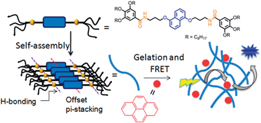
This paper reports self-assembly, photophysical studies and energy transfer in a bis-amide functionalized dialkoxynaphlane (DAN)-based organogel. Due to the synergistic effect of hydrogen bonding, π–π stacking and hydrophobic interaction, DAN showed long-range ordering in a non-polar solvent, methylcyclohexane (MCH), as evidenced by the fibrous morphology observed in TEM. It showed gelation in various organic solvents with very low critical gelation concentration (in some solvents as low as <0.1 wt%, suggesting very strong gelating propensity). UV/vis and photoluminescence studies of the gel indicated offset π-stacking between the DAN chromophores. Gel to sol transformation was noticed in presence of trace amount of H-bond competing solvent MeOH, indicating strong influence of H-bonds on gelation. Direct evidence for hydrogen bonding was further probed by a variable temperature NMR study in which the NH peak corresponding to the amide functionality was significantly upfield shifted at elevated temperature, signifying breakage of the assembly. Gelation induced enhanced emission was noticed which was attributed to offset π-stacking. As DAN and pyrene are known to form an efficient D–A pair for FRET, photoluminescence properties of the gel were tested in presence of pyrene. It was found that the fluorescence from the donor was completely quenched. In turn strong emission was observed from pyrene suggesting a very efficient FRET process possibly due to close proximity of the pyrene acceptor and the DAN chromophore in the gel state.


Introduction

π-conjugated chromophores are promising candidates for next-generation low-cost and flexible electronic devices.1 For such applications, however, the major challenge lies in achieving the desired supramolecular ordering by tuning molecular-level interactions which can control the resulting photophysical properties.2 In this regard, low molecular weight organic gelators3 based on pi-conjugated building blocks4 are particularly relevant since they are known to form elongated one-dimensional (1-D) fibers on gelation. One of the major advantages of these ordered supramolecular chromophoric arrangements is their ability to facilitate fast and efficient transport of charges (electrons and holes) over a long range,5 which is highly desirable in many device applications such as bulk-heterojunction solar cell, organic thin-film-transistors etc. Furthermore, pi-conjugated organic gelators also exhibit rich photophysical properties in the gel state such as aggregation induced enhanced emission.6 Organogels can also act as a media for co-assembly of donor and acceptor dyes for efficient energy transfer process.7 For example, organogel obtained from oligo(phenylenevinylene) (OPV) and doped with suitable energy acceptor organic dye, was proved to be an excellent candidate for artificial light harvesting material.8 Since supramolecular organization is very critical in all of these photophysical processes, it is foremost important to control the inter-chromophoric interaction at the molecular level and possibly correlate this interaction further with the macroscopic properties such as gelation, morphology and energy or electron transfer processes. With this aim, we have studied the self-assembly of a dialkoxynaphthalene gelator,9 which is an interesting and well-studied electron donor chromophore in the context of donor–acceptor interaction.9 Herein we report H-bonding mediated gelation of DAN-1 (Scheme 1)10 and elaborate on its photophysical properties in the gel state. Further we show highly efficient FRET from DAN-1 chromophore to a non-covalently co-assembled pyrene guest in the gel state.
image file: c4ra08920k-s1.tif
Scheme 1 Structure of DAN-1.

Results and discussion

Gelation study

Gelation property of DAN-1 (C = 5.0 mM) was examined in aliphatic hydrocarbon (cyclohexane, methylcyclohexane, n-octane), aromatic (benzene, toluene), and chlorinated solvents (carbon-tetrachloride, tetrachloroethylene). Interestingly, DAN-1 was able to gelate all investigated hydrocarbon- as well as chlorinated-solvents giving transparent or opaque gels (Fig. 1). In contrast, no gelation was observed for aromatic solvents even at relatively higher gelator concentration (10.0 mM). Critical gelation concentration (CGC) was found to be <0.2 wt% for all the cases. In aliphatic hydrocarbon medium, the values were even lower (∼0.1 wt%) and the gel-to-sol melting temperature (Tgel) values were rather high indicating very strong gelation property (Table 1).
image file: c4ra08920k-f1.tif
Fig. 1 Gelation test (c = 5 mM) in different organic solvents.
Table 1 Gelation data (c = 5 mM) for DAN-1
Solvent Observation Tgelc (°C) CGC (wt%)
MCH Gel 75 0.09
CH Gel 77 0.07
TCE Gel 51 0.19
CCl4 Gel 46 0.3
nOctane Gel >95 0.13
Benzene Sol N/A N/A
Toluene Sol N/A N/A


The enthalpy of melting for gel-to-sol transition was determined by analyzing the concentration-dependent Tgel using Schroeder–van Laar equation (eqn (1)).11

 
image file: c4ra08920k-t1.tif(1)
where c is the concentration of the gelator, ΔHm is the enthalpy of melting, R is the universal gas constant (value of R taken as 8.314 J mol−1 K−1) and Tgel is the temperature (in K) at which the gel transforms into sol. Firstly, Tgel was determined as a function of gelator concentration in MCH. As expected, a sharp decrease in Tgel was observed as the gelator concentration decreased (Fig. 2a). From the slope of the Arrhenius plot of ln[thin space (1/6-em)]c vs. 1/Tgel (Fig. 2b), the enthalpy of melting was estimated as 41 kJ mol−1, which indicates a moderately strong gel in comparison to other hydrogen bonding driven organo-gelators reported in the literature.12


image file: c4ra08920k-f2.tif
Fig. 2 (a) Variation in Tgel as a function of gelator concentration; (b) plot of ln[gelator] vs. 1/Tgel.

The morphology of the gel sample examined by TEM (Fig. 3) appeared as a fibrillar network. The elongated fibers were found to be micrometer long, which is typical for a good gelator and the width of the fibrils was found to be in the range of 30–50 nm.


image file: c4ra08920k-f3.tif
Fig. 3 TEM images of DAN-1 gel; width of the fibril shown by arrow (∼28 nm).

The rigidity and the flow behavior of the gel were investigated in MCH (5.0 mM concentration) by rheological studies. Fig. 4 shows that initially G′ (storage modulus, 112 Pa) was higher than G′′ (loss modulus, 29.6 Pa), indicating presence of a gel phase.13 With a gradual increase in the applied stress, G′ and G′′ remained almost invariant up to a certain extent until they crossed each other at the yield stress of 1.9 Pa, suggesting moderate stability of the gel.


image file: c4ra08920k-f4.tif
Fig. 4 Rheological data for DAN-1 gel in MCH (5.0 mM concentration at 25 °C).

Mode of chromophore assembly in gel state

UV/vis studies. To understand the chromophore packing in the gel state, absorption spectra of the MCH gel (c = 2.0 mM) was compared with that of the sol in CHCl3. Going from sol to the gel-state, distinct differences were noticed in the absorption spectrum (Fig. 5a). For example, the major absorption peaks at 327 and 314 nm exhibited hyperchromic shift with concomitant red shift of ∼1 nm in the gel compared to the monomeric unit. Furthermore, the absorption bands became sharper in the aggregated state as indicated by the reduction of the full width at half maxima by 0.5 nm. These observations indicate an offset π-stacking,10 which was predicted as one of the favorable mode of assembly for electron-rich systems.14 Noteworthy that self-assembly of DAN-1 under a different experimental condition (solution in 90[thin space (1/6-em)]:[thin space (1/6-em)]10 MCH/THF) did not show10 such red-shifted absorption indicating difference in nature of inter-chromophoric interaction in solution and gel state. To determine the thermal stability of the assembly, variable temperature UV-vis spectroscopy studies was carried out (Fig. 5a). The intensity of the absorption bands at 327 nm and 314 nm gradually decreased as the temperature was raised from 25 °C to 75 °C. The absorption spectrum at 75 °C almost overlapped with the monomeric spectrum in CHCl3, suggesting reversible self-assembly and also the melting temperature obtained from UV/vis studies corroborates very well with the observed Tgel (Table 1).
image file: c4ra08920k-f5.tif
Fig. 5 (a) Temperature variable UV-vis spectra of DAN-1 gel in MCH (c = 2.0 mM) and in CHCl3 (for comparison); (b) variation of mole fraction of aggregates with temperature.

From the variable temperature absorption spectra, we further calculated the mole fraction of the aggregates (αagg) as a function of temperature (Fig. 5b) by using eqn (2).4n

 
image file: c4ra08920k-t2.tif(2)
where αagg(T) is the mole fraction of the aggregate at temperature T (in °C), A(T), Amon and Aagg are the absorbance at temperature T (at 327 nm), absorbance in the monomeric form (taken from the spectrum in CHCl3), and the absorbance at the fully aggregated state at 25 °C, respectively. From the above plot α50 (T) (the temperature at which αagg = 0.5) was estimated as 62 °C.

1H NMR studies. In order to probe that hydrogen bonding is indeed responsible for the self-assembly, 1H-NMR was recorded at variable temperatures (VT-NMR) in the gel state and was compared with that in CDCl3 in which the chromophore remains in the monomeric form. For comparison, only the aromatic region of the spectra is shown in Fig. 6. In CDCl3, sharp and distinct signals corresponding to the aromatic peaks were observed. The triplet at δ = 6.5 ppm can be attributed to the N–H proton with JN–H = 4.4 Hz, typically consistent with the 14N–H coupling. To elucidate the nature of the aggregate, we attempted to record VT-NMR of the gel in MCH (10% benzene-d6 was added for locking the signal). However, no peak corresponding to either aromatic protons or amide NH were observed at low temperature, signifying very high relaxation time due to strong gelation.12 Then VT-NMR was recorded in TCE (5.0 mM concentration with 10% benzene-d6 for locking) in which the self-assembly was found to be weaker compared to that in MCH (see Table 1 for CGC and Tgel of DAN-1 in these two solvents). While going from CDCl3 to TCE at 30 °C, significant upfield shifts (Hb: 6.86 to 6.52 ppm; Hc: 7.28 to 7.05 ppm; and Hd: 7.77 to 7.64 ppm) were observed for all the signals corresponding to the DAN-aromatic protons, supporting self-assembly in TCE. Moreover, the peak corresponding to the NH proton appeared as a broad singlet at δ = 6.61 ppm, in contrast to the clear triplet peak pattern in CDCl3, indicating hydrogen bonding in TCE. To probe this point further, we recorded NMR spectra of the gel at elevated temperatures. The peak corresponding to the amide proton (NH) experienced significant upfield shift as the temperature was raised gradually from 30 °C (δ = 6.60 ppm) to 75 °C (δ = 6.12 ppm).
image file: c4ra08920k-f6.tif
Fig. 6 Variable temperature 1H NMR spectra of DAN-1 gel in TCE (5.0 mM) (10% benzene-d6 was used for locking). For comparison 1H NMR spectrum in CDCl3 was recorded at room temperature.

Also the peak pattern changed from a broad singlet (at 30 °C) to a clear triplet (at 75 °C), indicating disruption of the hydrogen bonding at elevated temperature. Concurrently, with gradual increase in temperature, the signals corresponding to the DAN-1 aromatic protons (Hb, Hc and Hd) moved downfield, clearly illustrating the dissociation of the self-assembled structure.

Additional evidence of hydrogen bonding in the gel network was established by FT-IR spectroscopy. Going from CHCl3 to MCH, the peak corresponding to N–H stretching shifted from 3454 cm−1 to 3284 cm−1 (Fig. 7). Also clear shifts of the peaks corresponding to amide-I (1653 cm−1 in CHCl3 and 1628 cm−1 in MCH) and amide-II (1600 cm−1 in CHCl3 and 1591 cm−1 in MCH) were observed. These spectroscopic data are unambiguous proof of hydrogen bonding arising from the amide functionalities and in line with the reported literature.15


image file: c4ra08920k-f7.tif
Fig. 7 Selected region of FT-IR spectra of DAN-1 in CHCl3 (black) and in MCH (red) (DAN-1 c = 5.0 mM).
Proposed model. Based on UV-vis experiments and the variable temperature NMR and FT-IR studies, we propose that the chromophore assembly prefers an arrangement that would synergize both pi–pi stacking and hydrogen-bonding (Fig. 8). This results in self-assembled fiber which bundles up to produce thicker fiber (from TEM the width was noticed 30–50 nm). They further entangle to form network structure which entraps solvent molecules to produce organogel. Further to elucidate the fact that hydrogen bonding is indeed major cause for such facile assembly and gelation, the effect of a hydrogen bond disrupting solvent methanol (MeOH) was examined. It was found that even in the presence of only 2% (v/v) MeOH, the gel was completely destroyed (Fig. 8) confirming H-bonding as the key force for gelation. It is assumed that the possibility of such extended hydrogen bonding among the amide groups in case of off-set stacking of the DAN chromophores does not allow the system from attaining edge-to-face arrangement, which is another preferable mode of arrangement for electron rich chromophores.14 In order to support this model, we carried out an X-ray diffraction study of the xerogel of DAN-1 prepared from MCH. The diffraction pattern showed a very sharp primary peak at 2θ = 2.74° (Fig. 9), corresponding to a length of d = 32.2 Å, matching very well with the molecular length (32.0 Å) of a fully stretched DAN-1 molecule (Fig. 8) as obtained from the energy minimized structure using Chem3D MM2 level molecular modeling (Fig. S1). The presence of second- and third-order peaks at periodic distance of d/2 and d/3 suggests a lamellar structure with a repeat unit of 32.2 Å.
image file: c4ra08920k-f8.tif
Fig. 8 Proposed model for self-assembly and gelation of DAN-1 showing synergistic effect of π–π interaction and H-bonding and gel-to-sol transition in presence of 2% MeOH in MCH suggesting strong influence of H-bonding on gelation.

image file: c4ra08920k-f9.tif
Fig. 9 Powder X-ray diffraction of DAN-1 xerogel.

Photoluminescence properties

Further we carried out the photoluminescence study of the DAN-1 gel in MCH (c = 2.0 mM) and compared the emission property with that of the monomeric form in CHCl3 or THF (Fig. 10a). In sharp contrast to solution state, where the emission is almost quenched (peaks are not visible in Fig. 10a), the emission intensity in the gel state increased significantly (Fig. 10a). Further, in the gel state the emission spectrum appeared with small Stoke's shift (18 nm), showed mirror-image relationship with the absorption spectrum. These features in addition to the observations made in UV-vis studies show similar trend that was observed for J-aggregating perylene-derivatives.16 To gain further insight into the emission property of the self-assembly, time-correlated single photon counting (TCSPC) experiments were carried out with the gel in MCH (Fig. 10b) and compared with the emission in the sol state (in THF, 2.0 mM concentration) (Fig. 10c). For both sol and gel, the emission profiles were monitored at 346 nm after exciting at 295 nm. Fluorescence decay time in the gel state was found to be 5.1 ns (Table S1), which was faster compared to the same in THF (7.2 ns). The fast decay profile in gel can be attributed to fast delocalization of the exciton in the aggregated state, matching well with the previous observation on self-assembled perylene16 and naphthalene-diimide17 chromophores. Further we checked the emission properties of the gel as a function of temperature (Fig. 11a).
image file: c4ra08920k-f10.tif
Fig. 10 (a) Absorption (dotted lines) and normalized (absorption and emission intensity in the gel state were normalized to 1 and intensity of rest of the spectra are adjusted proportionately) emission spectra (solid line) of DAN-1 in gel (MCH, blue line) and sol (THF, black line; CHCl3, wine color) state. c = 2.0 mM; λex = 295 nm; time resolved fluorescence decay of DAN-1 in (b) gel in MCH and (c) sol in THF. λex = 295 nm, λem = 346 nm.

image file: c4ra08920k-f11.tif
Fig. 11 (a) Temperature dependent emission spectra of DAN-1 gel (c = 2 mM, MCH); (b) absorption (solid line) and emission (dotted line) of DAN-1 gel (black, MCH) and sol (red line) in presence of 2% MeOH, λex = 295 nm.

It was found that the emission intensity remained almost invariant up to the temperature 70 °C. Beyond 75 °C (where the gel melting was observed before) a sharp decrease in the emission intensity was noted, confirming the enhanced emission was indeed due to gelation. We have shown (Fig. 8) in presence of a protic solvent MeOH gelation could be destroyed. Thus we checked effect of MeOH on emission of the gel and noticed (Fig. 11b) in presence of 2% MeOH the emission intensity reduced by more than 60% reconfirming gelation induced enhancement in the emission.

Co-assembly with pyrene and FRET study

In contrary to common observation that aggregation quenches emission, the present system shows enhanced emission in gel state. This prompted us to explore utilizing this luminescent gel scaffold as a light harvesting system7 by FRET from the DAN donor to a pyrene (py) acceptor,18 which was non-covalently assembled in the gel matrix. Prior to energy transfer studies, the heterogel of DAN-1 + py (1[thin space (1/6-em)]:[thin space (1/6-em)]1.1, DAN c = 5.0 mM) was characterized in MCH and was compared with the DAN-1 gel alone. The CGC of the mixed gel was found to be 0.06 wt% and Tgel = 79 °C, indicating improved gelating ability of DAN-1 in presence of py. Further rheological studies (Fig. 12a) revealed a yield stress value of 4.1 Pa which is significantly more than that of DAN-1 gel confirming more robust gelation in presence of py. In broader sense, enhanced gelation propensity of DAN-1 in presence of py is similar to well known phenomenon of salt induced lowering of critical gelation concentration of a surfactant molecule. The morphology of the hetero-gel sample was further examined by TEM. TEM image of the mixed gel shows (Fig. 12b) entangled fibrillar network (width ∼30–50 nm) similar to that observed for DAN-1 and thus suggests that the gel-network remains unperturbed in presence of pyrene. In order to gain further insight about the nature of the hetero-gel, its 1H NMR was recorded in MCH (DAN c = 5.0 mM, 10% benzene-d6 was added for locking the signal) (Fig. 12d) and compared with the 1H NMR of DAN-gel alone in MCH at similar concentration (Fig. 12c). At room temperature, peaks were observed only at δ = 7.99 to 7.77 ppm, corresponding to pyrene protons while all the signals corresponding to the DAN-aromatic protons were absent, signifying different relaxation time for pyrene and DAN-protons in the gel matrix. At elevated temperature, peaks corresponding to DAN-aromatic protons and amide proton (NH) appeared along with the pyrene peaks and all the peaks were assigned according to the NMR spectra in Fig. 6. It is noteworthy that no upfield (amide proton) or downfield (DAN-aromatic and pyrene protons) shifts of the peaks were observed even at 70 °C, suggesting stronger gelation in MCH than TCE. Increasing the temperature further resulted in minor downfield shift of the amide proton at 75 °C indicating onset of disassembly. Then UV/vis absorption spectrum of the hetero-gel DAN-1 + py (1[thin space (1/6-em)]:[thin space (1/6-em)]1.1, DAN c = 0.5 mM) was recorded and compared with the individual spectrum of DAN-1 and py at similar concentrations (Fig. 12e). The spectrum of the mixture exhibits peaks in the region of 250–340 nm, corresponding to both DAN-1 and py chromophores. The spectrum of DAN-1 + py matches well to the spectrum obtained from mathematical summation of the individual spectrum of DAN-1 and py chromophores (Fig. 12e), suggesting no ground state interaction between DAN and pyrene chromophores as also indicated by their 1H-NMR spectrum in the mixed gel which showed sharp pyrene peaks but no DAN peak at rt. Further FT-IR of DAN-1 + py mixed-gel shows identical spectral features compared to the FT-IR spectrum of DAN-1 gel alone in MCH (Fig. 12f), which unambiguously proves the hydrogen bonding network among amide functionalities remains unperturbed even after inclusion of py. Based on these observations it is conceivable that py chromophores do not intercalate between the stacked DAN-chromophores, rather are located in the peripheral region (within Förster distance) of the 1D stack.
image file: c4ra08920k-f12.tif
Fig. 12 (a) Rheological study for DAN-1 + py gel (DAN c = 5.0 mM); (b) TEM image of DAN-1 + py gel; (c) and (d) variable temperature 1H NMR of DAN-1 and DAN-1 + py gel, respectively (DAN c = 5.0 mM), 10% C6D6 was used for locking, X denotes peak from C6D6; (e) UV-vis spectrum of DAN-1 (red), py (black), DAN-1 + py (blue) and mathematical summation of DAN-1 and py (dotted line) (DAN-1:py = 1[thin space (1/6-em)]:[thin space (1/6-em)]1.1, DAN c = 0.5 mM, path-length = 1 mm); (f) comparative FT-IR spectra of DAN-1 (black) and DAN-1 + py (red) (DAN c = 5.0 mM) in MCH.

After successful characterization of the hetero-gel, FRET from the DAN donor and pyrene acceptor was tested. For FRET to happen, it is prerequisite that the absorption band of the acceptor should have significant overlapping with the emission spectrum of the donor, which is satisfied for the DAN–pyrene pair (Fig. S2). Moreover, the supramolecular alignment and the distance between the donor- and acceptor-chromophores also play very crucial role in dictating the efficacy of FRET process. To check whether all these criteria are fulfilled in the present gel scaffold we studied emission properties of a gel made from DAN-1 + py (1[thin space (1/6-em)]:[thin space (1/6-em)]1.1) in MCH (Fig. 13).


image file: c4ra08920k-f13.tif
Fig. 13 Emission spectra (λex = 295 nm) of DAN-1 gel (MCH, c = 2 mM) in the absence (red) and presence (black) of pyrene (2.2 mM); the blue line (inset shows same data in zoomed Y-axis scale) represents the emission of pyrene (2 mM) in MCH solution (λex 295 nm).

When the mixture was excited at DAN absorption (λex = 295 nm) a complete quenching of the emission bands ∼350 nm corresponding to the DAN-1 chromophore was noticed with a concomitant appearance of the intense emission bands in the range of 375–425 nm corresponding to py chromophore suggesting extremely efficient energy transfer with maximum possible FRET ratio. When py chromophore alone was excited at λex = 295 nm where its absorption is very negligible (black line, Fig. 12e), it showed negligible fluorescence (blue line, Fig. 13), indicating intense emission band that was observed in the hetero-gel is solely due to energy transfer from DAN to py. The broad band present at λem = 472 nm possibly corresponds to the excimer formation of py in the gel state. Such a high FRET efficiency can possibly indicate that the pyrene acceptor is located near the peripheral hydrophobic alkyl chains; which brings them well within the Förster radius facilitating efficient energy transfer.

Experimental

Materials and characterization

All the chemicals were purchased from Sigma Aldrich Chemical Corporation and used without further purification. Solvents were purchased from commercial sources and purified by reported protocol.19 Spectroscopic grade solvents were used for physical studies. All 1H and 13C NMR spectra were recorded on Bruker DPX 500 MHz machine and calibrated against TMS peak. Chemical shift (δ) and coupling constant (J) are reported in parts per million and Hertz, respectively. The abbreviations for splitting patterns are s, singlet; bs, broad singlet; d, doublet; t, triplet; q, quartet; dd, doublet of doublets, and m, multiplet. Variable temperature NMR was recorded on Bruker DPX 300 MHz machine. UV-visible absorption spectra were recorded on a PerkinElmer Lamda 25 spectrometer and photoluminescence studies were performed on a Horiba Fluoromax-3 spectrometer. Rheological studies were performed on an AR 2000 advanced Rheometer (TA instrument) by cone and plate method. FT-IR data were recorded on a PerkinElmer Spectrum 100 FT-IR spectrometer. XRD data were recorded on a Seifert XRD3000P diffractometer having a Cu Kα radiation source (wavelength λ = 0.1541 nm) operating at a voltage and current of 40 kV and 300 mA, respectively.

Synthesis

DAN-1 was synthesized according to the literature-reported procedure.10

Gelation test

The gelation property of DAN-1 was tested in a series of solvents such as MCH, CH, CCl4, TCE, n-octane, and toluene. In a typical experiment, a stock solution of the gelator was made from a good solvent like CHCl3 at a fixed concentration and measured amount of the stock solution was further taken in a screw-capped sample vial and the solvent was evaporated with a hot air gun. To the solid film, measured amount of gelating solvent was added and the contents were heated in closed condition to obtain a homogeneous solution. After cooling to room temperature and standing for 30 minutes, gelation property was tested by standard ‘stable-to-inversion of a vial’ method.

To determine the critical gelation concentration of DAN-1 in MCH, the gel was gradually diluted with known amount of MCH and each time the vial was heated to obtain a homogeneous solution and further cooled down to room temperature before the gelation was tested. The concentration below which no gelation was observed even after waiting for hours is reported as the critical gelation concentration.

By using above-mentioned procedure, Tgel of DAN-1 in MCH was obtained in five different gelator concentrations and ΔHm was determined using Schroeder–van Larr equation.

Similar procedure was used to check the gelation data of DAN-1 + py.

UV-visible and photoluminescence studies

DAN-1 gel (2.0 mM) was prepared and equilibrated for 2 h before performing the experiments. For variable temperature UV-vis experiment, the gel (2.0 mM in MCH) was heated from the lowest temperature (25 °C) to the highest temperature (75 °C) with an increment of 5 °C and allowed to equilibrate for 10 min at each step before carrying out the measurement. Similar protocol was used for photoluminescence studies.

UV-vis spectrum of DAN-1 + py was recorded according to the procedure as describe above.

Variable temperature NMR

DAN-1 gel was prepared in TCE at 5.0 mM concentration. 10% C6D6 was added for locking NMR signal. NMR spectra at variable temperatures were recorded on a 300 MHz NMR instrument from 30 °C to 70 °C with an increment of 10 °C. The sample was allowed to equilibrate for 10 min at each temperature before recording the spectrum.

Variable temperature NMR experiments of DAN-1 in MCH and DAN-1 + py in MCH were carried out according to the procedure as describe above.

Rheological measurement

DAN-1 gel (5.0 mM) was prepared in MCH and equilibrated for 2 h before performing the experiment. Stress-amplitude sweep measurement was carried out using an Advanced Rheometer AR 2000 (TA instrument) with the cone diameter, cone angle and truncation of 40 mm, 4°0′22′′ and 121 μM, respectively. The runs were conducted at 25 °C with a constant oscillation frequency of 1 Hz.

Rheological measurement of DAN-1 + py was conducted using similar protocol.

FT-IR measurement

FT-IR spectra were recorded on a PerkinElmar Frontier FT-IR spectrometer (DAN-1 c = 2.0 mM).

TEM study

One drop of the gel sample (0.5 mM in MCH) was put on a copper grid coated with carbon and the grid was air-dried for 1 h before the experiment was performed.

TEM study of DAN-1 + py (1[thin space (1/6-em)]:[thin space (1/6-em)]1.1, DAN c = 0.5 mM) was carried out by similar method.

Powder X-ray diffraction studies

DAN-1 gel in MCH (2.0 mM) was repeatedly drop-casted on a glass slide to prepare a thick film, which was air dried for 12 h at room temperature for powder X-ray diffraction study. Data was recorded from 1°–30° with sampling interval of 0.02 Å per state.

Conclusion

In conclusion, we described the self-assembly and gelation of DAN-1 chromophore. DAN-1 was found to gelate a variety of organic solvents ranging from aliphatic hydrocarbon (cyclohexane, methylcyclohexane, n-octane), aromatic (benzene, toluene), to non-polar chlorinated solvents (carbon-tetrachloride, tetrachloroethylene) with very low critical gelation concentration. TEM study of gel showed fibrous morphology, indicating long-range ordering due to synergistic effect of π–π stacking and hydrogen bonding. DAN chromophores were found to form offset π-stacking in the gel state leading to enhanced emission. A highly efficient light harvesting system could be constructed by gelation in presence of py as an acceptor which did not perturb the self-assembly of DAN-1. Emission spectra of the mixed gel showed very efficient energy transfer suggesting the present gel is a highly promising soft scaffold for co-assembly of functional chromophores. Luminescent gelation and highly effective energy transfer process is encouraging to explore this building block and structurally related systems for co-assembly of more than one chromophore to realize cascade energy transfer producing tunable emission color.

Acknowledgements

SG thanks the Alexander von Humboldt Foundation for donating a spectro-fluorimeter. DB and AD thank CSIR, India for a research fellowship.

Notes and references

  1. (a) S. S. Babu, S. Prasanthkumar and A. Ajayaghosh, Angew. Chem., Int. Ed., 2012, 51, 1766 CrossRef CAS PubMed; (b) F. S. Kim, G. Ren and S. A. Jenekhe, Chem. Mater., 2011, 23, 682 CrossRef CAS.
  2. S. S. Babu, K. K. Kartha and A. Ajayaghosh, J. Phys. Chem. Lett., 2010, 1, 3413 CrossRef CAS.
  3. (a) J. W. Steed, Chem. Commun., 2011, 1379 RSC; (b) S. Banerjee, R. K. Das and U. Maitra, J. Mater. Chem., 2009, 19, 6649 RSC.
  4. (a) S. H. Kim and J. R. Parquette, Nanoscale, 2012, 4, 6940 RSC; (b) H. Engelkamp, S. Middelbeek and R. J. M. Nolte, Science, 1999, 284, 785 CrossRef CAS; (c) A. Ajayaghosh and V. K. Praveen, Acc. Chem. Res., 2007, 40, 644 CrossRef CAS PubMed; (d) J. Puigmartí-Luis, Á. P. Pino, V. Laukhin, L. N. Feldborg, C. Rovira, E. Laukhina and D. B. Amabilino, J. Mater. Chem., 2010, 20, 466 RSC; (e) X. Q. Li, V. Stepanenko, Z. C. P. Prins, L. D. A. Siebbeles and F. Würthner, Chem. Commun., 2006, 3871 RSC; (f) A. Jain, K. V. Rao, C. Kulkarni, A. George and S. J. George, Chem. Commun., 2012, 1467 RSC; (g) H. Shao and J. R. Parquette, Chem. Commun., 2010, 4285 RSC; (h) P. Mukhopadhyay, Y. Iwashita, M. Shirakawa, S.-i. Kawano, N. Fujita and S. Shinkai, Angew. Chem., Int. Ed., 2006, 45, 1592 CrossRef CAS PubMed; (i) S. Diring, F. Camerel, B. Donnio, T. Dintzer, S. Toffanin, R. Capelli, M. Muccini and R. Ziessel, J. Am. Chem. Soc., 2009, 131, 18177 CrossRef CAS PubMed; (j) W. A. Velema, M. C. A. Stuart, W. Szymanski and B. L. Feringa, Chem. Commun., 2013, 49, 5001 RSC; (k) S. K. Samanta and S. Bhattacharya, Chem. Commun., 2013, 1425 RSC; (l) S. Yagai, M. Ishii, T. Karatsu and A. Kitamura, Angew. Chem., Int. Ed., 2007, 46, 8005 CrossRef CAS PubMed; (m) S. Yagai, T. Kinoshita, M. Higashi, K. Kishikawa, T. Nakanishi, T. Karatsu and A. Kitamura, J. Am. Chem. Soc., 2007, 129, 13277 CrossRef CAS PubMed; (n) S. Ghosh, X.-Q. Li, V. Stepanenko and F. Würthner, Chem.–Eur. J., 2008, 14, 11343 CrossRef CAS PubMed.
  5. (a) L. Magginia and D. Bonifazi, Chem. Soc. Rev., 2012, 41, 211 RSC; (b) J. Puigmartí-Luis, V. Laukhin, Á. P. Pino, J. V. Gancedo, C. Rovira, E. Laukhina and D. B. Amabilino, Angew. Chem., Int. Ed., 2007, 46, 238 CrossRef PubMed; (c) S. Prasanthkumar, A. Gopal and A. Ajayaghosh, J. Am. Chem. Soc., 2010, 132, 13206 CrossRef CAS PubMed; (d) D. A. Stone, A. S. Tayi, J. E. Goldberger, L. C. Palmer and S. I. Stupp, Chem. Commun., 2011, 5702 RSC; (e) F. S. Schoonbeek, J. H. Van Esch, B. Wegewijs, D. B. A. Rep, M. P. De Haas, T. M. Klapwijk, R. M. Kellogg and B. L. Feringa, Angew. Chem., Int. Ed., 1999, 38, 1393 CrossRef CAS; (f) P. Pratihar, S. Ghosh, V. Stepanenko, S. Patwardhan, F. C. Grozema, L. D. A. Siebbeles and F. Würthner, Beilstein J. Org. Chem., 2010, 6, 1070 CrossRef CAS PubMed; (g) A. Wicklein, S. Ghosh, M. Sommer, F. Würthner and M. Thelakkat, ACS Nano, 2009, 3, 1107 CrossRef CAS PubMed.
  6. (a) Y. Hong, J. W. Y. Lam and B. Z. Tang, Chem. Soc. Rev., 2011, 40, 5361 RSC; (b) Y. Hong, J. W. Y. Lama and B. Z. Tang, Chem. Commun., 2009, 4332 RSC.
  7. A. Ajayaghosh, V. K. Praveen and C. Vijayakumar, Chem. Soc. Rev., 2008, 37, 109 RSC.
  8. (a) A. Ajayaghosh, C. Vijayakumar, V. K. Praveen, S. S. Babu and R. Varghese, J. Am. Chem. Soc., 2006, 128, 7174 CrossRef CAS PubMed; (b) V. K. Praveen, S. J. George, R. Varghese, C. Vijayakumar and A. Ajayaghosh, J. Am. Chem. Soc., 2006, 128, 7542 CrossRef CAS PubMed; (c) A. Ajayaghosh, S. J. George and V. K. Praveen, Angew. Chem., Int. Ed., 2003, 42, 332 CrossRef CAS PubMed; (d) S. Bhowmik, S. Banerjee and U. Maitra, Chem. Commun., 2010, 46, 8642 RSC; (e) S. Bhowmik and U. Maitra, Chem. Commun., 2012, 48, 4624 RSC.
  9. (a) R. S. Lokey and B. L. Iverson, Nature, 1995, 375, 303 CrossRef CAS; (b) S. Ghosh and S. Ramakrishnan, Angew. Chem., Int. Ed., 2005, 44, 5441 CrossRef CAS PubMed; (c) A. Das and S. Ghosh, Angew. Chem., Int. Ed., 2014, 53, 1092 CrossRef CAS PubMed; (d) K. Liu, C. Wang, Z. Li and X. Zhang, Angew. Chem., Int. Ed., 2011, 50, 4952 CrossRef CAS PubMed; (e) K. Liu, Y. Yao, Y. Liu, C. Wang, Z. Li and X. Zhang, Langmuir, 2012, 28, 10697 CrossRef CAS PubMed; (f) M. R. Molla, A. Das and S. Ghosh, Chem.–Eur. J., 2010, 16, 10084 CrossRef CAS PubMed.
  10. For the first report on this molecule by us see: A. Das and S. Ghosh, Chem.–Eur. J., 2010, 16, 13622 CrossRef CAS PubMed . In this article the molecule was used for a control experiment but no detail studies were performed.
  11. (a) U. K. Das, D. R. Trivedi, N. N. Adarsh and P. Dastidar, J. Org. Chem., 2009, 74, 7111 CrossRef CAS PubMed; (b) D. J. Abdallah, L. Lu and R. G. Weiss, Chem. Mater., 1999, 11, 2907 CrossRef CAS; (c) D. J. Abdallah and R. G. Weiss, Langmuir, 2000, 16, 352 CrossRef CAS.
  12. (a) W. Edwards and D. K. Smith, J. Am. Chem. Soc., 2013, 135, 5911 CrossRef CAS PubMed; (b) J. R. Moffat and D. K. Smith, Chem. Commun., 2009, 316 RSC; (c) A. R. Hirst, I. A. Coates, T. R. Boucheteau, J. F. Miravet, B. Escuder, V. Castelletto, I. W. Hamley and D. K. Smith, J. Am. Chem. Soc., 2008, 130, 9113 CrossRef CAS PubMed.
  13. (a) P. Sollich, Molecular Gels: Materials with Self-Assembled Fibrillar Networks, Springer, Netherlands, 2006 Search PubMed; (b) F. M. Menger and A. V. Peresypkin, J. Am. Chem. Soc., 2003, 125, 5340 CrossRef CAS PubMed.
  14. C. A. Hunter and J. K. M. Sanders, J. Am. Chem. Soc., 1990, 112, 5525 CrossRef CAS.
  15. S. Diring, F. Camerel, B. Donnio, T. Dintzer, S. Toffanin, R. Capelli, M. Muccini and R. Ziessel, J. Am. Chem. Soc., 2009, 131, 18177 CrossRef CAS PubMed.
  16. (a) T. E. Kaiser, H. Wang, V. Stepanenko and F. Würthner, Angew. Chem., Int. Ed., 2007, 46, 5541 CrossRef CAS PubMed; (b) F. Würthner, T. E. Kaiser and C. R. Saha-Möller, Angew. Chem., Int. Ed., 2011, 50, 3376 CrossRef PubMed; (c) X.-Q. Li, X. Zhang, S. Ghosh and F. Würthner, Chem.–Eur. J., 2008, 14, 8074 CrossRef CAS PubMed.
  17. (a) A. Das and S. Ghosh, Macromolecules, 2013, 46, 3939 CrossRef CAS; (b) H. Kar, M. R. Molla and S. Ghosh, Chem. Commun., 2013, 49, 4220 RSC.
  18. H. J. Kim, S. Y. Park, S. Yoon and J. S. Kim, Tetrahedron, 2008, 64, 1294 CrossRef CAS PubMed.
  19. D. D. Perrin, W. L. F. Armarego and D. R. Perrin, Purification of Laboratory Chemicals, Oxford, Pergamon, 1980 Search PubMed.

Footnote

Electronic supplementary information (ESI) available: Energy minimized structure and fluorescence lifetime data. See DOI: 10.1039/c4ra08920k

This journal is © The Royal Society of Chemistry 2014