Synthesis of different crystallographic Al2O3 nanomaterials from solid waste for application in dye degradation

Seema Singha, Vimal Chandra Srivastava*a, Tapas Kumar Mandalb and Indra Deo Malla
aDepartment of Chemical Engineering, Indian Institute of Technology Roorkee, Roorkee-247667, Uttarakhand, India. E-mail: vimalcsr@yahoo.co.in; vimalfch@iitr.ac.in; Fax: +91-1332-276535; Tel: +91-1332-285889
bDepartment of Chemistry, Indian Institute of Technology Roorkee, Roorkee-247667, Uttarakhand, India

Received 18th August 2014 , Accepted 30th September 2014

First published on 1st October 2014


Abstract

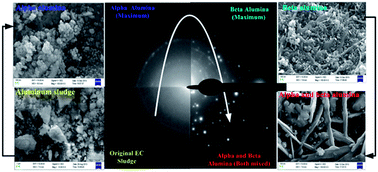
Recycling of solid wastes (sludge) generated during electrochemical (EC) treatment is one of the biggest technological challenges for scientists. Here we propose a facile approach for the preparation of active aluminium oxide nanomaterials (NMs) in different crystalline forms by recycling the sludge generated during EC treatment of textile dye wastewater. Thermogravimetric (TGA/DTA) analysis, powder X-ray diffraction (PXD), field emission scanning electron microscopy (FE-SEM) and energy-dispersive X-ray analysis (EDX) analyses of the samples incinerated at different temperatures demonstrate formation of nanocrystalline α-, β- and γ- aluminas (Al2O3). Moreover, FE-SEM together with trans-mission electron microscopy (TEM) and atomic force microscopy (AFM) showed morphological variations of the α-, β- and γ-Al2O3 NMs but largely with uniform size and shape for respective crystallographic forms. Brunauer–Emmett–Teller (BET) surface area measurement indicated fairly good surface areas for the prepared Al2O3 NMs. Catalytic activity tests revealed β-Al2O3 to be most active among all other crystallographic forms reported here. The present study offers a novel and green method for the recycling of sludge (solid waste) into NMs for environmental catalysis applications.


1. Introduction

The unique physical properties and potential application of metal oxide nanomaterials have received much attention.1,2 Therefore, nanomaterials (NMs) preparation with unique functional properties is of great interest and is actively being pursued.3,4 Oxides of aluminum, iron and titanium are the most commonly used nanomaterials for the treatment of wastewater. Each oxide nano-material has its own advantages and disadvantages when compared with each other.3 Al2O3 nanomaterials have low cost, high surface area and good thermal stability.5 Among metal oxides, polymorphic crystalline aluminium oxide (Al2O3) nanomaterials have been used for various applications.5,6 Several production techniques such as solution-based routes and vapour-phase processes, sol–gel process and Pechini's method have been reported in the open literature for the preparation of aluminium nano oxides.7–9 However, NMs preparation from solid waste is a big challenge for environmentalist and scientists.10,11 Various chemical based routes are reported in the literature which are often time-consuming and hazardous for environment. Hence, it still remains a challenge to develop a facile and environmental friendly green route for preparing NMs with different morphologies from solid waste.

Most of the conventional treatment methods used in wastewater treatment generate some amount of solid waste. Disposal and recycling these solid wastes is a big challenge. Very few studies are reported in open literature which are based on the NM preparation from different types of solid waste which have been used further in different useful applications.12–15 Sewage sludge-based composite materials have been used as an adsorbent for interacting with NO2.16 Municipal sludge has been used for biodiesel production proposing a promising technique for replacement of petroleum-based diesel fuel.15

Alumina oxide NMs with different morphology possessing meso- and micro-pores have received much attention in wastewater treatment.2 Various methods have been used in the literature utilizing well known techniques employing costly, toxic and hazardous chemicals and sophisticated instruments for the preparation of different Al2O3 NMs. However, a facile and green approach has been proposed in this paper for the preparation of different crystallographic Al2O3 NMs from the EC solid waste. There are two general classes of Al2O3. One α-Al2O3 or corundum which has low surface area, and β-Al2O3 and γ-Al2O3 are more porous alumina and are more of catalytic interest.8 Few studies are reported on the use of α-Al2O317 and γ-Al2O318 as catalysts, however, studies on use of β-Al2O3 are very scarce.

In our previous studies,19–23 detailed mechanism including changes in the zeta potential, schemes of dye degradation and contribution of zeta potential with particles size distribution during EC treatment of dye bath effluent with different electrodes and various combination of aluminum and stainless steel electrodes are reported. EC method generates residues (sludge and scum) whose composition depends on type of electrode material being used and characteristics of wastewater being treated. This sludge containing the metals needs to be disposed off.

In the present work, a new method for preparation of aluminium oxide NMs in different crystallographic form (α-, β-, and γ-Al2O3) and morphology by heating the EC solid waste has been developed. Changes in characteristic physical properties and phase evolution were confirmed by thermal analysis (TG/DTA) and powder X-ray diffraction (PXD) analysis. After optimisation of the thermal treatment conditions, the resulting materials were characterized scanning and transmission electron microscopy (SEM and TEM), selected area electron diffraction (SAED), atomic force microscopy (AFM) and Fourier-transform infrared (FTIR) analysis. Newly prepared NMs were further used as catalysts (for degradation of basic green dye by wet per-oxidation method) in order to explore the potential of these recycled materials in wastewater treatment.

2. Experimental section

2.1. Materials

All chemicals used in present study were of analytical grade and were used without further purification. Basic green 4 dye was purchased from Yogesh Dyestuff Products Pvt., Ltd, India and 30 wt% H2O2 was procured from Ranken, India.

2.2. Experimental methodology for generation of EC sludge

EC thermostatically controlled batch reactor was made of plexiglass having 1.0 L working volume. Two aluminium electrodes of 99.5% purity and having 10 cm × 8.5 cm × 0.15 cm dimensions with one electrode acting as an anode and other as cathode were used for basic green dye degradation. Inter-electrode spacing between the electrodes was 1.0 cm and they were connected in parallel mode with direct current power supply (0–20 V, 0–5 A) equipment either in galvanostatic mode with constant current density being 117.64 A m−2. Magnetic stir was used keeping the solution homogenous inside the reactor. The conductivity and pH of solution were adjusted by adding NaCl and 0.1 N NaOH or 0.1 N H2SO4, respectively, before start of the experiments. After desired level of treatment, the slurry was allowed to settle for some time. Solid residue (sludge) was separated, dried and analysed for various characteristics. It was further used for preparation of alumina oxides NMs with different morphology. Scheme 1 shows the graphical representation of the preparation of aluminium oxide NMs in different morphology and their application in dye degradation.
image file: c4ra08842e-s1.tif
Scheme 1 Schematic representation of Al2O3 nanomaterials preparation in different crystallographic form and morphology from EC solid waste and their application in dye degradation.

2.3. Preparation of aluminium oxides nano-materials

Schematic representation of EC treatment technology is described as first step (step a) in Scheme 1. For the preparation of aluminium oxide based NMs from EC sludge, first the sludge was sun-dried for two-three days.24 Then, ∼10 g of dried sludge was taken in a silica crucible and calcined in a muffle furnace at different temperatures ranging from 100–1000 °C and further at higher temperatures upto 1200 °C for different durations (1–18 h). The resulting sample obtained at 100 °C (Al-100), were subjected to sequential heating at 200 °C (Al-200), 400 °C (Al-400), 500 °C (Al-500), 600 °C (Al-600), 700 °C (Al-700) and 1000 °C (Al-1000). Besides, we have further heat treated the 1000 °C sample at higher temperatures for various durations, such as, 1150 °C/1 h, 1150 °C/6 h, 1150 °C/18 h, 1200 °C/3 h and longer to observe the phase transformations of alumina.

2.4. Characterisation

Thermogravimetric analyser (Thermo Cahn, model Versa Therm High Sensitivity Series 81547) was used for the analysis of different sludge sample from ambient temperature to 1000 °C with a heating rate of 10 °C min−1 under air flow. Various crystallographic parameters were investigated by using powder X-ray diffraction (Brueker AXS, Diffractometer D8, Germany) with Cu Kα radiation (1.542 Å) and 40 kV and 30 mA, accelerating voltage and emission current, respectively, at 1° min−1 scanning rate with 0.02 step size. Bragg angle range of 25–75° was used to determine the diffraction spectra of all samples. Lattice parameters were least-square refined using the PROSZKI program.25 Scherrer equation and the X-ray broadening analysis were used to determine the crystalline size of different samples with the help of joint committee on powder diffraction standards (JCPDS). Micromeritics ASAP-2020 was used for recording the adsorption–desorption N2 isotherms at 77 K in the range of 4 × 10⁻³ to 84 kPa and sample was degassed at 300 °C for about 6 h before analysis. Brunauer–Emmet–Teller (BET), Horvath–Kawazoe (HK) and Harkins–Jura (HJ) methods were used to determine the surface area, mean pore width and micropore volume. HJ25 and HK26 methods were employed for calculating pore size distribution in different samples. Morphology of different samples was determined using field emission-scanning electron microscopy (FE-SEM) (LEO, Model 438 VP, England). All the samples were coated with gold sputter (Edwards S150) for providing the conductivity to the sample. Images were taken at 20 kV acceleration voltage at low vacuum then EDX was used to determine the elemental composition (with maximum ±10% error) of the samples. Nicolet Avatar 370 CSI spectrometer (Thermo-Electron Corporation, USA) was used for analysing the Fourier transform infrared (FTIR) spectra over a range of 4000–400 cm−1 by using KBr pellet technique.

2.5. Dye degradation

Catalytic activity of different Al2O3 NMs was determined by monitoring the degradation of basic green 4 dye in wet per-oxidation method. All the alumina oxide NMs were tested for their catalytic ability at 45 °C to decolourise and degrade 100 mg L−1 basic green dye solution at natural pH 5.4 with catalyst dose of 1 g L−1 in presence of H2O2 as an initiator. The concentration of basic green dye was determined by comparing the absorbance of unknown sample with the absorbance of solution of known concentration at λmax = 619 nm. Double beam UV-visible spectrophotometer (HACH, DR 5000, USA) and digestion unit (DRB 200, HACH, USA) were used for determining the initial and final chemical oxygen demand (COD). Percentage removal efficiencies were determined by using the following relationship:
 
image file: c4ra08842e-t1.tif(1)
where, Co and Cf are the initial and final dye or COD concentration.

3. Results and discussion

3.1. Thermogravimetric analysis

Thermal analysis profile of aluminium sludge samples (calcined at 100 and 1000 °C coded as Al-100 and Al-1000, respectively) are shown in Fig. S1a and b (in ESI). For Al-100, dehydration and decarboxylation of alumina sludge takes place from 200 to 450 °C in primary and secondary combustion zone. However, mass loss was continuous up to 1000 °C. Two clear endothermic peaks at 350 and 410 °C indicate the elimination of water and decomposition of carbonaceous substances of sludge (Fig. S1b).12,24 Mass loss of 65% was observed in the second degradation zone. Rest of mass loss from 400 to 1000 °C was very small.

The weight loss of Al-1000 sample is negligible as the sample exists in pure form. Well defined exothermic peak around 800 °C was obtained in both of the samples and it can be associated with transformation of Al-sludge into nano alumina without any weight loss. The position, intensity and width of endothermic and exothermic peaks are the main differences between the Al-100 and Al-1000 sludge (Fig. S1a and b). These differences are related to the change in morphology of initial compound Al-100 to Al-1000 heated compound. The diffuse broad exothermic peak position of Al-100 shifts from 350 °C towards 210 °C and becomes single large broad in Al-1000 sample. TGA/DTA analysis results show that the crystallisation of different phases takes place when aluminum electrode generated EC sludge is heated at higher temperature.26,27

3.2. Powder X-ray diffraction (PXD) analysis

The phase identity and (hkl) indexing of various crystallographic forms of Al2O3 along with respective crystal structures are shown in Fig. 1. Analysis of the PXD data obtained for calcined samples at 1000 °C shows presence of hexagonal Al2O3 (γ-Al2O3, JCPDS-PDF #50-0741) phase (Fig. S1). The indexed PXD data for γ-Al2O3 phase is given in Table S1. On further heating beyond 1000 °C, an admixture of corundum type Al2O3 (α-Al2O3, JCPDS-PDF #46-1222) and another hexagonal Al2O3 (β-Al2O3: a stoichiometric compound of sodium aluminates, JCPDS-PDF #46-1222) is formed (Fig. 1). The indexed PXD data of α-Al2O3 and β-Al2O3 is given in Tables S1 and S2, respectively. By carefully monitoring the phase evolution by PXD studies as a function of temperature and duration, different crystallographic forms of Al2O3 could be stabilized under standardized conditions. An Al2O3 NM could be stabilized with β-Al2O3 as the major phase having small quantities of α-Al2O3 admixture by treating at 1150 °C for 1 h. Similarly, stabilisation of α-Al2O3 as the major phase with small amounts of β-Al2O3 could also be achieved by heating the sludge at 1150 °C for 6 h. Further, with a heat treatment at 1150 °C for18 h and beyond, a mixed phase of α- and β-Al2O3, with the α- and β-forms in nearly equal proportions, is also stabilized. This was confirmed from the X-ray intensity data.
image file: c4ra08842e-f1.tif
Fig. 1 Indexed PXD pattern having α-Al2O3, β-Al2O3 and α-β-Al2O3 both (b) hexagonal beta alumina type phase and (c) rhombohedral alpha alumina type phase.

All the diffraction peaks observed at 1000 °C in the PXD pattern arising due to the γ-Al2O3 type phase are indexable in the cubic Fd[3 with combining macron]m space group with a least-square refined lattice parameter of 7.8943(3) Å. For the admixed phase with α-Al2O3 as major constituent, all the diffraction peaks due to α-Al2O3 are indexable in the rhombohedral R[3 with combining macron]c space group with least-square refined lattice parameters, a = 4.7528(3) and c = 12.992(8) Å. Similarly, in the admixture with β-Al2O3 as the major phase, all the diffraction peaks responsible for β-form are indexable in the hexagonal P63/mmc space group with the least-square refined lattice parameters, a = 5.64(8) Å and c = 22.65(5) Å.28

3.3. Textural analysis

Surface area and pore volume analysis of solid samples is an important characterisation technique for understanding the solid structure, and potential application of different phases of alumina NMs. The textural analysis of various alumina NMs is shown in Fig. 2. The variation of pore volume, pore area and pore diameter is reported in Table 1. The order of BET surface area of EC sludge and different phases of alumina NMs was found to be in the following order: Al sludge (390.92 m2 g−1) > γ-Al2O3 (126.76 m2 g−1) > α-Al2O3 (52.1 m2 g−1) > β-Al2O3 (41 m2 g−1) > α-Al2O3 and β-Al2O3 (22.11 m2 g−1).
image file: c4ra08842e-f2.tif
Fig. 2 Pore size distributions (a) pore volume and (b) pore area of original aluminium sludge and different phases of alumina nano-materials.
Table 1 Textural analysis of the EC generated sludge of aluminium at different temperature and different phases
Parameters Original sludge Gamma phase Alpha phase Beta phase Alpha & beta phase
BET surface area (m2 g−1) 390.9 126.7 52.1 41.7 22.1
External surface area (m2 g−1) 369 119 62 44 24
BJH cumulative surface area of pores (m2 g−1) 385 132 56 43 23
BJH cumulative pores volume (cm3 g−1) 0.32 0.27 0.10 0.07 0.04
BJH pore diameter (Å) 33.6 81.9 74.6 69.0 64.4


Industrial application of the EC sludge generated alumina NMs is likely to be highly dependent upon its pore size distribution analysis. The internal structure and structure heterogeneity of EC sludge and alumina NMs can be determined in terms of pore size distribution with the help of Barret–Joyer–Halenda (BJH) analysis.8,29 BJH pore volume and pore area was found to be in the following order: EC sludge (0.323 cm3 g−1) > γ-Al2O3 (0.269 cm3 g−1) > α-Al2O3 (0.104 cm3 g−1) > β-Al2O3 (0.072 cm3 g−1) > α-Al2O3 and β-Al2O3 (0.042 cm3 g−1). Similarly, BJH pore diameter was found to be in the following order: γ-Al2O3 (81.89 Å) > α-Al2O3 (79.44 Å) > β-Al2O3 (67.99 Å) > α-Al2O3 and β-Al2O3 (64.62 Å) > Al-Sludge (33.59 Å), respectively. IUPAC classifies various types of pores based on pore diameter (d) as: super-micropores (d < 7 Å) < ultra-micropores (7 Å < d < 20 Å) < micropores (d < 20 Å) < mesopores (20 Å < d < 500 Å) < macropores (d > 500 Å). Based on textural analysis, it can be said that all the alumina NPs and EC sludge are meso-porous in nature.

3.4. FE-SEM and TEM analysis

FE-SEM was used to identify the morphology of aluminium NMs. SEM images of original sludge S(Al-100) shows agglomerated particles of 50–200 μm size (Fig. 3).30 After heating from 700 °C, the crystallinity of sludge increases as shown by PXD and the average particle size decreases to 30–35 nm as evidenced by SEM images (Fig. 3). SEM image analysis of alumina NMs at different temperature i.e. 1000 °C (Al-1000), 1100 °C/1 h, 1100 °C/6 h and 1100 °C/18 h shows different morphology due to presence of different nano-particles (γ-Al2O3, α-Al2O3 maximum, β-Al2O3 maximum) phases having different shape and sizes.
image file: c4ra08842e-f3.tif
Fig. 3 SEM image of aluminium sludge and different morphology of alumina.

SEM images of the actual EC sludge and various NMs have different morphologies. Al2O3 shows that γ-Al2O3 does not possess any well defined porous structure and only few pores are present on its surface (Fig. 3). However, α-Al2O3 NM has small round ball shape with quite uniform surface. Similarly, β-Al2O3 NM has rods like shape in SEM image with diameters 35–70 nm. NM having both α-Al2O3 and β-Al2O3 has non-uniform mixture of rod and small round ball shaped particles spread in its structure.

Element dispersion in different alumina NMs is shown in Fig. S3 and S4. The elemental dispersion of α-Al2O3 NM (Fig. S3) indicates that Al (61.73%) and oxygen (38.27%) distributed on the surface non-uniformly, with lower and higher elements distribution. β-Al2O3, having 62.76% Al and 37.24% oxygen, both elements are cover most of the surface of NMs. However, small humps are also observed. Fig. S4 shows 68.08% Al and 31.92% oxygen in mixed α-Al2O3 and β-Al2O3 phase NMs. Elements are uniformly and smoothly distributed on the surface.

TEM images of different alumina NMs are shown in Fig. 4. It may be seen in Fig. 4a that the γ-Al2O3 NM form aggregates lower than 100 nm in size. SAED pattern confirmed presence of small crystalline particles in γ-Al2O3. SAED indexing pattern confirmed the image with PXD (hkl) of γ-Al2O3.29,31 SAED indexing pattern also shows that it is in amorphous form and not in completely crystalline form. For α-Al2O3, spherical agglomerated particles with particle size around 60 nm can be observed in Fig. 4a and b. SAED pattern of α-Al2O3, inset in Fig. 4b, indicates that the phases are almost crystalline, aggregates are very small round plates with some of them being arranged in a plane.10,32 Similarly, β-Al2O3 indicates that most of the particles are tiny small square plate shaped having 30–40 nm particles size. SAED indexing pattern, inset in Fig. 4c, confirms the lattice pattern observed with PXD. It was found that most of β-Al2O3 have a tendency to present a hexagonal shape which was confirmed by the lattice plane. The SAED indexing pattern of different phases in TEM images was confirmed by comparing the PXD pattern of different phases of alumina (γ-Al2O3, α-Al2O3 and β-Al2O3) (Fig. 1 and S2).10,31 Crystallinity is very flexible in alumina powder as compared to other ceramic compounds. It was found to vary with change in calcination temperature and duration of heating.


image file: c4ra08842e-f4.tif
Fig. 4 TEM images (a) γ-Al2O3 (b) α-Al2O3 and (c) β-Al2O3 nanomaterials.

3.5. Atomic force microscopy (AFM) analysis

SEM does not give an idea of quantitative information about height distributions, roughness and particles after drying in dispersion solution.33 AFM analysis provides better observation of the aggregation after drying the suspension in solid state. Three-dimensional information of AFM provides information about alumina nano clusters34 and material surface.35 It is also helpful for the finding out the topography, shape and size of the nano structure surface.

Fig. S5a shows 2D AFM images of Al-100. It shows non-uniform distribution of bright spots with less heterogeneous distribution of bright spots. 3D AFM image shown in Fig. S5a′ shows that the average maximum surface roughness (roughness distribution) was 4.5 nm which decreased with an increase the temperature because of sintering. 2D image of Al-1000 (γ-Al2O3 as confirmed by PXD) shown in Fig. S5b indicates that the particles are flat, compact and in regular arrangement. All the particles having almost 60 nm constant height and corresponding to the average diameter of the alumina NPs (Fig. S5b′) with 11.46 nm average roughness (inset Fig. S4b′). However, as the temperature increases, change in phase and morphology of NPs takes place which was confirmed by SEM and TEM analysis. Fig. 5 depict NPs aggregates have fibrillar structure. It may due to the anisotropic physical properties of alumina NPs. The solid state deposited on the surface indicates quite compact arrangement of the spherical particles in the α-Al2O3. 3D image confirmed the 8.0 nm average roughness and 1.6 nm average height of the particles (Fig. 5a and a′). AFM analysis of β-Al2O3 was also confirmed in Fig. 5b with small agglomeration between the particles. 3D AFM image showed the 4.5 nm average roughness and 0.34 nm average height of the particles (Fig. 5b′). The topography of β-Al2O3 in 2D image indicates that the particles could not arrange on a flat layer.35 Fig. 5c and c′ show that the rounded shape particles overlap to each other in mixed α-Al2O3 and β-Al2O3 phase NM. 3D image of Fig. 5c′ shows that maximum height of the particles is 0.74 nm with average surface roughness of 8.0 nm.


image file: c4ra08842e-f5.tif
Fig. 5 AFM analysis of alumina nanoparticles at different morphology (a) 2-D image of alpha alumina, (b) 2-D image beta alumina, (c) 2-D image of alpha and beta alumina (a′) 3-D image of alpha alumina, (b′) 3-D image of beta alumina and (c′) 3-D image of alpha and beta alumina.

3.6. FTIR analysis

FTIR spectra of EC treated aluminium sludge at different temperature Al-100, Al-1100 (α-Al2O3) and Al-1150 (β-Al2O3) is shown in Fig. 6. Band in the range of 2923–2847 cm−1 arises due to the stretching vibration of –CH3 group of dye molecules attached in side chain of benzene ring.22,23 Broad peak at ≈3450 cm−1 indicates the presence of –OH and –NH2 group which come from water molecules as moisture and the amine group of dye present in the EC sludge samples. The presence of peaks within the range of 3000 to 3600 cm−1 are related to the water molecule present in the lattice of alumina powder due to the presence of moisture in KBr pellet. However, some new characteristic peaks get generated after thermal treatment. Most of the characteristic peaks of nano-alumina were obtained in finger-print region (1500–400 cm−1) of FTIR spectrum.8 In alumina, most of the characteristic peaks of alumina were observed around 802, 760 and 580 cm−1 due to OH, Al–OH, and Al–O bonds.20,36,37 FTIR stretching and bending vibration bands at 610, 650 and 460 cm−1 indicate the presence of Al–O presence in each spectra of alumina sample.18 The peaks in the fingerprint region present a number of peaks (Fig. 6) due to the fundamental modes of vibration of amorphous or nanocrystalline nature of alumina powder.10 These peaks are complex and are very sensitive to the crystallinity of samples. PXD pattern (Fig. 1 and S2) confirmed the existence of different crystallography form in alumina powder. Some of the weak band around 1100–1200 cm−1 are due to the Al–O bonds.38
image file: c4ra08842e-f6.tif
Fig. 6 FTIR analysis of thermally treated electrochemical sludge: sludge at 100 °C (original sludge), sludge calcined at 1000 °C (gamma alumina), 1150 °C/6 h (alpha alumina), 1150 °C/1 h (beta alumina) and 1150 °C/18 h (alpha and beta alumina).

3.7. Catalytic activity of NMs

Wastewater treatment using catalytic methods has gained significant attention in recent years.39 However, very few studies are available in the literature where alumina NMs derived from solid waste has been used as nano catalyst for dye wastewater treatment. All phases of alumina NMs were tested for their catalytic ability at 45 °C for decolourisation and degradation of basic green 4 (BG4) dye solution (100 mg L−1) at natural pH with catalyst dose of 1.0 g L−1 in presence of H2O2 as an initiator. Results are shown in Fig. 7. During catalytic oxidation, alumina NPs provide the catalyst surface (having various functional groups) for initiating the H2O2 decomposition into free radicals which subsequently help in dye decomposition via free radical pathways.40 Dye degradation begins via H2O2 decomposition on the active surface of alumina NMs which generates OH˙ radicals and helps in mineralisation of dyes. However, at high dosage of H2O2, OH˙ radicals react to form hydro-peroxyl radicals (HO2˙) and water. HO2˙ is an effective oxidant by itself but its oxidation potential is much lower as compared to OH˙ radical and promotes radical chain reactions.41 Hence, HO2˙ does not contribute to the oxidative degradation of dye molecule, which occurs only by reaction with OH˙.42,43 Expected chemical reactions during the treatment with alumina NMs are as follows:
 
image file: c4ra08842e-t2.tif(2)
 
image file: c4ra08842e-t3.tif(3)
 
image file: c4ra08842e-t4.tif(4)
 
image file: c4ra08842e-t5.tif(5)
 
image file: c4ra08842e-t6.tif(6)

image file: c4ra08842e-f7.tif
Fig. 7 (a) Decolorisation efficiency (b) COD removal efficiency of BG-4 dye with different Al2O3 NMs at different time intervals.

It may be seen that in Fig. 7 that the catalytic reactivity order of different morphology of alumina nano-materials is as follow: β-Al2O3 > α-Al2O3> α-β Al2O3> γ-Al2O3. However, the BET surface area (γ-Al2O3 (126.76 m2 g−1) > α-Al2O3 (52.1 m2 g−1) > β-Al2O3 (41 m2 g−1) > α-β Al2O3 (22.11 m2 g−1)) and pore size distribution (γ-Al2O3 (81.89 Å) > α-Al2O3 (79.44 Å) > β-Al2O3 (67.99 Å) > α-Al2O3 and β-Al2O3 (64.62 Å) > Al-Sludge (33.59 Å)) are found in different order. The reason for higher reactivity of β-Al2O3 may be due to its structure (Fig. 1c) which shows more vacant lattice space and highly crystalline nature (Fig. 1a and Fig. S5c) as compared to α-Al2O3 (Fig. 1a, b and S5a) and γ-Al2O3 (Fig. S2 and S5a). This may also be due to acidic nature of β-Al2O3.

4. Conclusions

One step calcination of the sludge at higher temperature aimed at sintering and growing crystalline metal oxide NMs, as proposed in this study, provides a new avenue for recycling and reuse of the sludge generated during EC treatment of a wastewater in the form of NMs via a simple and green route. The conversion of the solid waste into NMs is achieved by removal of volatiles and non-volatile organic contaminants present in the solid waste by the oxidation and reduction of the constituents via exothermic and endothermic reactions. The proposed methodology of NMs preparation not only solves the environmental issue of sludge disposal but also provides a research path for reuse of solid waste as NMs for wastewater treatment. Pure alumina NMs within the size range from 30–65 nm were prepared by the calcination of the EC sludge. The Al2O3 NMs exhibited different crystalline phases such as alpha, beta, gamma and alpha–beta both, depending on the calcination temperature (1000–1150 °C) and duration. The oxidative catalytic activity for the basic green dye degradation with different phases of alumina NMs was determined.

Acknowledgements

Authors are thankful to Council of Scientific and Industrial Research (CSIR), India for providing financial help for carrying out this work.

References

  1. E. Saputra, S. Muhammad, H. Sun, M. O. Tade and S. Wang, Environ. Sci. Technol., 2013, 47, 5882–5887 CrossRef CAS PubMed.
  2. I. Ali, Chem. Rev., 2012, 112, 5073–5091 CrossRef CAS PubMed.
  3. P. R. Potti and V. C. Srivastava, Ind. Eng. Chem. Res., 2012, 51, 7948–7956 CrossRef CAS.
  4. R. J. Gupta, D. Y. Kusuma, P. S. Lee and M. P. Srinivasan, ACS Appl. Mater. Interfaces, 2011, 3, 4619–4625 CAS.
  5. Y. C. Sharma, V. Srivastava, S. N. Upadhyay and C. H. Weng, Ind. Eng. Chem. Res., 2008, 47, 8095–8100 CrossRef CAS.
  6. A. Boumaza, L. Favaro, J. Ledion, G. Sattonnay, J. B. Brubach, P. Berthet, A. M. Huntz, P. Roy and R. J. Tetot, Solid State Chem., 2009, 182, 1171–1176 CrossRef CAS PubMed.
  7. Y. K. Park, E. H. Tadd, M. Zubris and R. Tannenbaum, Mater. Res. Bull., 2005, 40, 1506–1512 CrossRef CAS PubMed.
  8. D. S. Maciver, H. H. Tobin and R. T. Barth, Catalytic aluminas I. Surface chemistry of eta and gamma alumina, J. Catal., 1963, 2, 485–487 CrossRef CAS.
  9. Y. Chen, Y. Su, X. Zheng, H. Chen and H. Yang, Water Res., 2012, 46, 4379–4386 CrossRef CAS PubMed.
  10. A. Lopez-Delgado, L. Fillali, J. A. Jimenez and S. Lopez-Andres, J. Sol-Gel Sci. Technol., 2012, 64, 162–169 CrossRef CAS.
  11. C. S. K. Lin, L. A. Pfaltzgraff, L. Herrero-Davila, E. B. Mubofu, S. Abderrahim, J. H. Clark, A. A. Koutinas, N. Kopsahelis, K. Stamatelatou, F. Dickson, S. Thankappan, Z. Mohamed, R. Brocklesby and R. Luque, Energy Environ. Sci., 2013, 6, 426–464 CAS.
  12. H. K. Shon, S. I. Vigneswaran, N. S. Kim, J. Chog, J. Kim, J. B. Kim and J.-H. Kim, Environ. Sci. Technol., 2007, 41, 1372–1377 CrossRef CAS.
  13. Y. Fan, F. S. Zhang and Y. J. Feng, J. Hazard. Mater., 2008, 159, 313–318 CrossRef CAS PubMed.
  14. S. R. K. Pandiana, V. Deepaka, K. Kalishwaralal, J. Muniyandi, N. Rameshkumar and S. Gurunathan, Colloids Surf., B, 2009, 74, 266–273 CrossRef PubMed.
  15. D. M. Kargbo, Energy Fuels, 2010, 24, 2791–2794 CrossRef CAS.
  16. R. Pietrzak and T. J. Bandosz, J. Hazard. Mater., 2008, 154, 946–953 CrossRef CAS PubMed.
  17. S. Lan, N. Guo, L. Liu, X. Wu, L. Li and S. Gan, Appl. Surf. Sci., 2013, 283, 1032–1040 CrossRef CAS PubMed.
  18. T. Zaki, K. I. Kabel and H. Hassan, Ceram. Int., 2012, 38, 4861–4866 CrossRef CAS PubMed.
  19. B. Mondal, V. C. Srivastava and I. D. Mall, J. Environ. Sci. Health, Part A: Toxic/Hazard. Subst. Environ. Eng., 2012, 47, 2040–2051 CrossRef CAS PubMed.
  20. B. Mondal, V. C. Srivastava, J. P. Kushwaha, R. Bhatnagar, S. Singh and I. D. Mall, Sep. Purif. Technol., 2013, 109, 135–143 CrossRef CAS PubMed.
  21. S. Singh, V. C. Srivastava and I. D. Mall, RSC Adv., 2013, 3, 16426–16439 RSC.
  22. S. Singh, V. C. Srivastava and I. D. Mall, J. Phys. Chem. C, 2013, 117, 15229–15240 CAS.
  23. S. Singh, V. C. Srivastava and I. D. Mall, Ind. Eng. Chem. Res., 2014, 53(26), 10743–10752 CrossRef CAS.
  24. J. Matos, M. Rosales, A. Garcia, C. Nieto-Delgado and J. R. Rangel-Mendez, Green Chem., 2011, 13, 3431–3439 RSC.
  25. P. Alphonse and M. Courty, Thermochim. Acta, 2005, 425(2), 75–89 CrossRef CAS PubMed.
  26. G. d. C. Cunh, L. Piment, C. Romao and S. Z. Macedo, Powder Technol., 2014, 254, 344–351 CrossRef PubMed.
  27. P. P. Nampi, S. Ghosh and W. G. Krishna, Ceram. Int., 2011, 37(8), 3329–3334 CrossRef CAS PubMed.
  28. W. Losoch and K. J. Lewinski, Appl. Crystallogr., 1994, 27, 437 CrossRef.
  29. S. Lan, N. Guo, L. Liu, X. Wu, L. Li and S. Gan, Appl. Surf. Sci., 2013, 283, 1032–1040 CrossRef CAS PubMed.
  30. R. Ahlawat, V. C. Srivastava, I. D. Mall and S. Sinha, Clean: Soil, Air, Water, 2008, 36(10–11), 863–869 CrossRef CAS.
  31. P. Alphonse and M. Courty, Thermochim. Acta, 2005, 425(1–2), 75–89 CrossRef CAS PubMed.
  32. V. Jayaraman, G. Periaswami and T. R. N. Kutty, Mater. Chem. Phys., 1998, 52(1), 46–53 CrossRef CAS.
  33. W. M. Zeng, L. Gao and J. K. Guo, Nanostruct. Mater., 1998, 10(4), 543–550 CrossRef CAS.
  34. J. Sukmanowski, J.-R. Viguie, B. N. olting and F. X. Royer, J. Appl. Phys., 2005, 97, 104332–104338 CrossRef PubMed.
  35. J.-R. Viguie, J. Sukmanowski, N. Bengt and F.-X. Royer, Colloids Surf., A, 2007, 302, 269–275 CrossRef CAS PubMed.
  36. A. Vazquez, T. Lopez, R. Gomez, M. Bokhimi and O. Novarot, J. Solid State Chem., 1997, 128, 161–168 CrossRef CAS.
  37. Y. C. Sharma, V. Srivastava and A. K. Mukherjee, J. Chem. Eng. Data, 2010, 55, 2390–2398 CrossRef CAS.
  38. S. Ram, Infrared Phys. Technol., 2001, 42(6), 547–560 CAS.
  39. X. H. Wang, G. Z. Lu, Y. Guo, Y. S. Wang and Y. L. Guo, Mater. Chem. Phys., 2005, 90(2–3), 225–229 CrossRef CAS PubMed.
  40. H.-L. Wang, J. Dong and W.-F. Jiang, J. Hazard. Mater., 2010, 183, 347–352 CrossRef CAS PubMed.
  41. Y. Iida, T. Kozuka, T. Tuziuti and K. Yasui, Ultrasonics., 2004, 42(1–9), 635–639 CrossRef CAS PubMed.
  42. B. H. Hameed and T. W. Lee, J. Hazard. Mater., 2009, 164, 468–472 CrossRef CAS PubMed.
  43. A. Rathinam, N. F. Nishtar, R. R. Jonnalagadda and U. N. Balachandran, J. Hazard. Mater., 2006, B138, 152–159 Search PubMed.

Footnote

Electronic supplementary information (ESI) available: “Table of powder XRD data for γ-Al2O3 and α-Al2O3”, “powder XRD data for β-Al2O3” “thermogravimetric (a) TGA (b) DTA analysis of electrochemical sludge of S(Al-100) and S(Al-1000)” “PXD patterns of thermally treated EC alumina sludge at different temperature and different duration”, “FE-SEM/EDX elemental dispersion analysis of alpha and beta maximum alumina NMs”, “FE-SEM/EDX and elemental dispersion analysis of both alpha and beta mixed alumina NMs.”, and “AFM analysis of alumina sludge (a) 2-D image of aluminium sludge, (a′) 3-D image of aluminium sludge, (b) 2-D image of gamma alumina (b′) 3-D image of gamma alumina”. See DOI: 10.1039/c4ra08842e

This journal is © The Royal Society of Chemistry 2014
Click here to see how this site uses Cookies. View our privacy policy here.