Fabrication of a novel magnetic yolk–shell Fe3O4@mTiO2@mSiO2 nanocomposite for selective enrichment of endogenous phosphopeptides from a complex sample

Hao Wanab, Jinan Lib, Wenguang Yub, Zheyi Liub, Quanqing Zhangb, Weibing Zhang*a and Hanfa Zou*b
aShanghai Key Laboratory of Functional Materials Chemistry, East China University of Science and Technology, Shanghai 200237, China. E-mail: weibingzhang@ecust.edu.cn
bCAS Key Laboratory of Separation Sciences for Analytical Chemistry, National Chromatographic R&A Center, Dalian Institute of Chemical Physics, Chinese Academy of Sciences (CAS), Dalian 116023, China. E-mail: hanfazou@dicp.ac.cn

Received 15th August 2014 , Accepted 12th September 2014

First published on 12th September 2014


Abstract

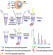
A yolk–shell nanocomposite composed of a magnetic mesoporous anatase TiO2 (Fe3O4@mTiO2) core, a medium cavity and an outermost mesoporous silica (mSiO2) shell was successfully fabricated. Due to a combination of the strong magnetic response, improved diffusion of peptides, numerous affinity sites towards phosphopeptides and the size-exclusion effect, the nanocomposite demonstrated high enrichment efficacy and selectivity towards endogenous phosphopeptides from human serum.


Phosphorylation is one of the most important and ubiquitous post-translational modifications. The phosphorylation of endogenous peptides is involved in lots of biological and pathological variations, which makes endogenous phosphopeptides potential biomarkers with high clinical sensitivity and specificity. However, due to the interference and cover of signal by high-abundance proteins and endogenous non-phosphopeptides in the complex sample, the mass spectrometry (MS) detection of endogenous phosphopeptides still remains challenging.1

Selective enrichment of endogenous phosphopeptides exists as an effective way to exclude the interference and improve the precision and sensitivity of MS detection. Compared with other enrichment methods, metal oxide affinity chromatography (MOAC) has been reported to demonstrate higher binding selectivity and sensitivity to phosphate group. Among these metal oxides, anatase TiO2 has been most widely utilized for selective enrichment of phosphopeptides from various enzymatic digests due to the higher affinity and more tolerance of many biochemical reagents.2 However, because of the occupation and cover of affinity sites on anatase TiO2 by some large-molecule phosphoproteins and non-phosphoproteins in the complex sample, the enrichment selectivity and efficacy towards endogenous phosphopeptides would be highly affected.3 Appearing as a molecular sieve, mesoporous SiO2 has been applied in different systems to differentiate small and large molecules based on the size-exclusion effect.4 But due to the low-efficiency and time-consuming centrifugation separation, the practical application of mesoporous SiO2 in the exclusion of proteins beneficial for efficient and selective enrichment of endogenous phosphopeptides is limited. Recently, the magnetic yolk–shell structure has been attracting more and more attentions. On one hand, the magnetic response would simplify the process of sample separation. On the other hand, the cavity would be beneficial for the diffusion of guest molecules and also enhance the contact area between the substrate and guest molecules.5 Attributed to all above unique properties, an integration of mesoporous SiO2, anatase TiO2 and a magnetic yolk–shell structure should better fulfil the requirement for selective enrichment of endogenous phosphopeptides from the complex sample.

Herein, for highly efficient and selective enrichment of endogenous phosphopeptides from the complex sample, we elaborately fabricated a novel magnetic yolk–shell nanocomposite (Scheme 1). Starting from Fe3O4 nanoparticles, we directly coated a layer of amorphous TiO2, followed by solvothermal reaction assisted pore formation and crystallization of amorphous TiO2 into anatase type (Fe3O4@mTiO2). Subsequently, with the help of PVP the Fe3O4@mTiO2 was encapsulated by a dense SiO2 shell (Fe3O4@mTiO2@nSiO2), onto which another outmost mesoprosous SiO2 shell was assembled by using CTAB as a structure director (Fe3O4@mTiO2@nSiO2@mSiO2–CTAB). After the selective etching of the dense SiO2 shell by Na2CO3 solution and calcination of CTAB and PVP, the final nanocomposite (YS Fe3O4@mTiO2@mSiO2) was obtained. The YS Fe3O4@mTiO2@mSiO2 was suitable for selective enrichment of endogenous phosphopeptides from the complex sample: (1) the outmost mSiO2 shell would act as a size-dependent sieve to exclude large-molecule proteins out; (2) the medium cavity would improve the diffusion of endogenous phosphopeptides and enhance the contact area between endogenous phosphopeptides and the Fe3O4@mTiO2 core; (3) the Fe3O4@mTiO2 core with high surface area would not only provide numerous affinity sites to capture endogenous phosphopeptides but also simplify the process of sample separation.


image file: c4ra08692a-s1.tif
Scheme 1 The schematic illustration of the fabrication process of YS Fe3O4@mTiO2@mSiO2.

Shown in Fig. 1, the diameter of as-synthesized Fe3O4@mTiO2 core distributed around 200–300 nm (Fig. 1c). After encapsulation with the dense SiO2 shell, a well-defined core–shell structure was obtained (Fig. 1d). Under the direction of CTAB, another outmost mSiO2 shell was successfully assembled and the corresponding size increased to 500–600 nm (Fig. 1e). The yolk–shell structure appeared after the selective etching process, which was based on the unexpected differentiated dissolution behaviours, stemming from stabilization of the outmost mSiO2 shell by CTAB (Fig. 1f).6 Through the experiment, it was found out the mass ratio of the Fe3O4@mTiO2@nSiO2@mSiO2–CTAB and Na2CO3 was an indispensable factor for the successful fabrication of the final yolk–shell structure. Fixing the amount of Na2CO3 at 212 mg, we varied the added amount of Fe3O4@mTiO2@nSiO2@mSiO2–CTAB. Based on the TEM results (Fig. 2), if the added amount of Fe3O4@mTiO2@nSiO2@mSiO2–CTAB was limited, an etching of both dense SiO2 and outmost mSiO2–CTAB shells happened. On the contrast, when the added amount of Fe3O4@mTiO2@nSiO2@mSiO2–CTAB was too much, the Na2CO3 solution was not strong enough to completely etch the dense SiO2 shell away, leaving a small gap. So only when the added amount of Fe3O4@mTiO2@nSiO2@mSiO2–CTAB was appropriate, can a well-defined yolk–shell structure be successfully developed. According to the XRD analysis, all diffraction peaks of original magnetic nanoparticles can be perfectly indexed to the magnetite phase of Fe3O4 (JCPDS 19-629) and the final nanoparticles showed additional diffraction peaks of anatase TiO2 (JCPDS 21-1272) (Fig. S1, ESI). The EDS element mapping illustrated the exact distribution of fundamental compositions, further identifying the successful fabrication of YS Fe3O4@mTiO2@mSiO2 (Fig. 3).


image file: c4ra08692a-f1.tif
Fig. 1 The SEM image of (a) YS Fe3O4@mTiO2@mSiO2; the TEM images of (b) Fe3O4, (c) Fe3O4@mTiO2, (d) Fe3O4@mTiO2@nSiO2, (e) Fe3O4@mTiO2@nSiO2@mSiO2–CTAB and (f) YS Fe3O4@mTiO2@mSiO2.

image file: c4ra08692a-f2.tif
Fig. 2 TEM images of nanoparticles after the selective etching process by Na2CO3 solution when the added amount of Fe3O4@mTiO2@nSiO2@mSiO2–CTAB was (a) 50 mg, (b) 100 mg, (c) 150 mg and (d) 250 mg.

image file: c4ra08692a-f3.tif
Fig. 3 The EDS element mapping of YS Fe3O4@mTiO2@mSiO2.

Then, the N2 adsorption experiment was used to probe the detailed structure information about YS Fe3O4@mTiO2@mSiO2. The adsorption–desorption isotherms exhibited the typical IV pattern of the mesoporous material with a distinct H3 hysteresis loop, proving the ink-bottle-type pores in which large cavities are connected by narrow windows.7 The pore distribution showed three main pore diameters of 2.7, 3.9 and 9.1 nm, which could be ascribed to pores distributing within the outmost mSiO2 shell and inner mesoporous anatase TiO2, respectively (Fig. S2, ESI). Besides, the Fe3O4@mTiO2 was also conducted through the BET and BJH analyses. The pores resulting from the solvothermal reaction centralized at 3.7 and 9.5 nm, matching the above result well and the surface area reached 169.79 g m−2, providing numerous affinity sites to capture endogenous phosphopeptides. The original Fe3O4 exhibited a saturation magnetization value of 58.51 emu g−1. While after coating with another three shells, the value decreased to 10.76 emu g−1. Attributed to subsequent processes of selective etching and calcination, the saturation magnetization value increased to 15.7 emu g−1, which would favour the easy separation of YS Fe3O4@mTiO2@mSiO2 (Fig. S3, ESI).

The YS Fe3O4@mTiO2@mSiO2 was supposed to efficiently and selectively enrich endogenous phosphopeptides from the complex sample (Scheme 2). At first, the enrichment effect was evaluated with simple samples. Without enrichment the tryptic digests of β-casein showed lots of high-density non-phosphopeptides and the signal of phosphopeptides was suppressed. However, after enrichment with YS Fe3O4@mTiO2@mSiO2, three significant peaks corresponding to phosphopeptides were identified. Compared with the commercial TiO2 (surface area: 61.25 g m−2, Fig. S8, ESI), the YS Fe3O4@mTiO2@mSiO2 demonstrated the higher enrichment efficacy and selectivity (Fig. 4a–c), which may be a result of the higher surface area and the special yolk–shell structure. Inspired by this, a detection limit experiment was subsequently conducted. The intensity strength of enriched phosphopeptides was positively related to the primitive concentration of tryptic digests of β-casein. And the detection limit could reach as low as 3 fmol, confirming the high enrichment efficacy (Fig. 4c–e). To further identify the high selectivity of YS Fe3O4@mTiO2@mSiO2, different amounts of tryptic digests of BSA as the interference were spiked into the determined amount of tryptic digests of β-casein. The direct analysis of the mixture showed no phosphopeptides but numerous non-phosphopeptides. Brightly, after enrichment with YS Fe3O4@mTiO2@mSiO2, only phosphopeptides existed. Even if the molar ratio was 1[thin space (1/6-em)]:[thin space (1/6-em)]1000 (β-casein[thin space (1/6-em)]:[thin space (1/6-em)]BSA), phosphopeptides could still be selectively captured (Fig. S4, ESI). For YS Fe3O4@mTiO2@mSiO2, the outmost mSiO2 shell would act as a size-dependent sieve to exclude the large-molecule proteins out beneficial for efficient and selective enrichment of phosphopeptides. To test this functionality, we applied YS Fe3O4@mTiO2@mSiO2 and Fe3O4@mTiO2 in parallel for the enrichment of a mixture of α-casein (model phosphoprotein) and tryptic digests of β-casein (a mimic of the complex sample, mass ratio: 100[thin space (1/6-em)]:[thin space (1/6-em)]1). After enrichment, the elution of YS Fe3O4@mTiO2@mSiO2 contained very little α-casein with respect to high-abundance α-casein in the elution of Fe3O4@mTiO2. On the contrast, as for the case of small-molecule peptides, the situation was opposite (Fig. 5). In detail, although both YS Fe3O4@mTiO2@mSiO2 and Fe3O4@mTiO2 could capture the phosphopeptides, the signal intensity of phosphopeptides in the elution of Fe3O4@mTiO2 was much weaker. It was speculated α-casein would cover and occupy lots of affinity sites on the Fe3O4@mTiO2 in view of the large amount, which mostly affected the enrichment efficacy of phosphopeptides. However, the diffusion of large-molecule α-casein (23[thin space (1/6-em)]690 Da, radius of gyration is about 4.5 nm) through the narrow mesopore channels (2.7 nm) distributing within the outmost mSiO2 shell on the YS Fe3O4@mTiO2@mSiO2 was relatively difficult, resulting in the size-exclusion effect to improve the enrichment efficacy.


image file: c4ra08692a-s2.tif
Scheme 2 The process of selective enrichment of endogenous phosphopeptides from the complex sample with YS Fe3O4@mTiO2@mSiO2.

image file: c4ra08692a-f4.tif
Fig. 4 The MALDI-TOF-MS analysis of tryptic digests of β-casein. (a) Direct analysis; (b) after enrichment with commercial TiO2; after enrichment with YS Fe3O4@mTiO2@mSiO2 when the added amount was (c) 50 fmol, (d) 10 fmol and (e) 3 fmol. * indicates phosphopeptides and # indicates dephosphorylated peptides.

image file: c4ra08692a-f5.tif
Fig. 5 The MALDI-TOF-MS analysis of the elution after enrichment of a mixture of α-casein protein and tryptic digests of β-casein with (a and c) Fe3O4@mTiO2 and (b and d) YS Fe3O4@mTiO2@mSiO2.

After all, we applied the YS Fe3O4@mTiO2@mSiO2 in the selective enrichment of endogenous phosphopeptides from human serum. The direct analysis of human serum showed none of endogenous phosphopeptides, accompanied with lots of high-density endogenous non-phosphopeptides and proteins. However, after enrichment only four endogenous phosphopeptides with high signal-to-noise were detected, which showed better enrichment selectivity and efficacy than previous studies.8 In the high-molecular range, two obvious proteins (e.g. human serum albumin, 67 kDa, 5 × 7 × 7 nm) were found out in the supernatant with respect to nothing in the elution, all of which hinted the effective capacity of YS Fe3O4@mTiO2@mSiO2 for selective enrichment of endogenous phosphopeptides from the complex sample (Fig. 6).


image file: c4ra08692a-f6.tif
Fig. 6 The MALDI-TOF-MS analysis of human serum. (a) Direct analysis; (b) the endogenous phosphopeptides in the elution, the residual proteins in the (c) elution and (d) supernatant after enrichment with YS Fe3O4@mTiO2@mSiO2. # indicates endogenous phosphopeptides and * indicates residual proteins in human serum.

In summary, a novel magnetic yolk–shell Fe3O4@mTiO2@mSiO2 nanocomposite was successfully developed. The nanocomposite exhibited a strong magnetic response, high enrichment selectivity and efficacy towards phosphopeptides and the size-exclusion effect, which was suitable for selective enrichment of endogenous phosphopeptides from the complex sample. With all these advances, it is believed the nanocomposite would be applied in many other fields in the future.

Acknowledgements

This work was supported by the China State Key Basic Research Program Grant (2013CB911202, 2012CB910604), the Creative Research Group Project of NSFC (21321064), the National Natural Science Foundation of China (21235006, 81161120540), National Key Special Program on Infection diseases (2012ZX10002009-011), Analytical Method Innovation Program of MOST (2012IM030900) to H. F. Zou.

Notes and references

  1. (a) G. Cheng, Y. Wang, Z. Wang, X. Sui, J. Zhang and J. Ni, RSC Adv., 2014, 4, 7694–7702 RSC; (b) L. Zhao, H. Qin, Z. Hu, Y. Zhang, R. a. Wu and H. Zou, Chem. Sci., 2012, 3, 2828 RSC; (c) Y. Yan, Z. Zheng, C. Deng, Y. Li, X. Zhang and P. Yang, Anal. Chem., 2013, 85, 8483–8487 CrossRef CAS PubMed; (d) J. C. Smith, M. A. Duchesne, P. Tozzi, M. Ethier and D. Figeys, J. Proteome Res., 2007, 6, 3174–3186 CrossRef CAS PubMed.
  2. (a) Y. Yan, X. Sun, C. Deng, Y. Li and X. Zhang, Anal. Chem., 2014, 86, 4327–4332 CrossRef CAS PubMed; (b) Y. Liang, X. He, L. Chen and Y. Zhang, RSC Adv., 2014, 4, 18132–18135 RSC; (c) W. F. Ma, Y. Zhang, L. L. Li, L. J. You, P. Zhang, Y. T. Zhang, J. M. Li, M. Yu, J. Guo, H. J. Lu and C. C. Wang, ACS Nano, 2012, 6, 3179–3188 CrossRef CAS PubMed; (d) T. E. Thingholm, T. J. Jorgensen, O. N. Jensen and M. R. Larsen, Nat. Protoc., 2006, 1, 1929–1935 CrossRef CAS PubMed; (e) Y. Li, X. M. Zhang and C. H. Deng, Chem. Soc. Rev., 2013, 42, 8517–8539 RSC; (f) W. F. Ma, Y. T. Zhang, M. Yu, J. X. Wan and C. C. Wang, RSC Adv., 2014, 4, 9148–9151 RSC.
  3. (a) Z. Lu, M. Ye, N. Li, W. Zhong and Y. Yin, Angew. Chem., Int. Ed., 2010, 49, 1862–1866 CrossRef CAS PubMed; (b) G. Cheng, Z. G. Wang, Y. L. Liu, J. L. Zhang, D. H. Sun and J. Z. Ni, ACS Appl. Mater. Interfaces, 2013, 5, 3182–3190 CrossRef CAS PubMed; (c) Y. Zhang, W. F. Ma, C. Zhang, C. C. Wang and H. J. Lu, ACS Appl. Mater. Interfaces, 2014, 6, 6290–6299 CrossRef CAS PubMed.
  4. (a) R. Tian, H. Zhang, M. Ye, X. Jiang, L. Hu, X. Li, X. Bao and H. Zou, Angew. Chem., Int. Ed., 2007, 46, 962–965 CrossRef CAS PubMed; (b) A. Bouamrani, Y. Hu, E. Tasciotti, L. Li, C. Chiappini, X. Liu and M. Ferrari, Proteomics, 2010, 10, 496–505 CrossRef CAS PubMed; (c) Y. Hu, A. Bouamrani, E. Tasciotti, L. Li, X. W. Liu and M. Ferrari, ACS Nano, 2010, 1, 439–451 CrossRef PubMed; (d) Z. Y. Gu, Y. J. Chen, J. Q. Jiang and X. P. Yan, Chem. Commun., 2011, 47, 4787–4789 RSC; (e) N. R. Sun, C. H. Deng, Y. Li and X. M. Zhang, ACS Appl. Mater. Interfaces, 2014, 6, 11799–11804 CrossRef CAS PubMed.
  5. (a) H. Wan, H. Qin, Z. Xiong, W. Zhang and H. Zou, Nanoscale, 2013, 5, 10936–10944 RSC; (b) W. Zhao, H. Chen, Y. Li, L. Li, M. Lang and J. Shi, Adv. Fun. Mater., 2008, 18, 2780–2788 CrossRef CAS; (c) Y. Chen, H. R. Chen, L. M. Guo, Q. J. He, F. Chen, J. Zhou, J. W. Feng and J. L. Shi, ACS Nano, 2010, 1, 529–539 CrossRef PubMed.
  6. (a) X. Fang, C. Chen, Z. Liu, P. Liu and N. Zheng, Nanoscale, 2011, 3, 1632–1639 RSC; (b) Y. Fang, G. Zheng, J. Yang, H. Tang, Y. Zhang, B. Kong, Y. Lv, C. Xu, A. M. Asiri, J. Zi, F. Zhang and D. Zhao, Angew. Chem., Int. Ed., 2014, 53, 5366–5370 CrossRef CAS PubMed; (c) J. Yang, D. Shen, L. Zhou, W. Li, J. Fan, A. M. El-Toni, W. X. Zhang, F. Zhang and D. Zhao, Adv. Healthcare Mater., 2014 DOI:10.1002/adhm.201400053.
  7. (a) Y. Yang, X. Liu, X. Li, J. Zhao, S. Bai, J. Liu and Q. Yang, Angew. Chem., Int. Ed., 2012, 51, 9164–9168 CrossRef CAS PubMed; (b) H. Wan, Y. Zhang, Z. Y. Liu, G. J. Xu, G. Huang, Y. S. Ji, Z. C. Xiong, Q. Q. Zhang, J. Dong, W. B. Zhang and H. F. Zou, Nanoscale, 2014, 6, 8743–8753 RSC; (c) Y. Yang, J. Liu, X. Li, X. Liu and Q. Yang, Chem. Mater., 2011, 23, 3676–3684 CrossRef CAS.
  8. (a) G. Cheng, X. Yu, M. D. Zhou and S. Y. Zheng, J. Mater. Chem. B, 2014, 2, 4711–4719 RSC; (b) Z. Lu, J. C. Duan, L. He, Y. X. Hu and Y. D. Yin, Anal. Chem., 2010, 82, 7249–7258 CrossRef CAS PubMed; (c) Z. G. Wang, G. Cheng, Y. L. Liu, J. L. Zhang, D. H. Sun and J. Z. Ni, J. Mater. Chem. B, 2013, 1, 1491–1500 RSC.

Footnote

Electronic supplementary information (ESI) available: Experimental details and additional figures. See DOI: 10.1039/c4ra08692a

This journal is © The Royal Society of Chemistry 2014
Click here to see how this site uses Cookies. View our privacy policy here.