Fabrication of self-assembling peptide nanofiber hydrogels for myocardial repair

Xiao Yuan a, Bin He b, Zi Lv c and Suxin Luo *a
aDepartment of Cardiology, The First Affiliated Hospital of Chongqing Medical University, Chongqing, China. E-mail: luosuxin0204@163.com
bDepartment of Orthopedics, The First Affiliated Hospital of Chongqing Medical University, Chongqing, China
cWest China School of Medicine, Sichuan University, Chengdu, China

Received 13th August 2014 , Accepted 13th October 2014

First published on 13th October 2014


Abstract

Myocardial infarction (MI) has become widely spread in clinical medicine, and results in massive loss of myocardium, ventricular remodeling and ultimately heart failure. Many strategies for repairing injured myocardium are under extensive investigation with some early encouraging effects, but cannot effectively prevent disease progression and heart failure. Cardiac tissue engineering has emerged as an increasingly important approach to repair infarcted myocardium. Self-assembling peptide nanofiber hydrogels are regarded as tailored biomaterial scaffolds with the aim to develop cardiac tissue engineering. Their fabrication is based on the principle of molecular self-assembly which allows tailoring of the biological features (e.g. modification with functional motifs and controlled release of signal molecules) of supramolecular structures for cell behaviors and tissue regeneration. This review will outline the potential of combinational treatments of peptide nanofiber hydrogels with cells, growth factors, or both together in myocardial repair.
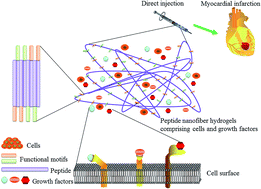

image file: c4ra08582e-p1.tif

Xiao Yuan

Xiao Yuan is a Master degree candidate under the supervision of Prof. Suxin Luo in the Department of Cardiology, The First Affiliated Hospital of Chongqing Medical University, Chongqing, China. Her current research concentrates on the fabrication and properties of self-assembled peptide nanofiber scaffolds and their application in drug release and cardiac tissue engineering.

image file: c4ra08582e-p2.tif

Bin He

Bin He is a Master degree candidate under the supervision of Prof. Dianming Jiang in the Department of Orthopedics, The First Affiliated Hospital of Chongqing Medical University, Chongqing, China. His current research concentrates on the design and fabrication of self-assembled peptide nanofiber scaffolds and their application in drug release and tissue engineering.

image file: c4ra08582e-p3.tif

Zi Lv

Zi Lv is an undergraduate degree student in West China School of Medicine/West China Hospital, Sichuan University, Chengdu, China. She has become a member of the West China Student Society of Digestive System Diseases, the West China Student Society of Emergency Medicine, and The Chinese Cochrane Center, West China Student Society of Evidence-based Medicine.

image file: c4ra08582e-p4.tif

Suxin Luo

Suxin Luo is a Professor in The First Affiliated Hospital of Chongqing Medical University, Chongqing, China, and the Department Director of Cardiology. Her current research interests focus on basic and clinical studies of coronary heart diseases. Suxin Luo was supported by the National Natural Science Foundation Item (8127021), the Nature Science Foundation of Chongqing (CSTC, 2009BB5412 and CSTC, 2012JJA0911), a 2009 program for oversea scholars in Chongqing, Chongqing city health bureau project (2009-2-421) and the Education Teaching Reform Project of Chongqing (2013, 132087). She has published more than 30 original papers.


1. Introduction

Myocardial infarction (MI) has played the dominant role in the incidence of heart failure which is thought to be one of the leading causes of morbidity and mortality in many countries.1–6 It is mainly caused by coronary artery diseases and ischemia followed by cardiomyocyte necrosis and apoptosis.7,8 Myocardial injury can stimulate a series of pathological events including inflammation, granulation tissue formation and eventual scar tissue formation.9,10 Especially, extensive fibrosis serves as an important barrier to cardiac regeneration, and results in the loss of contractile function, ventricular remodeling, scar tissue formation as well as increased susceptibility to arrhythmias.11–13 However, the heart has a very limited regenerative capacity following the injury.14,15 For successful myocardial regeneration, it is very important and crucial not only to promote cardiomyocyte protection/regeneration and vasculature formation, but also to prevent cardiac fibrosis and ventricular remodeling for retaining cardiac function.16–18

Treatment strategies for MI are grossly divided into three groups: pharmacological therapies, interventional therapies (e.g. coronary angioplasty and stenting) and surgical therapies (e.g. coronary artery bypass grafting and heart transplantation). Pharmacological therapies (e.g. angiotensin receptor blockers, angiotensin-converting enzyme inhibitors and β-acceptor blockers) are intended to reduce cardiac workload, improve systolic performance and offer protection from the toxic humoral factors derived from heart failure.19–21 It is believed that coronary artery bypass grafting, coronary angioplasty and stenting can significantly promote the revascularization of the myocardium.20,22 However, these treatment methods cannot effectively prevent ventricular remodeling and disease progression, and the incidence of heart failure following MI remains high.23–25 Heart transplantations are widely accepted as the gold standard for patients with terminal heart failure, but suffer from some limitations including inadequate organ donors and some complications caused by immune suppressive treatments.19,20,26 Thus, there is a great need to develop novel strategies to repair and regenerate the injured heart.

Tissue engineering has emerged as a promising and exciting approach not only to rebuild the structure of damaged tissues or organs, but also to recover their biological function.27–30 Tissue engineering strategies include cell therapies, acellular scaffold implantation, and combination of scaffolds with cells, growth factors or genes.31,32 Although cell-based therapies (e.g. adult stem and pro-genitor cells) have shown some potential in treating MI, they are limited by poor cell retention and difficulties in controlling cell functions (e.g. survival, differentiation and proliferation) and lack an ideally conducive microenvironment for cell behaviors.7,33,34 Injectable biomaterial scaffolds have evolved as an increasingly significant element for cardiac tissue engineering with some advantages (e.g. tailored microenvironment for cell behaviors, minimal invasiveness and easiness to implant in vivo), and have performed some encouraging results independently as well as in combination with cells when treating MI.20,35,36 There are mainly three biomaterial approaches to restore or regenerate injured myocardium, including polymeric left ventricular restraints with the aim to prevent heart failure, engineered cardiac tissue in vitro that can be subsequently transplanted in vivo with or without active substances (e.g. cells and growth factors), as well as direct injection of material scaffold with or without active substances into the myocardium to construct in situ engineered cardiac tissue.37,38 Generally, it is important but difficult to optimize the symbiosis of biomaterial scaffolds, cells and signal molecules (e.g. growth factors, chemokines and cytokines) for cardiac regeneration.11,39–41

Peptide nanofiber scaffolds have become a novel and promising material for myocardial repair.31,42 Their fabrication is based on the principle of molecular self-assembly by which macromolecular architectures can be formed by enormous molecules.39,43 Self-assembling peptide nanofiber scaffolds derived from natural amino acids have the properties of biological self-recognition, good biocompatibility and non-toxic degradation products.44,45 Their microstructures and microenvironments highly mimic the natural extracellular matrices (ECMs), and the similarity can be increased via modification with peptide epitopes (i.e. functional motifs) and controlled release of signal molecules.41 With these advantages, peptide nanofiber scaffolds have been widely applied in the fields of tissue engineering, regenerative medicine and pharmacotherapeutics.39,46–48 This review will focus on the design of self-assembling peptide nanofiber scaffolds, and their capability in repairing injured myocardium in combination with cells, growth factors or both together (Fig. 1).


image file: c4ra08582e-f1.tif
Fig. 1 Schematic representation of self-assembling peptide nanofiber hydrogels in combination with cells, growth factors, or both together for repairing infarcted myocardium through direct injection. Two important approaches to design self-assembling peptide nanofiber scaffolds are modification with functional motifs and controlled release of signal molecules. There have been many self-assembling peptide sequences that can be modified with functional motifs (e.g. RGD and IKVAV). These self-assembling peptides have the ability to form stable 3D nanofiber structures with functional motifs. The density of these motifs can be changed through mixing self-assembling peptides and peptides with functional motifs in different ratios. In addition, it is feasible to store, retain and release various signal molecules (e.g. growth factors) within such 3D nanofiber scaffolds. These released growth factors and functional motifs can significantly promote cell function and tissue regeneration via binding to their corresponding receptors on cell surfaces.48 Adapted and reprinted with permission from ref. 48. Copyright 2014, Cambridge University Press.

2. Hydrogel materials for cardiac tissue engineering

Hydrogel materials have attracted an increasing attention as scaffolds for cardiac repair due to their viscoelastic nature, amenability to chemical and physical modifications, as well as minimal invasiveness.49,50 Their synthesis relies on chemical or physical cross-linking of water-soluble precursors (i.e. natural or synthetic polymers).51,52 Hydrogels materials have the features of swelling and increasing in dimensions while maintaining their shape through absorbing considerable amount of water or biofluids.51 The swelling–deswelling characteristics of hydrogels can be modulated though surface modification in response to some stimulus (e.g. pH, temperature, ion strength, molecules, electric or magnetic signals).53 Furthermore, the porosity and the adequate pore size of hydrogel materials facilitate the free diffusion of metabolites/water, exchange of oxygen and nutrients.52,54 These features enable hydrogel materials to provide a conducive microenvironment for cell behaviors and the delivery of signal molecules.40,49 For eliciting the best regenerative ability of injured tissues, it is important to tailor the interactions between cells and hydrogel materials at molecular level which mainly rely on choosing appropriate materials, functional modifications and controlled release of signal molecules.55

There have been many injectable materials favoring heart regeneration, including natural polymers (e.g. collagen,56,57 fibrin,55,58 alginate,59,60 hyaluronic acid (HA),61,62 chitosan,60,63 matrigel),64,65 synthetic polymers (e.g. polyethylene glycol (PEG),49,66,67 polyvinyl alcohol (PVA),68 polylactic-co-glycolic acid (PLGA),69 and poly(glycerol sebacate) (PGS)70,71) as well as hybrid polymers (e.g. poly-ethylene glycol-diacrylate (PEG-DA) supplemented with alginate72 and the combination of arginine-based unsaturated poly(ester amide) (Arg-UPEA) with Pluronic diacrylate (Pluronic-DA)73). However, there are still some limitations, including inflammatory response, poor cardiomyocytes retention and vascularization, difficulties in controlling cell functions (e.g. differentiation and proliferation) and physiological integration.31,74 It is also complicated for these materials to simultaneously obtain the properties of good bioactivity, appropriate elastic modulus and optimal degradation rate corresponding to myocardial ingrowth.20 In general, the design and fabrication of ideal hydrogel materials used for cardiac regeneration require the following features: (1) good biocompatibility beneficial to reduce inflammatory responses, (2) passive mechanical properties resembling myocardial ECM, (3) controlled release of signal molecules for cell functions (e.g. attachment, proliferation and differentiation), (4) appropriate viscoelasticity allowing the injection through long and narrow catheters, (5) rapid solidification capable to prevent the washout of injected cells, (6) non-toxic degradation products and controlled degradation rate corresponding to cardiac regeneration.39,41,42,48,75

3. Self-assembling peptide nanofiber scaffolds

3.1 Various self-assembling peptides

Self-assembling peptides has become increasingly useful building blocks for constructing macromolecular architectures (e.g. synthetic membranes, multilamellar structures, amphiphilic micelles, tubules and fibrillar networks) ready for biomedical applications,39,43,76,77 and they are featured by intrinsically self-biological recognition and non-toxic degradation products.41 There are three principal types of peptides for synthesizing the hierarchy, peptide-based biomaterial scaffolds, including β-sheet peptides, α-helical peptides and recently collagen-mimicking peptides.48,78–80 It is well known that β-sheet peptides have performed the dominant role in creating nanofiber nanostructures which hold great promise in cell culture, tissue engineering and regenerative medicine.41,81,82 In the β-sheet system, many peptides have been extensively developed to produce functional biomaterial scaffolds for repairing damaged tissues or organs,76,80 including ionic self-complementary peptides (e.g. EAK16-II, RAD16-I and RAD16-II),83–85 peptide amphiphile (PA, e.g. IKVAV-PA and RGD-PA),86–88 self-assembling β-hairpins peptide,89,90 β-sheet tape peptide,91,92 ABA-block copolymer,93,94 as well as various dipeptide and fluorenylmethoxycarbonyl (Fmoc)-conjugates.82,95,96

From peptides to 3D nanofiber scaffolds, one significant approach to favor these dynamic processes is molecular self-assembly by which well-organized and supramolecular architectures are spontaneously assembled molecule by molecule, as showed by the formation of natural substances such as DNA double helix and ribosomes.43,47,97 Chemical complementarity and structural compatibility are fundamental to the formation and structural stability of assembled supramolecular structures.43 During peptide self-assembly, several crucial non-covalent intermolecular forces are involved: (1) hydrogen bonds, (2) ionic interactions, (3) electrostatic interactions, (4) hydrophobic interactions, (5) van der Waals forces and (6) water-mediated hydrogen bonds.41,98 Although these non-covalent intermolecular forces in isolation are relatively weak, tremendous accumulation of them can exert strong forces to support the formation and structural stability of peptide-based biomaterials.43,44,86,97 It is confirmed that many factors (e.g. chemical design versatility of peptides, specific secondary structures, assembling environment (e.g. pH, temperature and ionic strength) and assembling kinetics) have important influence on peptide self-assembly and assembled material scaffolds with controllable structural and biological features at the nanoscale.41,99 This review will focus on using ionic self-complementary peptides and PAs to create nanofiber biomaterial scaffolds that have revealed exceptional capability to repair injured myocardium.

3.2 Ionic self-complementary peptides

Ionic self-complementary peptides have alternating hydrophobic sides (e.g. alanine, valine, leucine, isoleucine, and phenylalanine), and hydrophilic sides including positively charged amino acid (e.g. lysine, arginine, histidine) and negatively charged amino acids (e.g. aspartic acids and glutamic acids).44,45,47 Based on the hydrophilic molecular surface with alternating positively and negatively charged amino acid residues, complementary ionic sides are classified into modulus I, II, III, IV and mixed moduli: modulus I, − + − + − + − +; modulus II, − − + + − − + +; modulus III, − − − + + +; and modulus IV, − − − − + + + +.41,43 The reverse orientation to design charge orientation can yield entirely different molecules with distinct molecular behaviors.43,85 Since a natural protein motif Zuotin (i.e. EAK16-II) was found to spontaneously assemble into entangled nanofibers and 3D macromolecular structures,83 many ionic self-complementary peptides have been under active investigation to synthesize novel biomaterial scaffolds, including peptides RAD16-I, RAD16-II, EAK16-I, EAK16-II, ELK16-II, KLD12-I and d-EAK16 (Fig. 2).44,85,100,101
image file: c4ra08582e-f2.tif
Fig. 2 (a) Molecular model of several self-assembling peptide RAD16-I (top), and SEM image of RAD16-I nanofiber scaffold (PuraMatrix) with fiber diameter of ∼10–50 nm and pores size of ∼10–200 nm (bottom).100 (b) Molecular model of self-assembling peptide EAK16-II (top), and SEM image of EAK16-II nanofiber scaffold with the nanopores of ∼5–200 nm in diameter (bottom).44 (c) Molecular model of self-assembling peptide d-EAK16 (top), and SEM of d-EAK16 with fiber diameter of ∼10 nm and pore size of ∼20–500 nm (bottom).101 Adapted and reprinted with permission from ref. 44, 100 and 101. Ref. 44, Copyright 2006, Royal Society of Chemistry. Ref. 100, Copyright 2008, Schneider et al. Ref. 101, Copyright 2011, Elsevier.

Ionic self-complementary peptides have the preference for β-sheet secondary structure, and subsequently yield nanofibers that are further interwoven into nanofiber hydrogels containing high water content more than 99% (5–10 mg ml−1, w/v).48,102 These peptide hydrogels have entangled nanofibers with the diameter of ∼10–20 nm and pore sizes ranging from ∼5–200 nm.41,44 Tremendous studies have revealed that ion strength, temperature, pH of the solution and denaturing agents have some impact on the dynamic process of peptide self-assembly, but cannot elicit important changes to ionic self-complementary peptides-formed nanostructures.41,101,102 After the disruption of mechanical agitation (e.g. sonication), these peptides have the ability to reassembly and produce indistinguishable structures compared to the previous peptide-formed nanofiber structures.103 There is an increasing agreement that peptide self-assembly mainly relies on overlapping hydrophobic interactions favoring the slide diffusion of hydrophobic cohesive ends along the nanofiber, and intermolecular ionic interactions allowing positive and negative charges of adjacent peptides to pack together in a checkerboard-like manner.45,103 However, the detailed mechanisms to elucidate peptide self-assembly into nanofiber scaffolds remain elusive.

3.3 Peptide amphiphiles

The chemical structures of representative PA molecules are short peptide sequences attached to a hydrophobic tail, consisting of four important structural units (Fig. 3).99 One key component of PAs is the β-sheet forming region directly adjacent to the hydrophobic tail, and usually contains 4–8 amino acids able to yield hydrogen-bonding important for cylindrical nanofiber formation.86,88,104 This β-sheet forming region also has an influence on mechanical properties of PA gels, as evidenced by less twisted β-sheet structures assisting in increasing the stiffness of materials.105 One or more charged amino acid residues facilitate the solubility and gelation, as well as increase the sensitivity to the stimuli of the change in pH and electrolytes.99,106 Another important component is the hydrophobic alkyl tail (e.g. peptide epitopes) to present biological signals for interaction with cells or proteins.87,88 For instance, peptide epitopes RGD and IKVAV have displayed favorable impact on cell functions and tissue regeneration.88,99 It is generally accepted that self-assembly of PAs can form cylindrical nanofibers through hydrophobic collapse, while a hydrogen-bonding network can be formed down the long axis of the nanofiber based on simulations using a mixed Monte Carlo-stochastic dynamics method.107
image file: c4ra08582e-f3.tif
Fig. 3 Schematic representation of self-assemble of PAs into nanofibers: (a) Chemical structure of PA with four key chemical entities. (b) Molecular model of an IKVAV-containing PA, their self-assembly into nanofibers, as well as SEM image of IKVAV nanofibers after adding cell media (DMEM) to PA aqueous solution.99 Adapted and reprinted with permission from ref. 99. Copyright 2010, Wiley Periodicals, Inc.

3.4 Functionalized nanofiber structures

It is believed that nanofiber diameter, pore sizes and numbers within self-assembling peptide hydrogels have played an important role in cell immobilization and behaviors.42,108 Large interconnected pores of biomaterials facilitate cell colonization,109 but excessively large pores (>cell diameter) can prevent endothelial cells (ECs) to bridge pores.110 In contrast, pore sizes of <100 nm have some negative influence on the delivery of nutrients, oxygen, nutrients or even some signal molecules which are all indispensable for the survival and functions of implanted cell.74 However, peptide hydrogels have small pore sizes (∼5–200 nm) crucial to not only promote endothelial adhesion and capillary formation, but also to allow rapid migration of cells owing to their flexibility.41,111

Two important approaches (i.e. modification with functional motifs and controlled release of signal molecules) have been found to significantly increase the bioactivity of peptide nanofiber hydrogels and raise the possibility of emulating the regulatory mechanisms of natural ECMs (Fig. 1).41,48,102 Many functional motifs (e.g. RGD,112 IKVAV113 and YIGSR114) have shown some promise in modulating cell functions. RGD found in fibronectin and other ECM proteins, is beneficial to cell differentiation and migration via binding to α5β1 integrin receptor.112 RGD-modified biomaterials can significantly promote angiogenesis, myocardial regeneration, preservation and recovery of cardiac function when treating MI.115,116 While IKVAV-coated nanofibers assist in preventing astrogliosis and promoting neurite outgrowth.99 In addition, the release of functional proteins (e.g. lysozyme, trypsin inhibitor, BSA, IgG, basic fibroblast growth factor (βFGF), vascular endothelial growth factor (VEGF), and brain-derived neurotrophic factor (BDNF)) from self-assembling nanofiber hydrogels is not only sustained, but also has no influence to protein conformation and functionality based on secondary and tertiary structure analyses and biological assays.117,118 Many factors are revealed to affect this release dynamics, including physical hindrances to prevent proteins mobility, charge interactions between proteins and nanofibers, as well as electrostatic interactions between proteins and heparan sulphate proteoglycans beneficial to increase the local concentrations.27,118,119

4. Myocardial repair using self-assembling peptide nanofiber scaffolds

Functionalized self-assembling peptide hydrogels have obtained some progress for repair various damaged tissues or organs including heart,120,121 bone,122,123 cartilage48,124,125 and nerve.41,126,127 This review will highlight their potential in repairing infarcted myocardium from the aspects of in vitro cell culture and in vivo myocardial regeneration in combination with cells, growth factors, or both together.

4.1 In vitro cell culture

Fibroblasts (FBs) and ECs can induce the expression of angiogenic growth factors and matrix metalloproteinases, collagen deposition and vascularization. Human microvascular ECs, FBs or cocultures were seeded on RAD16-II peptide nanofiber scaffolds, resulting in substantial promotion to cell survival and migration, as well as capillary network formation. During the coculture induction, EC–FB interactions were discovered to attenuate FB matrix metalloproteinase-2 expression and increase collagen I deposition favorable to the greater structural stiffness and more stable microenvironment, indicating that cell–cell interactions and cell migration were supported within peptide nanofiber hydrogels and assisted in creating an optimal environment for cardiac regeneration (Fig. 4a).128 In another study, human microvascular ECs seeded on RAD16-II peptide hydrogels were revealed to promote long-term cell survival, capillary-like network formation, as well as reduced EC apoptosis and enhanced gene expression of VEGF compared to ECs-seeded on collagen type I. Furthermore, tailoring scaffold properties may be helpful to modulate capillary-like network formation as well as the size and spatial organization of cell networks, as demonstrated by a more rigid scaffold beneficial to form extended structures with a larger inter-structure distance, while a more compliant scaffold favorable to produce more dense structures with smaller size (Fig. 4b).111 Additionally, PA–YIGSR peptides were designed to develop native endothelium mimicking nanomatrices for culturing human umbilical vein endothelial cells (HUVECs) and aortic smooth muscle cells (AoSMCs). In contrast to PA scaffolds without cell adhesive ligands, PA–YIGSR peptide-formed scaffolds can notably stimulate the adhesion, spreading and proliferation of HUVEC, as well as AoSMC adhesion, suggesting the importance of modification with functional motifs for cell culture in vitro (Fig. 4c and d).129
image file: c4ra08582e-f4.tif
Fig. 4 Promotion to in vitro cell culture: (a) uniform distribution of endothelial cells (ECs) (lectin, green) and fibroblasts (FBs) (CellTracker, orange) at 24 h after cell seeding on RAD16-II peptide nanofiber hydrogels, and coordinated cell migration and formation of multicell networks was seen in cocultures, with FBs (CellTracker, orange) surrounding the nascent EC (lectin, green) networks.128 (b) Human microvascular endothelial cells (MVEC, actin staining) seeded on the surface of the RAD16-II peptide hydrogel after 16 h, showing capillary-like network formation.111 (c and d) F-actin staining of cells on the PA–YIGSR peptide hydrogel. Cells were stained with rhodamine-phalloidin at 4 hours. (c) Endothelial cells clearly showed great spread morphology and stress fiber formation, (d) AoSMCs showed good spreading and stress fiber formation.129 Adapted and reprinted with permission from ref. 111, 128 and 129. Ref. 111, Copyright 2005, Elsevier. Ref. 128, Copyright 2010, the American Physiological Society. Ref. 129, Copyright 2010, Elsevier.

4.2 In vivo myocardial regeneration

4.2.1 Combination of peptide hydrogels and cells. Many studies have focused on the combination of peptide hydrogels and cells to improve myocardial repair after MI, and produce some exciting results in relative to implantation with peptide hydrogels or cells alone. Autologous bone marrow mononuclear cell (MNC), peptide nanofibers or combined treatment of them were utilized for intramyocardial injection in a pig MI model. The results showed that MNCs injection alone benefited to ameliorate systolic function, while NF injection alone improved diastolic function and reduced ventricular remodeling. In contrast, injection of MNCs and peptide nanofibers simultaneously not only extensively augmented systolic and diastolic functions, but also increased transplanted cells retention and capillary density in the peri-infarct area (Fig. 5a–c).130 RADA16-II peptide hydrogels injected into the free wall of left ventricle in male C57BL/6 mice were found to recruit progenitor cells expressing endothelial markers, and vascular smooth muscle cells that ultimately led to the plausible formation of functional vascular structures. Furthermore, peptide hydrogels comprising neonatal cardiomyocytes were transplanted into the myocardium, resulting in not only good cell survival, but also the increase in endogenous cell recruitment. This study suggested that peptide hydrogels were capable to provide conducive microenvironments for cell retention and vascular cell recruitment which extensively reinforced vasculature formation and myocardial regeneration.42 In another study, selected mesenchymal stem cells (SMSCs) isolated from bone marrow of adult male rats were cultured in RADA16-II peptide nanofiber scaffolds, revealing good growth, survival and differentiation of SMSCs. Furthermore, SMSCs were combined with peptide hydrogels to inject into the infarcted area at 30 min after the establishment of MI in female rats, extensively contributing to reduce infarction size, as well as improve the systolic function indices, left ventricle ejection fraction and left ventricle fractional shortening.131 It is believed that clonal stem cell antigen-1 positive cardiac progenitors (cSca-1) can secrete VEGF and have the ability to differentiate into cardiomyocytes and vascular smooth muscle cells. They were transplanted with Puramatrix™ hydrogels to MI area and the border area of wild mice (C57Bl/6J), significantly benefiting to attenuate ventricular enlargement, increase capillary density and restore cardiac function.132
image file: c4ra08582e-f5.tif
Fig. 5 Combination of peptide nanofiber hydrogels and cells for myocardial repair in vivo: (a–c) transplantation with self-assembling peptide nanofiber hydrogel and bone marrow MNCs in the pig MI model, resulted in reduced collagen content (a), improved transplanted cell retention (b, representative pictures of Dil+ cells (red) in the area) and capillary density in the peri-infarct area (c, representative immunostaining of isolectin (green) overlapped with cardiomyocytes stained with cardiac tropomyosin (red) at the border zone). Nuclei were stained by 4,6-diamidino-2-phenylindole (blue).130 (d and e) MCSCs seeded on RGDSP peptide hydrogels were implanted into the infarcted myocardium of female rat, (d) AO/EB staining demonstrated good cell survival, and (e) Masson's trichrome staining showed reduced collagen deposition after 4 weeks.133 (f) FITC-dextran (green) in mice treated with subATDPCs and peptide hydrogel revealed a vessel connection between the mouse myocardium and the bioactive hybrid implant. Nuclei were counterstained with Hoechst 33342.135 Adapted and reprinted with permission from ref. 130, 133 and 135. Ref. 130, Copyright 2010, American Heart Association, Inc. Ref. 133, Copyright 2010, Elsevier. Ref. 135, Copyright 2014, American Journal of Translational Research.

Modification with functional motifs for peptide nanofiber hydrogels can substantially improve cell function and cardiac regeneration. Self-assembling peptide RADA16 was designed with an RGDSP cell-adhesion motif, and these RGDSP-modified peptide scaffolds mixed with marrow-derived cardiac stem cells (MCSCs) were transplanted in female rat MI model, leading to good cell survival and differentiation into cardiomyocytes, reduced collagen deposition and improved cardiac function compared to the treatment of non-modified RADA16 scaffolds containing MCSCs (Fig. 5d and e).133 In addition, cardiac progenitor cells (CPCs) treatment has produced promising outcomes for MI in phase I clinical trials. Notch1 signaling can stimulate the survival, cardiogenic lineage commitment and differentiation of cardiac stem/progenitor cells. RAD16-II peptides were functionalized with a peptide mimic of the Notch1 ligand Jagged1 (RJ) to form peptide hydrogels. Their therapeutic benefit was evaluated in a rat model of MI when in combination with CPCs delivery, resulting in significant improvement of myocardial retention and cardiac function indicative of the promotion of RJ-modified hydrogels for CPC-based cardiac repair.120

Furthermore, self-assembling peptide-based composite materials are also used to combine with transplanted cells for cardiac regeneration. Decellularized human pericardium-embedded RADA16-I peptide hydrogels were mixed with GFP-labeled mediastinal adipose tissue-derived progenitor cells (MATPCs) to develop a myocardial bioprosthesis for repairing ischemic myocardium in the swine model of MI. After one month, the results demonstrated 36% reduction in the infarct area, Ki67+-GFP+-MATPCs found at the infarct core and border zones, as well as bioprosthesis vascularization.134 Polycaprolactone methacryloyloxyethyl ester were filled with peptide hydrogels (PuraMatrix™) to synthesize bioactive hybrid implants for seeding subcutaneous adipose tissue-derived pro-genitor cells (subATDPCs). Subsequently, subATDPCs-seeded hybrid implants were transplanted to cover injured myocardium in a mouse model, consequently benefiting to form fully functional vessels traversing the myocardium–bioactive implant interface and improve cardiac function (Fig. 5f).135 In another study, biohybrid cardiac patches were synthesized by elastomer scaffolds and peptide hydrogels entrapping adipose stem cells in their pores, and were used to repair MI in a sheep model. It was founded that one optimal procedure for fabricating these biohybrid cardiac patches was the combination of peptide preloading, cell injection, and shaking favorable to significantly increase cell density and promote cell distribution for myocardial regeneration.136

4.2.2 Combination of peptide hydrogels and growth factors. Enormous experiments have shown the capability of peptide nanofiber hydrogels for controlled release of functional proteins, without influence on their conformation and functional activity. It is well known that ECs can protect cardiomyocytes from the injury through platelet-derived growth factor-BB (PDGF-BB) signaling in which PDGF-BB can induce cardiomyocyte Akt phosphorylation in a time- and dose-dependent manner and prevent cell apoptosis via PI3K/Akt signaling. Self-assembling peptide nanofibers mixed with PDGF-BB were utilized for treating MI of rats, displaying the ability to decrease cardiomyocyte death, preserve systolic function as well as decrease infarct size after the ischemia/reperfusion via stimulating PDGFR-β expression and Akt phosphorylation in cardiomyocytes (Fig. 6a and b).137 RAD16-II peptide hydrogels containing PDGF-BB allowed controlled release of growth factors within the myocardium when treating MI of Sprague-Dawley (SD) rats, extensively improving ventricular fractional shortening, end-systolic dimension, and end-diastolic dimension, as well as contributing to reduced infarcted myocardial volume and improved ventricular function without apparent pulmonary toxicity at 4 months.138 It is important to obtain the durability of the microvasculature and form new arterial vessels for effective revascularization when repairing injured myocardium. RADA16-II peptide hydrogels combined with recombinant human VEGF allowed the prolonged release of VEGF for up to 14 days within infarcted myocardium of rats and pigs, resulting in significantly improved angiogenesis, arteriogenesis, and cardiac performance 28 days after MI, as well as increased recruitment of endogenous myofibroblasts and cardiomyocyte-like cells to home to the injected sites.139 Furthermore, dual growth factors (i.e. FGF-2 and PDGF-BB) were allowed for sustained release within RADA16-II peptide scaffolds after injecting into infarcted myocardium of rats, suggesting substantially reduced infarct size and cardiomyocyte apoptosis, as well as increased capillary and arterial density (Fig. 6c and d).16
image file: c4ra08582e-f6.tif
Fig. 6 Combination of peptide nanofiber hydrogels and growth factors for myocardial repair in vivo: (a and b) implantation of peptide nanofibers and PDGF-BB into the rat MI model provided the local delivery of PDGF-BB, promoting the preservation of cardiac contractility through PI3K/Akt signaling as evidenced by immunofluorescence costaining of phospho-Akt (green) and cell-specific markers (red) for (a) cardiomyocytes (tropomyosin) and (b) endothelial cells (isolectin) after 14 days.137 (c and d) Dual growth factors (i.e. FGF-2 and PDGF-BB) mixed with self-assembly peptide scaffolds were transplanted in the rat of MI after 4 weeks. Representative immunostaining of vascular smooth muscle cells (VSMC) stained with (c) α-smooth muscle actin (α-SMC, green) and endothelial cells (EC) stained with (d) von Willebrand factor (vWF, red), suggesting significantly increased capillary and arterial density.16 (e and f) Tethered IGF-1 and RAD16-II peptide hydrogel were combined to treat rat MI, resulting in (e) increased activation of Akt showed by immunohistochemistry of phospho-Akt-specific staining (arrows denoted areas staining positive for phospho-Akt (brown staining)) and (f) increased cell size displayed by representative immunofluorescent staining (GFP-positive cells (green) costained with tropomyosin (red)).141 Adapted and reprinted with permission from ref. 16, 137 and 141. Ref. 16, Copyright 2011, Elsevier. Ref. 137, Copyright 2006, American Society for Clinical Investigation. Ref. 141, Copyright 2006, National Academy of Sciences.

Chemokine protein delivery has two important limitations (i.e. rapid diffusion and cleavage by proteases). For preventing the cleavage of matrix metalloproteinase-2 and exopeptidase, peptide S-SDF-1(S4V) was designed with chemotactic bioactivity retention and reduced neurotoxic potential of native stromal cell-derived factor-1 (SDF-1). Tethering S-SDF-1(S4V) to self-assembling peptide nanofibers allowed the local delivery of S-SDF-1(S4V) for repairing MI of rats, favorably supporting the recruitment of CXCR4+/c-Kit+ stem cells, the increase in capillary density and ejection fraction.140 RAD16-II peptide nanofibers were developed for prolonged delivery of insulin-like growth factor 1 (IGF-1) using a ‘‘biotin sandwich’’ approach. Biotinylated IGF-1 complexed with tetravalent streptavidin was bound to biotinylated self-assembling peptides and biotin sandwich strategy facilitated the binding of IGF-1 to peptides with no influence on their self-assembly. Biotinylated nanofibers provided the possibility of sustained delivery of IGF-1 for 28 days capable to increase the activation of Akt within rat myocardium (Fig. 6e). And sustained release of tethered IGF-1 also demonstrated the capability to increase myocyte cross-sectional area, decrease caspase-3 cleavage and improve systolic function after transplantation with cardiomyocytes in the rat MI model (Fig. 6f).141 Modifying RADA16-I with heparin-binding domain sequence LRKKLGKA facilitated the prolonged release of angiogenic factors. After injecting into infarcted myocardium of female SD rats, LRKKLGKA-coated peptide scaffolds benefited to the controlled release of VEGF for at least 1 month and markedly increased microvessel density and cardiac function with reduced scar size and collagen deposition.121

4.2.3 Combination of peptide hydrogels, cells and growth factors. Many studies have displayed that self-assembling peptide hydrogels can provide as an instructive environment for the delivery of cells and growth factors within infarcted heart in the animal model.142 Combination of CPCs, tethered IGF-1 and peptide nanofibers hydrogels was applied to treat MI in rats, and the results revealed that prolonged release of tethered IGF-1 from peptide nanofibers had important interaction with CPCs through the IGF-1–IGF-1 receptor system, favorably contributing to improve the length density of newly formed coronary arterioles, newly formed myocytes, as well as to increase the ratio of left ventricular mass to chamber volume, ejection fraction and diastolic wall stress.143 More studies are needed to evaluate the capability of combination of peptide hydrogels, cells and growth factors in repairing infarcted myocardium. It is very significant to obtain the symbiosis of peptide hydrogels, cells and growth factors, and this combinational treatment may have some special promise in myocardial repair.

5. Conclusion and future prospective

In summary, self-assembling peptide nanofiber hydrogels are revealed to hold great promise in repairing infarcted myocardium with minimal invasiveness due to their structural properties (e.g. viscoelasticity). Modification with functional motifs and controlled release of growth factors within peptide nanofiber scaffolds substantially raise the possibility to construct an ideally instructive microenvironment when in combination with cells, growth factors or both together, which have attained considerable progress for the regeneration and functional recovery of injured heat.

However, there are some limitations. Peptide nanofiber hydrogels have relatively weak mechanical forces. Ideal material scaffolds implanted into the myocardium are required to have sufficiently passive mechanical properties mimicking native myocardium to support the movement of hearts. It may be possible to combine peptide nanofiber hydrogels with other materials with good mechanical forces to fabricate hybrid material scaffolds which simultaneously obtain the properties of good bioactivity and mechanical forces for repairing infarcted myocardium. In addition, rats are the most common animal models selected to evaluate the potential of peptide nanofiber hydrogels in treating MI due to its ease of implementation and cost considerations. But these animal models are limited by lack of clinical relevance because of different left ventricular volume and structure, as well as injection volume of material in the rat, and large animal models in clinically relevant scenarios are demanded to study the regenerative ability of injured myocardium using peptide hydrogels. Furthermore, for stimulating the best regenerative capacity of damaged tissues or organs, synthetic biomaterial scaffolds should be designed through resembling the microstructures and regulatory mechanisms of natural ECMs, and establish the stable symbiosis of biomaterial scaffolds, cells, and signal molecules.

Conflict of interest

The authors declare no conflict of interest.

Acknowledgements

S. X. Luo was supported by National Natural Science Foundation of China (NSFC, 81270210) and the Nature Science Foundation of Chongqing (CSTC, 2012JJA0911). We apologize to all the scientists whose work we could not cite due to space restrictions.

References

  1. M. Jessup and S. Brozena, N. Engl. J. Med., 2003, 348, 2007–2018 CrossRef PubMed .
  2. A. D. Lopez, C. D. Mathers, M. Ezzati, D. T. Jamison and C. J. Murray, Lancet, 2006, 367, 1747–1757 CrossRef .
  3. H. Jawad, N. N. Ali, A. R. Lyon, Q. Z. Chen, S. E. Harding and A. R. Boccaccini, J. Tissue Eng. Regener. Med., 2007, 1, 327–342 CrossRef CAS PubMed .
  4. F. K. Swirski and M. Nahrendorf, Science, 2013, 339, 161–166 CrossRef CAS PubMed .
  5. H. M. Krumholz, Z. Lin, P. S. Keenan, J. Chen, J. S. Ross, E. E. Drye, S. M. Bernheim, Y. Wang, E. H. Bradley and L. F. Han, JAMA, J. Am. Med. Assoc., 2013, 309, 587–593 CrossRef CAS PubMed .
  6. P. K. Lindenauer, T. Lagu, M. B. Rothberg, J. Avrunin, P. S. Pekow, Y. Wang and H. M. Krumholz, BMJ, 2013, 346, f521 CrossRef PubMed .
  7. J. P. Karam, C. Muscari and C. N. Montero-Menei, Biomaterials, 2012, 33, 5683–5695 CrossRef CAS PubMed .
  8. P. Anversa, Circ. Res., 2000, 86, 121–124 CrossRef CAS .
  9. J. Leor, Y. Amsalem and S. Cohen, Pharmacol. Ther., 2005, 105, 151–163 CrossRef CAS PubMed .
  10. M. Xin, E. N. Olson and R. Bassel-Duby, Nat. Rev. Mol. Cell Biol., 2013, 14, 529–541 CrossRef CAS PubMed .
  11. M. A. Laflamme and C. E. Murry, Nature, 2011, 473, 326–335 CrossRef CAS PubMed .
  12. N. G. Frangogiannis, Pharmacol. Res., 2008, 58, 88–111 CrossRef CAS PubMed .
  13. E. C. Goldsmith, A. D. Bradshaw and F. G. Spinale, Am. J. Physiol.: Cell Physiol., 2013, 304, C393–C402 CrossRef CAS PubMed .
  14. M. Yacoub, K. Suzuki and N. Rosenthal, Nat. Clin. Pract. Cardiovasc. Med., 2006, 3(suppl. 1), S133–S135 CrossRef PubMed .
  15. Y. J. Nam, K. Song and E. N. Olson, Nat. Med., 2013, 19, 413–415 CrossRef CAS PubMed .
  16. J. H. Kim, Y. Jung, S. H. Kim, K. Sun, J. Choi, H. C. Kim and Y. Park, Biomaterials, 2011, 32, 6080–6088 CAS .
  17. K. Song, Y. J. Nam, X. Luo, X. Qi, W. Tan, G. N. Huang, A. Acharya, C. L. Smith, M. D. Tallquist, E. G. Neilson, J. A. Hill, R. Bassel-Duby and E. N. Olson, Nature, 2012, 485, 599–604 CrossRef CAS PubMed .
  18. F. G. Welt, R. Gallegos, J. Connell, J. Kajstura, D. D'Amario, R. Y. Kwong, O. Coelho-Filho, R. Shah, R. Mitchell, A. Leri, L. Foley, P. Anversa and M. A. Pfeffer, Circ.: Heart Failure, 2013, 6, 99–106 CrossRef PubMed .
  19. Q.-Z. Chen, S. E. Harding, N. N. Ali, A. R. Lyon and A. R. Boccaccini, Mater. Sci. Eng., R, 2008, 59, 1–37 CrossRef PubMed .
  20. D. M. Nelson, Z. Ma, K. L. Fujimoto, R. Hashizume and W. R. Wagner, Acta Biomater., 2011, 7, 1–15 CrossRef CAS PubMed .
  21. A. Dominguez-Rodriguez, P. Abreu-Gonzalez and R. J. Reiter, World J Cardiol., 2014, 6, 100–106 CrossRef PubMed .
  22. S. Bangalore, S. Pursnani, S. Kumar and P. G. Bagos, Circulation, 2013, 127, 769–781 CrossRef PubMed .
  23. E. G. Nabel and E. Braunwald, N. Engl. J. Med., 2012, 366, 54–63 CrossRef CAS PubMed .
  24. J. N. Cohn, R. Ferrari and N. Sharpe, J. Am. Coll. Cardiol., 2000, 35, 569–582 CrossRef CAS .
  25. E. Daskalopoulos, K. Hermans, B. Janssen and W. Blankesteijn, Eur. Heart J., 2013, 34, P3292 Search PubMed .
  26. T. Dvir, M. Bauer, A. Schroeder, J. H. Tsui, D. G. Anderson, R. Langer, R. Liao and D. S. Kohane, Nano Lett., 2011, 11, 4411–4414 CrossRef CAS PubMed .
  27. T. Dvir, B. P. Timko, D. S. Kohane and R. Langer, Nat. Nanotechnol., 2011, 6, 13–22 CrossRef CAS PubMed .
  28. R. S. Tuan, Nat. Rev. Rheumatol., 2013, 9, 74–76 CrossRef CAS PubMed .
  29. Y. Liu, J. Lim and S. H. Teoh, Biotechnol. Adv., 2013, 31, 688–705 CrossRef CAS PubMed .
  30. S. Fleischer and T. Dvir, Curr. Opin. Biotechnol., 2013, 24, 664–671 CrossRef CAS PubMed .
  31. M. E. Davis, P. C. Hsieh, A. J. Grodzinsky and R. T. Lee, Circ. Res., 2005, 97, 8–15 CrossRef CAS PubMed .
  32. N. F. Huang, J. Yu, R. Sievers, S. Li and R. J. Lee, Tissue Eng., 2005, 11, 1860–1866 CrossRef CAS PubMed .
  33. J. Tongers, D. W. Losordo and U. Landmesser, Eur. Heart J., 2011, 32, 1197–1206 CrossRef CAS PubMed .
  34. J. Radhakrishnan, U. M. Krishnan and S. Sethuraman, Biotechnol. Adv., 2014, 32, 449–461 CrossRef CAS PubMed .
  35. M. Plotkin, S. R. Vaibavi, A. J. Rufaihah, V. Nithya, J. Wang, Y. Shachaf, T. Kofidis and D. Seliktar, Biomaterials, 2014, 35, 1429–1438 CrossRef CAS PubMed .
  36. S. B. Seif-Naraghi, J. M. Singelyn, M. A. Salvatore, K. G. Osborn, J. J. Wang, U. Sampat, O. L. Kwan, G. M. Strachan, J. Wong, P. J. Schup-Magoffin, R. L. Braden, K. Bartels, J. A. DeQuach, M. Preul, A. M. Kinsey, A. N. DeMaria, N. Dib and K. L. Christman, Sci. Transl. Med., 2013, 5, 173ra125 Search PubMed .
  37. K. L. Christman and R. J. Lee, J. Am. Coll. Cardiol., 2006, 48, 907–913 CrossRef CAS PubMed .
  38. A. A. Rane and K. L. Christman, J. Am. Coll. Cardiol., 2011, 58, 2615–2629 CrossRef CAS PubMed .
  39. H. Hosseinkhani, P. D. Hong and D. S. Yu, Chem. Rev., 2013, 113, 4837–4861 CrossRef CAS PubMed .
  40. M. P. Lutolf and J. A. Hubbell, Nat. Biotechnol., 2005, 23, 47–55 CrossRef CAS PubMed .
  41. B. He, X. Yuan and D. Jiang, RSC Adv., 2014, 4, 23610–23621 RSC .
  42. M. E. Davis, J. P. Motion, D. A. Narmoneva, T. Takahashi, D. Hakuno, R. D. Kamm, S. Zhang and R. T. Lee, Circulation, 2005, 111, 442–450 CrossRef CAS PubMed .
  43. S. Zhang, Nat. Biotechnol., 2003, 21, 1171–1178 CrossRef CAS PubMed .
  44. X. Zhao and S. Zhang, Chem. Soc. Rev., 2006, 35, 1105–1110 RSC .
  45. Y. Yanlian, K. Ulung, W. Xiumei, A. Horii, H. Yokoi and Z. Shuguang, Nano Today, 2009, 4, 193–210 CrossRef PubMed .
  46. A. Altunbas and D. J. Pochan, Top. Curr. Chem., 2012, 310, 135–167 CrossRef CAS .
  47. Z. Luo and S. Zhang, Chem. Soc. Rev., 2012, 41, 4736–4754 RSC .
  48. B. He, X. Yuan, A. Zhou, H. Zhang and D. Jiang, Expert Rev. Mol. Med., 2014, 16, e12 CrossRef PubMed .
  49. B. V. Slaughter, S. S. Khurshid, O. Z. Fisher, A. Khademhosseini and N. A. Peppas, Adv. Mater., 2009, 21, 3307–3329 CrossRef CAS PubMed .
  50. J. M. Singelyn and K. L. Christman, J. Cardiovasc. Transl. Res., 2010, 3, 478–486 CrossRef PubMed .
  51. B. Jeong, S. W. Kim and Y. H. Bae, Adv. Drug Delivery Rev., 2002, 54, 37–51 CrossRef CAS .
  52. C. Wang, R. R. Varshney and D. A. Wang, Adv. Drug Delivery Rev., 2010, 62, 699–710 CrossRef CAS PubMed .
  53. J. Kopecek, Biomaterials, 2007, 28, 5185–5192 CrossRef CAS PubMed .
  54. D. Q. Wu, F. Qiu, T. Wang, X. J. Jiang, X. Z. Zhang and R. X. Zhuo, ACS Appl. Mater. Interfaces, 2009, 1, 319–327 CAS .
  55. M. S. Shoichet, Macromolecules, 2009, 43, 581–591 CrossRef .
  56. J. R. Frederick, J. R. Fitzpatrick, III, R. C. McCormick, D. A. Harris, A. Y. Kim, J. R. Muenzer, N. Marotta, M. J. Smith, J. E. Cohen, W. Hiesinger, P. Atluri and Y. J. Woo, Circulation, 2010, 122, S107–S117 CrossRef PubMed .
  57. D. Simpson, H. Liu, T. H. Fan, R. Nerem and S. C. Dudley, Jr, Stem Cells, 2007, 25, 2350–2357 CrossRef PubMed .
  58. J. Liu, Q. Hu, Z. Wang, C. Xu, X. Wang, G. Gong, A. Mansoor, J. Lee, M. Hou, L. Zeng, J. R. Zhang, M. Jerosch-Herold, T. Guo, R. J. Bache and J. Zhang, Am. J. Physiol.: Heart Circ. Physiol., 2004, 287, H501–H511 CrossRef CAS PubMed .
  59. N. Landa, L. Miller, M. S. Feinberg, R. Holbova, M. Shachar, I. Freeman, S. Cohen and J. Leor, Circulation, 2008, 117, 1388–1396 CrossRef CAS PubMed .
  60. S. Van Vlierberghe, P. Dubruel and E. Schacht, Biomacromolecules, 2011, 12, 1387–1408 CrossRef CAS PubMed .
  61. S.-Y. Choh, D. Cross and C. Wang, Biomacromolecules, 2011, 12, 1126–1136 CrossRef CAS PubMed .
  62. H. Tan, C. M. Ramirez, N. Miljkovic, H. Li, J. P. Rubin and K. G. Marra, Biomaterials, 2009, 30, 6844–6853 CrossRef CAS PubMed .
  63. B. Dhandayuthapani, U. M. Krishnan and S. Sethuraman, J. Biomed. Mater. Res., Part B, 2010, 94, 264–272 Search PubMed .
  64. T. Kofidis, D. R. Lebl, E. C. Martinez, G. Hoyt, M. Tanaka and R. C. Robbins, Circulation, 2005, 112, I173–I177 Search PubMed .
  65. L. Ou, W. Li, Y. Zhang, W. Wang, J. Liu, H. Sorg, D. Furlani, R. Gabel, P. Mark, C. Klopsch, L. Wang, K. Lutzow, A. Lendlein, K. Wagner, D. Klee, A. Liebold, R. K. Li, D. Kong, G. Steinhoff and N. Ma, J. Cell. Mol. Med., 2011, 15, 1310–1318 CrossRef CAS PubMed .
  66. A. J. Rufaihah, S. R. Vaibavi, M. Plotkin, J. Shen, V. Nithya, J. Wang, D. Seliktar and T. Kofidis, Biomaterials, 2013, 34, 8195–8202 CrossRef CAS PubMed .
  67. S. Dobner, D. Bezuidenhout, P. Govender, P. Zilla and N. Davies, J. Card. Failure, 2009, 15, 629–636 CrossRef CAS PubMed .
  68. S. B. Seif-Naraghi, D. Horn, P. J. Schup-Magoffin and K. L. Christman, Acta Biomater., 2012, 8, 3695–3703 CrossRef CAS PubMed .
  69. J. M. Lu, X. Wang, C. Marin-Muller, H. Wang, P. H. Lin, Q. Yao and C. Chen, Expert Rev. Mol. Diagn., 2009, 9, 325–341 CrossRef CAS PubMed .
  70. C. L. E. Nijst, J. P. Bruggeman, J. M. Karp, L. Ferreira, A. Zumbuehl, C. J. Bettinger and R. Langer, Biomacromolecules, 2007, 8, 3067–3073 CrossRef CAS PubMed .
  71. R. Ravichandran, J. R. Venugopal, S. Sundarrajan, S. Mukherjee, R. Sridhar and S. Ramakrishna, Nanotechnology, 2012, 23, 385102 CrossRef PubMed .
  72. L. A. Hockaday, K. H. Kang, N. W. Colangelo, P. Y. Cheung, B. Duan, E. Malone, J. Wu, L. N. Girardi, L. J. Bonassar, H. Lipson, C. C. Chu and J. T. Butcher, Biofabrication, 2012, 4, 035005 CrossRef CAS PubMed .
  73. J. Wu, D. Wu, M. A. Mutschler and C. C. Chu, Adv. Funct. Mater., 2012, 22, 3815–3823 CrossRef CAS .
  74. W. H. Zimmermann, I. Melnychenko and T. Eschenhagen, Biomaterials, 2004, 25, 1639–1647 CrossRef CAS .
  75. V. F. Segers and R. T. Lee, Circ. Res., 2011, 109, 910–922 CrossRef CAS PubMed .
  76. J. D. Hartgerink, E. R. Zubarev and S. I. Stupp, Curr. Opin. Solid State Mater. Sci., 2001, 5, 355–361 CrossRef CAS .
  77. Y. Loo, S. Zhang and C. A. Hauser, Biotechnol. Adv., 2012, 30, 593–603 CrossRef CAS PubMed .
  78. C. E. MacPhee and D. N. Woolfson, Curr. Opin. Solid State Mater. Sci., 2004, 8, 141–149 CrossRef CAS PubMed .
  79. S. E. Paramonov, V. Gauba and J. D. Hartgerink, Macromolecules, 2005, 38, 7555–7561 CrossRef CAS .
  80. N. Stephanopoulos, J. H. Ortony and S. I. Stupp, Acta Mater., 2013, 61, 912–930 CrossRef CAS PubMed .
  81. M. C. Branco and J. P. Schneider, Acta Biomater., 2009, 5, 817–831 CrossRef CAS PubMed .
  82. F. Zhao, M. L. Ma and B. Xu, Chem. Soc. Rev., 2009, 38, 883–891 RSC .
  83. S. Zhang, T. Holmes, C. Lockshin and A. Rich, Proc. Natl. Acad. Sci. U. S. A., 1993, 90, 3334–3338 CrossRef CAS .
  84. F. Gelain, A. Horii and S. Zhang, Macromol. Biosci., 2007, 7, 544–551 CrossRef CAS PubMed .
  85. S. Zhang, Biotechnol. Adv., 2002, 20, 321–339 CrossRef CAS .
  86. J. D. Hartgerink, E. Beniash and S. I. Stupp, Proc. Natl. Acad. Sci. U. S. A., 2002, 99, 5133–5138 CrossRef CAS PubMed .
  87. G. A. Silva, C. Czeisler, K. L. Niece, E. Beniash, D. A. Harrington, J. A. Kessler and S. I. Stupp, Science, 2004, 303, 1352–1355 CrossRef CAS PubMed .
  88. J. D. Hartgerink, E. Beniash and S. I. Stupp, Science, 2001, 294, 1684–1688 CrossRef CAS PubMed .
  89. J. P. Schneider, D. J. Pochan, B. Ozbas, K. Rajagopal, L. Pakstis and J. Kretsinger, J. Am. Chem. Soc., 2002, 124, 15030–15037 CrossRef CAS PubMed .
  90. D. J. Pochan, J. P. Schneider, J. Kretsinger, B. Ozbas, K. Rajagopal and L. Haines, J. Am. Chem. Soc., 2003, 125, 11802–11803 CrossRef CAS PubMed .
  91. J. H. Collier and P. B. Messersmith, Bioconjugate Chem., 2003, 14, 748–755 CrossRef CAS PubMed .
  92. J. P. Jung, J. L. Jones, S. A. Cronier and J. H. Collier, Biomaterials, 2008, 29, 2143–2151 CrossRef CAS PubMed .
  93. H. Dong, S. E. Paramonov, L. Aulisa, E. L. Bakota and J. D. Hartgerink, J. Am. Chem. Soc., 2007, 129, 12468–12472 CrossRef CAS PubMed .
  94. L. Aulisa, H. Dong and J. D. Hartgerink, Biomacromolecules, 2009, 10, 2694–2698 CrossRef CAS PubMed .
  95. E. Gazit, Chem. Soc. Rev., 2007, 36, 1263–1269 RSC .
  96. R. V. Ulijn and A. M. Smith, Chem. Soc. Rev., 2008, 37, 664–675 RSC .
  97. L. C. Palmer and S. I. Stupp, Acc. Chem. Res., 2008, 41, 1674–1684 CrossRef CAS PubMed .
  98. J. Liu, H. Song, L. Zhang, H. Xu and X. Zhao, Macromol. Biosci., 2010, 10, 1164–1170 CrossRef CAS PubMed .
  99. H. Cui, M. J. Webber and S. I. Stupp, Biopolymers, 2010, 94, 1–18 CrossRef CAS PubMed .
  100. A. Schneider, J. A. Garlick and C. Egles, PloS One, 2008, 3, e1410 Search PubMed .
  101. Z. Luo, S. Wang and S. Zhang, Biomaterials, 2011, 32, 2013–2020 CrossRef CAS PubMed .
  102. C. A. Hauser and S. Zhang, Chem. Soc. Rev., 2010, 39, 2780–2790 RSC .
  103. H. Yokoi, T. Kinoshita and S. Zhang, Proc. Natl. Acad. Sci. U. S. A., 2005, 102, 8414–8419 CrossRef CAS PubMed .
  104. J. P. Jung, J. Z. Gasiorowski and J. H. Collier, Biopolymers, 2010, 94, 49–59 CrossRef CAS PubMed .
  105. J. B. Matson and S. I. Stupp, Chem Commun., 2012, 48, 26–33 RSC .
  106. M. A. Greenfield, J. R. Hoffman, M. O. de la Cruz and S. I. Stupp, Langmuir, 2010, 26, 3641–3647 CrossRef CAS PubMed .
  107. Y. S. Velichko, S. I. Stupp and M. O. de la Cruz, J. Phys. Chem. B, 2008, 112, 2326–2334 CrossRef CAS PubMed .
  108. E. A. Botchwey, M. A. Dupree, S. R. Pollack, E. M. Levine and C. T. Laurencin, J. Biomed. Mater. Res., Part A, 2003, 67, 357–367 CrossRef PubMed .
  109. T. S. Karande, J. L. Ong and C. M. Agrawal, Ann. Biomed. Eng., 2004, 32, 1728–1743 CrossRef .
  110. A. K. Salem, R. Stevens, R. G. Pearson, M. C. Davies, S. J. Tendler, C. J. Roberts, P. M. Williams and K. M. Shakesheff, J. Biomed. Mater. Res., 2002, 61, 212–217 CrossRef CAS PubMed .
  111. D. A. Narmoneva, O. Oni, A. L. Sieminski, S. Zhang, J. P. Gertler, R. D. Kamm and R. T. Lee, Biomaterials, 2005, 26, 4837–4846 CrossRef CAS PubMed .
  112. E. Ruoslahti and M. D. Pierschbacher, Cell, 1986, 44, 517–518 CrossRef CAS .
  113. J. Graf, R. C. Ogle, F. A. Robey, M. Sasaki, G. R. Martin, Y. Yamada and H. K. Kleinman, Biochemistry, 1987, 26, 6896–6900 CrossRef CAS .
  114. M. Jucker, H. K. Kleinman and D. K. Ingram, J. Neurosci. Res., 1991, 28, 507–517 CrossRef CAS PubMed .
  115. S. S. Mihardja, J. A. Gonzales, D. Gao, R. E. Sievers, Q. Fang, C. A. Stillson, J. Yu, M. Peng and R. J. Lee, Biomaterials, 2013, 34, 8869–8877 CrossRef CAS PubMed .
  116. J. Yu, K. T. Du, Q. Fang, Y. Gu, S. S. Mihardja, R. E. Sievers, J. C. Wu and R. J. Lee, Biomaterials, 2010, 31, 7012–7020 CrossRef CAS PubMed .
  117. S. Koutsopoulos, L. D. Unsworth, Y. Nagai and S. Zhang, Proc. Natl. Acad. Sci. U. S. A., 2009, 106, 4623–4628 CrossRef CAS PubMed .
  118. F. Gelain, L. D. Unsworth and S. Zhang, J. Controlled Release, 2010, 145, 231–239 CrossRef CAS PubMed .
  119. I. Freeman, A. Kedem and S. Cohen, Biomaterials, 2008, 29, 3260–3268 CrossRef CAS PubMed .
  120. A. V. Boopathy, P. L. Che, I. Somasuntharam, V. F. Fiore, E. B. Cabigas, K. Ban, M. E. Brown, Y. Narui, T. H. Barker, Y. S. Yoon, K. Salaita, A. J. Garcia and M. E. Davis, Biomaterials, 2014, 35, 8103–8112 CrossRef CAS PubMed .
  121. H. D. Guo, G. H. Cui, J. J. Yang, C. Wang, J. Zhu, L. S. Zhang, J. Jiang and S. J. Shao, Biochem. Biophys. Res. Commun., 2012, 424, 105–111 CrossRef CAS PubMed .
  122. T. Hou, Z. Li, F. Luo, Z. Xie, X. Wu, J. Xing, S. Dong and J. Xu, Biomaterials, 2014, 35, 5689–5699 CrossRef CAS PubMed .
  123. S. S. Lee, B. J. Huang, S. R. Kaltz, S. Sur, C. J. Newcomb, S. R. Stock, R. N. Shah and S. I. Stupp, Biomaterials, 2013, 34, 452–459 CrossRef CAS PubMed .
  124. J. Kisiday, M. Jin, B. Kurz, H. Hung, C. Semino, S. Zhang and A. J. Grodzinsky, Proc. Natl. Acad. Sci. U. S. A., 2002, 99, 9996–10001 CrossRef CAS PubMed .
  125. R. N. Shah, N. A. Shah, M. M. Del Rosario Lim, C. Hsieh, G. Nuber and S. I. Stupp, Proc. Natl. Acad. Sci. U. S. A., 2010, 107, 3293–3298 CrossRef CAS PubMed .
  126. V. M. Tysseling-Mattiace, V. Sahni, K. L. Niece, D. Birch, C. Czeisler, M. G. Fehlings, S. I. Stupp and J. A. Kessler, J. Neurosci., 2008, 28, 3814–3823 CrossRef CAS PubMed .
  127. T. C. Holmes, S. de Lacalle, X. Su, G. Liu, A. Rich and S. Zhang, Proc. Natl. Acad. Sci. U. S. A., 2000, 97, 6728–6733 CrossRef CAS .
  128. J. R. Hurley, S. Balaji and D. A. Narmoneva, Am. J. Physiol.: Cell Physiol., 2010, 299, C444–C453 CrossRef CAS PubMed .
  129. A. Andukuri, W. P. Minor, M. Kushwaha, J. M. Anderson and H. W. Jun, Nanomedicine, 2010, 6, 289–297 CrossRef CAS PubMed .
  130. Y. D. Lin, M. L. Yeh, Y. J. Yang, D. C. Tsai, T. Y. Chu, Y. Y. Shih, M. Y. Chang, Y. W. Liu, A. C. Tang, T. Y. Chen, C. Y. Luo, K. C. Chang, J. H. Chen, H. L. Wu, T. K. Hung and P. C. Hsieh, Circulation, 2010, 122, S132–S141 CrossRef CAS PubMed .
  131. X. J. Cui, H. Xie, H. J. Wang, H. D. Guo, J. K. Zhang, C. Wang and Y. Z. Tan, Tohoku J. Exp. Med., 2010, 222, 281–289 CrossRef CAS PubMed .
  132. M. Tokunaga, M. L. Liu, T. Nagai, K. Iwanaga, K. Matsuura, T. Takahashi, M. Kanda, N. Kondo, P. Wang, A. T. Naito and I. Komuro, J. Mol. Cell. Cardiol., 2010, 49, 972–983 CrossRef CAS PubMed .
  133. H. D. Guo, G. H. Cui, H. J. Wang and Y. Z. Tan, Biochem. Biophys. Res. Commun., 2010, 399, 42–48 CrossRef CAS PubMed .
  134. C. Prat-Vidal, C. Galvez-Monton, V. Puig-Sanvicens, B. Sanchez, I. Diaz-Guemes, P. Bogonez-Franco, I. Perea-Gil, A. Casas-Sola, S. Roura, A. Llucia-Valldeperas, C. Soler-Botija, F. M. Sanchez-Margallo, C. E. Semino, R. Bragos and A. Bayes-Genis, Int. J. Cardiol., 2014, 174, 654–661 CrossRef PubMed .
  135. C. Soler-Botija, J. R. Bago, A. Llucia-Valldeperas, A. Valles-Lluch, C. Castells-Sala, C. Martinez-Ramos, T. Fernandez-Muinos, J. C. Chachques, M. M. Pradas, C. E. Semino and A. Bayes-Genis, Am. J. Transl. Res., 2014, 6, 291–301 CAS .
  136. C. Martinez-Ramos, E. Rodriguez-Perez, M. P. Garnes, J. C. Chachques, D. Moratal, A. Valles-Lluch and M. Monleon Pradas, Tissue Eng., Part C, 2014, 20, 817–827 CrossRef CAS PubMed .
  137. P. C. Hsieh, M. E. Davis, J. Gannon, C. MacGillivray and R. T. Lee, J. Clin. Invest., 2006, 116, 237–248 CrossRef CAS PubMed .
  138. P. C. Hsieh, C. MacGillivray, J. Gannon, F. U. Cruz and R. T. Lee, Circulation, 2006, 114, 637–644 CrossRef CAS PubMed .
  139. Y. D. Lin, C. Y. Luo, Y. N. Hu, M. L. Yeh, Y. C. Hsueh, M. Y. Chang, D. C. Tsai, J. N. Wang, M. J. Tang, E. I. Wei, M. L. Springer and P. C. Hsieh, Sci. Transl. Med., 2012, 4, 146ra109 Search PubMed .
  140. V. F. Segers, T. Tokunou, L. J. Higgins, C. MacGillivray, J. Gannon and R. T. Lee, Circulation, 2007, 116, 1683–1692 CrossRef CAS PubMed .
  141. M. E. Davis, P. C. Hsieh, T. Takahashi, Q. Song, S. Zhang, R. D. Kamm, A. J. Grodzinsky, P. Anversa and R. T. Lee, Proc. Natl. Acad. Sci. U. S. A., 2006, 103, 8155–8160 CrossRef CAS PubMed .
  142. A. V. Boopathy and M. E. Davis, Methods Mol. Biol., 2014, 1141, 159–164 Search PubMed .
  143. M. E. Padin-Iruegas, Y. Misao, M. E. Davis, V. F. Segers, G. Esposito, T. Tokunou, K. Urbanek, T. Hosoda, M. Rota, P. Anversa, A. Leri, R. T. Lee and J. Kajstura, Circulation, 2009, 120, 876–887 CrossRef CAS PubMed .

This journal is © The Royal Society of Chemistry 2014
Click here to see how this site uses Cookies. View our privacy policy here.