Shuijin Lei*a,
Chuanning Wanga,
Donghai Guoa,
Xijie Gaoa,
Di Chenga,
Jianliang Zhoub,
Baochang Chenga and
Yanhe Xiaoa
aSchool of Materials Science and Engineering, Nanchang University, Nanchang, Jiangxi 330031, P. R. China. E-mail: shjlei@ncu.edu.cn; Fax: +86-791-83969329
bDepartment of Cardiothoracic Surgery, The Second Affiliated Hospital of Nanchang University, Nanchang, Jiangxi 330006, P. R. China
First published on 13th October 2014
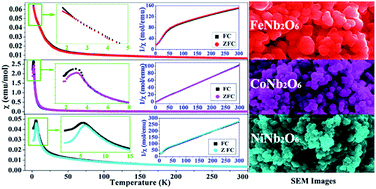
Considerable efforts have been exerted on the controllable synthesis of columbite niobate ceramics due to their fascinating properties and applications. Especially, it is still a great challenge to fabricate nanostructures of the niobate series. In this research, FeNb2O6, CoNb2O6 and NiNb2O6 nanoparticles have been successfully prepared via a facile hydrothermal route followed by heat treatment. X-ray powder diffraction patterns show that all the products have the typical orthorhombic columbite structure. The electron microscopy analyses reveal that the obtained nanoparticles have diameters of 50–100 nm. The magnetic property results demonstrate that the magnetically ordered state is hard to observe down to 1.8 K for the FeNb2O6 sample, while the magnetic transition temperatures of TN = 3 K and TN = 6 K can be obtained for CoNb2O6 and NiNb2O6 samples, respectively. A weak ferromagnetic moment can be detected below 5 K for both CoNb2O6 and NiNb2O6 samples. Furthermore, the NiNb2O6 sample even exhibits a metamagnetic transition at 1.8 K due to the spin flipping of the ferromagnetic chains.
Among the columbite niobate family, the ANb2O6 (A = Fe, Co, Ni) compounds, in which A refers to the magnetic transition metal, have greatly stimulated considerable research interest because of their prominent magnetic, dielectric, optical and catalytic properties.1,4–6 In the recent two decades, the low-dimensional magnetic systems have been the subject of intense theoretical and experimental research. Actually, these ANb2O6 (A = Fe, Co, Ni) niobates are considered as prototype materials since they exhibit quasi-one-dimensional magnetic characteristics.7–11 In general, ANb2O6 compounds exhibit magnetic ordering at rather low temperatures (typically below 10 K), and all the ordered phases have been found to be antiferromagnetic. Their magnetic structures have been investigated and identified by theoretical calculations and experimental measurements based on both polycrystalline and single-crystal samples. Weitzel first studied the magnetism for FeNb2O6 powder by neutron diffraction and reported the antiferromagnetic order at 4.2 K with a collinear spin arrangement.3 Later, according to the magnetization and susceptibility of FeNb2O6 single crystal, an antiferromagnetic ordering was observed at about 5.5 K by Yaeger et al.12 Even later, Heid et al. reinvestigated the magnetism in FeNb2O6 through a very detailed research based on both powder sample and single crystal.7 The magnetic structure of CoNb2O6 powder sample and single crystal was also first studied by Weitzel13 and Yaeger14 group, respectively, and later deeply investigated by Scharf et al.,15 Heid et al.,8,9 and Kobayashi et al.16,17 An intermediate incommensurate magnetic phase was observed below the Néel temperature of 2.95 K. Recently, Sarvezuk and co-workers performed a new investigation of the magnetic structure of CoNb2O6, in which the magnetic ordering at 2.5 K was found.18 As for NiNb2O6, it has a similar magnetic structure with FeNb2O6 and CoNb2O6, which has also been systematically studied by Weitzel et al.,19 Yaeger et al.,20 and Heid et al.7 It exhibits antiferromagnetic order below about 6 K with a canted magnetic structure.
Besides the magnetic behavior, the microwave dielectric properties of the ANb2O6 (A = Fe, Co, Ni) compounds have also drawn great attention for many research groups, such as Pullar,21–24 Hong,25,26 and Belous.27,28 These ceramic materials usually have the high dielectric constant and the low dielectric losses at microwave frequency making them suitable as dielectric resonator and filter for use in the field of mobile and satellite communication. Additionally, these dielectric ceramics have lower sintering temperatures than the perovskites. Arroyo y de Dompablo and co-workers presented a computational study on the stability and electronic properties of the reduced (anion vacancy type) ANb2O6 compounds.29 What's more, the electrochemical insertion properties of CoNb2O6 and NiNb2O6 as cathodes in lithium batteries were also studied.30 Zhou et al. have explored the photoluminescence of NiNb2O6 nanoparticles, which show a blue emission at 440 nm due to the distorted edge-shared NbO6 groups.5 The band gaps of NiNb2O6 was estimated to be 2.2 eV, which can be served as the solid photocatalyst in water splitting.31
There are various methods for the preparation of ANb2O6 (A = Fe, Co, Ni) compounds. Among them, the solid state combination has always been the most popular technique for the synthesis of polycrystalline ANb2O6 powders using stoichiometric mixtures of Nb2O5 and metal elements or oxides at high temperatures.3,4,7–11,13,18,19,21–28,30,31 However, multiple heating and regrinding steps are generally essential to overcome the solid state diffusion barrier, and a control of the atmosphere conditions is usually necessary. Additionally, the products always have irregular morphology and large particle size. NiNb2O6 nanoparticles have been synthesized by a sol–gel combustion method using citric acid as fuel and nitrates as oxidants at a relatively low temperature compared to the solid-state reaction method.5 Ravi group have prepared CoNb2O6 and NiNb2O6 ceramics by a coprecipitation technique using the mixture of ammonium carbonate and ammonium hydroxide as the precipitants under basic conditions.32,33 Recently, Ma et al. have fabricated CoNb2O6 with rutile structure via a hydrothermal route.34 As for the single-crystalline ANb2O6 (A = Fe, Co, Ni), the common growth methods include flux growth,12,14–17,20 and floating zone method.2
Motivated by the promising physicochemical properties and broad potential applications of ANb2O6 (A = Fe, Co, Ni) materials, it is interesting and challenging to explore a simple synthetic approach to fabricate ANb2O6 series, especially their nanostructures. In this research, a convenient and green hydrothermal process followed by heat treatment has been successfully employed for the preparation of ANb2O6 (A = Fe, Co, Ni) nanoparticles and the magnetic properties of the products have also been investigated.
Fig. 1 XRD patterns of the prepared columbite niobate ceramics via a general hydrothermal approach at 180 °C followed with annealing at 800 °C for (a) FeNb2O6, (b) CoNb2O6, and (c) NiNb2O6. |
It should be mentioned that all these three hydrothermal products without annealing are amorphous according to the XRD results (as shown in Fig. 2d–f). To understand the thermal behaviour of them, TG-DTA curves were investigated as displayed in Fig. 3. The TG-DTA curves of the three samples have a similar profile. From the TG curves (solid line), it can be observed that there are two steps of weight loss. The first weight loss below 300 °C is due to the evaporation of physically adsorbed water on the surface of the samples. The second weight loss of about 3% occurs in the range of 300–600 °C. If the samples are speculated as the amorphous hydrates, then the second weight loss may be ascribed to the dehydration of coordinated water. After 600 °C, then no obvious weight loss can be detected. As for the DTA curves (dash line), the initial endothermic peak is definitely resulted from the evaporation of water. However, the second endothermic peak corresponding to the dehydration process is absent probably due to the limited sensitivity of DTA. Additionally, at about 650–700 °C, a broad exothermic peak also can be observed, which implies the initiation of crystallization.
Fig. 3 TG (solid line) and DTA (dash line) curves of the hydrothermal products of FeNb2O6, CoNb2O6, and (c) NiNb2O6 without annealing. |
Fig. 4a and b respectively presents the SEM and TEM image of the FeNb2O6 sample after annealing at 800 °C in N2 atmosphere. From the SEM image, it can be seen that the sample consists of large-scale quasi-round nanoparticles with diameters of about 100 nm. The corresponding size distribution graph (inset of Fig. 4a) reveals that most nanoparticles have the sizes no more than 100 nm. However, there exists an obvious agglomeration of these nanoparticles to form the larger particles probably owing to the sintering effect during annealing. To further explore the microstructure of the obtained FeNb2O6 sample, the HRTEM and SAED analyses were undertaken from a single constituent nanoparticle. As shown in Fig. 4c, the HRTEM image shows clearly resolved two-dimensional atomic lattice fringes, suggesting a good crystallinity of these nanoparticles. The observed interplanar spacings can be measured to be 0.3 nm and 0.48 nm with a separation angle of about 70°, which match well with the (311) and (01) planes of orthorhombic columbite structure of FeNb2O6 phase.36 Fig. 4d presents the corresponding SAED patterns of the FeNb2O6 nanoparticle, which also confirms the good single-crystalline structure. These results further demonstrate that the prepared product consists of FeNb2O6 pure phase. The EDS spectrum taken from the nanoparticles is displayed in Fig. 4e, which shows the presence of Fe, Nb, O, Cu, C and Cr. The element Cu and C is derived from the copper grid and carbon film, respectively, while the element Cr should be originated from the TEM sample holder. The EDS spectrum shows that the atomic ratio of Fe:Nb:O is 1:2.03:8.16. The ratio of Fe to Nb is very close to 1:2 and the excessive O may be from the adsorbed O2 and/or H2O. So, it also confirms the formation of FeNb2O6 phase.
The SEM and TEM images of the CoNb2O6 sample after annealing are shown in Fig. 5a and b. It is also made up of round-like nanoparticles in the size range 50–100 nm, which is a little smaller than that of FeNb2O6 nanoparticles. In the HRTEM image as presented in Fig. 5c, the interplanar spacings can be measured to be 0.29 nm and 0.26 nm with a separation angle of about 75°, which can be indexed to the (311) and (20) planes of columbite CoNb2O6 phase.36 The corresponding SAED patterns are displayed in Fig. 5d, which also reveal the good single-crystalline nature of these CoNb2O6 nanoparticles. The EDS spectrum (Fig. 5e) indicates the presence of Co, Nb, O, Cu, C and Cr with the atomic ratio of Co:Nb:O = 1:2.1:7.28. All these results confirm the formation of columbite CoNb2O6 phase.
As to the NiNb2O6 sample, according to its SEM and TEM images presented in Fig. 6a and b, it is interesting that the further smaller nanoparticles with an average diameter of about 50 nm are obtained. From the TEM image, it can be observed that most nanoparticles have the diameter of about 50 nm and below, but there are still few particles with very large size. As displayed in Fig. 6c, the HRTEM image of the individual NiNb2O6 nanoparticle also reveals a good crystallinity, in which the observed interplanar spacings can be measured to be 0.29 nm and 0.23 nm with a separation angle of about 65°, which are reasonably consistent with the (311) and (12) planes of NiNb2O6 columbite structure.36 Accordingly, its SAED patterns are shown in Fig. 6d revealing the single-crystalline characteristic of the NiNb2O6 nanoparticles. The elemental analysis based on EDS spectrum presented in Fig. 6e demonstrates that the sample is composed of Ni, Nb, and O elements (Cu and C signals arise from the TEM grid, and Cr signal from the TEM sample holder), and the atomic ratio of Ni:Nb:O is 1:2.02:7.15, indicating the production of NiNb2O6 phase.
To further investigate the magnetic properties of the synthesized FeNb2O6 nanoparticles, the magnetization as a function of applied magnetic field was measured at different temperatures (1.8, 5, and 10 K) displayed in Fig. 7b. For the curve measured at 10 K, it can be found that the magnetization linearly increases with the applied field, signifying the typical paramagnetic phase, while the curve at 5 K slightly deviates from the linearity. When the measurement temperature is decreased to 1.8 K, the hysteresis loop tends to be an S-shaped line rather than the straight one, implying a possible magnetic order. Since there is still no opening of the magnetization hysteresis loop (as shown in the inset), i.e. no coercivity and remanence, it means that no ferromagnetic moment occurs and accordingly the system is approaching a magnetically ordered state at a still lower temperature, but also may be caused by the short-range correlations in a disordered state due to suppression of long-range order by frustration.10
Fig. 8a shows the thermal variation of the magnetic susceptibility for obtained CoNb2O6 nanoparticles. Both ZFC and FC curves have nearly the same shape. In the temperature region above 15 K, a very small magnetization associated with the existence of paramagnetic and spin-disorder states can be observed. After then, the magnetization steeply increases. At the very low temperature region, a close-up view of the ZFC and FC curves are displayed in the left inset panel. It should be noted that the ZFC and FC curves will reach a maximum and then decrease giving rise to a hump of magnetization at about 3 K suggesting a magnetic transition from paramagnetic to antiferromagnetic behavior, which should be denoted as the Néel temperature (TN = 3 K) of the prepared CoNb2O6 nanoparticles, consistently with what reported in literatures.13–18 The corresponding χ−1–T curves of the CoNb2O6 sample are presented in the right inset panel of Fig. 8a. Based on the Curie–Weiss law in the paramagnetic state, the Curie constant of C = 2.93 and the Weiss temperature of Θ = −2.72 K can be obtained, further confirming the antiferromagnetic exchange in the product. The effective magnetic moment can be calculated to be μeff = 4.86μB, which is in good accordance with the theoretical moment (4.8μB) for Co2+ based on both spin and orbital contribution.
The isothermal magnetization curves of CoNb2O6 nanoparticles measured at 1.8, 5, and 10 K are gathered in Fig. 8b. All the three M–H curves have an S-shaped profile. However, actually, the curves at 1.8 and 5 K show the opening hysteresis loops according to the corresponding zoom view at low magnetic fields as shown in the inset, which confirms the antiferromagnetic structure and the presence of a ferromagnetic moment. The coercivity (HC) is very small and can be determined as about 35 and 18 Oe for the hysteresis loop at 1.8 and 5 K, respectively. The magnetization increases steeply at low field region and rises relatively slowly above 10 kOe, but is not completely saturated even up to 60 kOe, further suggesting the predominant antiferromagnetic ordering. For the M–H curve at 10 K, the absence of opening in hysteresis loop, i.e. the zero coercivity and remanence, suggests the presence of superparamagnetic state. Accordingly, considering the finite size of the constituent nanoparticles, the possibility of superparamagnetic blocking may be expected in the sample.
The χ–T curves under both ZFC and FC conditions of the synthesized NiNb2O6 nanoparticles are shown in Fig. 9a. Similar with FeNb2O6 and CoNb2O6, the ZFC and FC curves undergo almost the same behavior and show a small magnetization due to the paramagnetic state in the high temperature region. With decreasing the temperature, the ZFC and FC curves maintain overlapped and rise sharply until a maximum magnetization in ZFC curve. Subsequently, the ZFC curve decreases suddenly causing a peak of magnetization at 6 K, while the FC curve reaches a maximum value at about 5.5 K. The separation between ZFC and FC magnetization curves at 6 K indicates a characteristic irreversibility, corresponding to the transition from paramagnetic to antiferromagnetic order. Therefore, the peak at 6 K in ZFC curve can be characterized as the Néel temperature (TN = 6 K) for the synthesized NiNb2O6 sample. This result is consistent with the values reported by Heid et al.7 and Yaeger et al.20 The susceptibility data also can be fitted by a least-squares method to the Curie–Weiss equation as given in the right inset panel of Fig. 9a. Accordingly, it is paramagnetic and obeys the Curie–Weiss law in the high temperature region above 25 K. As deduced from the fitting results, the value of C and Θ for NiNb2O6 sample is 1.31 and −42.78, respectively. The calculated effective magnetic moment is μeff = 3.25μB, which is quite close to the theoretical value (3.2μB) for Ni2+ considering both spin and orbital contribution.
Fig. 9b shows the magnetization vs. magnetic field plots for prepared NiNb2O6 nanoparticles measured at 1.8, 5, and 10 K. In all the three magnetization curves, no perfect saturation can be established even at a magnetic field of 6 T, corresponding to the antiferromagnetic ordering. However, it can be observed that in the M–H curve at the lower temperature, the magnetization exhibits a much slower linear increase with the applied magnetic field after 4 T. It is interesting that the magnetization curve at 1.8 K, as shown in Fig. 9c, not only shows the open hysteresis loop with a coercivity of about 200 Oe, but also exhibits a metamagnetic transition with a distinct change of slope at about 9 kOe, which is greatly similar to the results reported earlier by Heid et al.8 and Sarvezuk et al.10,11 Such a transition has been interpreted as the result from the spin flipping of the ferromagnetic chains with increasing the applied magnetic field, which are kept in an overall antiferromagnetic structure by very weak interchain interactions.10,11 Fig. 9d presents the close-up view of the hysteresis loops measured at 5 and 10 K. The evident opening can be observed in both curves indicating the existence of ferromagnetic moment, and the coercivity is about 125 and 85 Oe for the hysteresis loop at 5 and 10 K, respectively. This weak ferromagnetic component should arise from spin canting of the atomic magnetic moments and uncompensated spin at the surfaces of the NiNb2O6 nanoparticles.7,19,20 As Néel suggested, the magnetic moments tilt toward one another and the uncompensated surface spins possess a lower coordination number. Then a net magnetic moment can appear in antiferromagnetic nanoparticles owing to surface disorder and spin canting.39 Generally, the smaller size of the nanoparticles, the larger ratio of surface to volume, and then the more significant effects will be exerted on the magnetic behavior. To facilitate comparison, the magnetic data including the observed and reported Néel temperatures (TN), the observed, reported and theoretical effective magnetic moments (μB), and the coercivity fields (HC) at different temperatures of these prepared niobates are summarized in Table 1.
TN (K) (observed) | TN (K) (reported) | μeff(μB) (observed) | μeff(μB) (reported) | μeff(μB) (theoretical) | HC (Oe) (1.8 K) | HC (Oe) (5 K) | HC (Oe) (10 K) | |
---|---|---|---|---|---|---|---|---|
FeNb2O6 | <2 | 4.2 (ref. 3) | 5.42 | 5.42 (ref. 7 and 8) | 5.4 | — | — | — |
CoNb2O6 | 3 | 2.95 (ref. 5) | 4.86 | 4.40 (ref. 7) | 4.8 | 35 | 18 | — |
NiNb2O6 | 6 | 6 (ref. 17) | 3.25 | 3.30 (ref. 8) | 3.2 | 200 | 125 | 85 |
This journal is © The Royal Society of Chemistry 2014 |