Wentao Zhangab,
Shaoxuan Yua,
Wei Liua,
Daohong Zhanga,
Wenxin Zhua,
Yuhuan Zhanga,
Wanqiang Wua,
Lixue Zhang*b and
Jianlong Wang*a
aCollege of Food Science and Engineering, Northwest A&F University, Yangling 712100, Shaanxi, China. E-mail: wanglong79@yahoo.com; Fax: +86-29-87092275; Tel: +86-29-87092275
bQingdao Institute of Bioenergy and Bioprocess Technology, Chinese Academy of Sciences, No. 189 Songling Road, 266101 Qingdao, China. E-mail: zhanglx@qibebt.ac.cn; Fax: +86-532-80662744; Tel: +86-532-80662747
First published on 19th September 2014
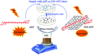
This work demonstrated that graphene oxide (GO) could not only be exploited as a nanovector to efficiently transfer π-conjugated polyene biomolecules from the organic phase to the aqueous phase, but also could enhance light-thermal stability and bioactivity of the transferred π-conjugated polyene biomolecules.
Astaxanthin (AST), a typical π-conjugated polyene biomolecule, is synthesized by phototrophic organisms and many non-phototrophic species (except animals). AST possesses various pharmacological activities both in vivo and in vitro, such as antioxidant activity, anti-inflammatory properties and enhanced cognitive function and immune response.12 As a result of its superior functions, AST has widespread applications in the medical, nutraceutical, cosmetic and aquaculture industries. However, the highly unsaturated structure of AST means that it is hydrophobic and susceptible to degradation during manufacture and storage, which severely debases its bioavailability and hampers its applications.13 In an attempt to solve this problem, several delivery systems, such as incorporation into emulsions, microencapsulation into chitosan matrix and encapsulation into polymeric nanospheres,12 have been proposed. However, these methods require complex synthesis progress of phase-transfer systems and have low transfer efficiency. Thus, an easy transfer system with high efficiency of AST loading for large-scale application is in demand.
Herein, we report a facile, low-cost and efficient approach, based on the phase-transfer capability of GO, to improve aqueous dispersity of AST. Although there are no reports of transfer of π-conjugated polyene biomolecules by GO, from a structural point of view, phase transfer is possible as GO can form π–π stacking interaction in the same way as nanotube encapsulates β-carotene,10 and there are hydrophobic interactions between them. Additionally, several –OH groups on biomolecules can form a strong hydrogen bond interaction with the –OH and –COOH groups on the GO sheet. Thus, it is possible to realize phase transfer of π-conjugated polyene biomolecules from organic phase to aqueous phase via non-covalent interaction between GO and AST to form a GO–AST complex (Fig. 1).
![]() | ||
Fig. 1 Schematic to illustrate the facile phase transfer of AST by GO via simple stirring to improve the properties of AST, such as poor water solubility, storage stability and antitumor activity. |
To demonstrate the feasibility of the proposed approach for phase transfer of π-conjugated polyene biomolecules, GO was prepared by oxidizing graphite using a modified Hummer's method.1 Then the phase transfer of AST was carried out by mixing AST dissolved in ethyl acetate with a GO water solution under a nitrogen atmosphere and stirring at 25 °C in the dark for 48 h. The mixing solution was left standing overnight for stratification (shown in Fig. S1b†), and then the aqueous phase was collected. The obtained solution was monitored by UV-vis spectroscopy, and the result is displayed in Fig. 2a. It reveals the characteristic AST peaks (near 491 nm) superimposing with the typical absorption of GO (at 230 nm), suggesting that AST was transferred by GO, forming a GO–AST phase-transfer complex. At the same time, different colors of AST, GO–AST and GO can be observed from the inset of Fig. 2a, visible evidence of the GO–AST phase-transfer complex.
Then, to further demonstrate formation of the GO–AST complex, a FTIR experiment was carried out. The FTIR spectrum of GO (Fig. 2b) reveals the characteristic bands of CO stretching vibrations from carbonyl and carboxyl groups around 1730 cm−1. The band at 1628 m−1 corresponds to C
C stretching and skeletal vibrations from un-oxidized graphitic domains. The band at 1380 cm−1 is the absorption band of O–H bending vibrations from hydroxyl groups. The peaks around 1229 cm−1 and 1073 cm−1 are attributed to C–OH stretching vibrations and stretching vibration of C–O in ethers and/or epoxides, respectively.14 The FTIR spectrum of pure AST shows characteristics of very strong absorption bands at about 1656 cm−1 for C
O stretching vibration, 1554 cm−1 for stretching vibration of C
C in the hexatomic ring, and 1281 and 1075 cm−1 for stretching vibrations of C–OH. The bands at 1367 cm−1 and 1037 cm−1 are attributed to symmetric deformation (CH deformation in plane) and rocking of methyl, respectively. The band at 966 cm−1 was assigned to C–H stretching vibration in π conjugate system and 671 cm−1 belongs to C–H stretching vibration in methyl.15 After GO interacted with AST, it could be seen that the FTIR spectrum of the GO–AST exhibited both the typical AST and GO absorption features. Additionally, FESEM and TEM were used to confirm fabrication of the complex. As presented in Fig. 2c, a number of flake-like nanostructures with smooth surface of GO are observed. For the GO–AST sample, however, the surface roughness increases (Fig. 2d), indicating that AST is loaded onto the surface of GO. A TEM image (Fig. 2e) illustrates the morphology of GO, which clearly exhibits the flake-like shapes of GO on the plane of which some corrugations and scrolling are observed. Whereas, when GO interacted with AST, as shown in Fig. 2f, some visual speckle-like nanostructures are clearly illustrated on the surface of GO. This could be because AST attached on the surface of GO sheet is carbonized at a high applied potential difference during the measurement process of FESEM and TEM.16,17 Taken together, all these results clearly confirm that AST molecules have been successfully transferred from ethyl acetate to water by the formation of a GO–AST complex.
After having successfully transferred AST from organic phase to aqueous phase by assembling GO–AST complex, the phase-transfer capacity of GO for AST was further evaluated. It should be pointed out that the amount of AST in the GO–AST complex could not be directly quantitated by the adsorption spectrum of GO–AST complex because of the severe background interference of GO. Thus, AST extraction from freeze-dried GO–AST (∼1.7 mg), as complete as possible, was conducted with dichloromethane prior to spectrometric analysis (Fig. S1c†). The extraction was then concentrated to dry under nitrogen, and redissolved in DMSO for UV-vis analysis of AST according to the standard curves of AST. Based on the regression equation and the absorbance value (0.520) of AST extracted from GO–AST complex in Fig. 3a, the concentration of AST in the final DMSO solution is 2.506 μg mL−1. Therefore, the concentration of AST in GO–AST stock solution is 50.12 μg mL−1, and 1 g GO can transfer ∼58.96 mg AST, which is similar to that of AST/β-cyclodextrin inclusion complexes prepared at 25 °C.18 Thus, AST can be efficiently transferred from the organic phase to the aqueous one by GO. The strong phase transfer of GO could be attributed to two aspects: AST molecules are largely carried on the surface of GO by π–π stacking interaction and hydrophobic interactions; and abundant –COOH and –OH on the surface of GO mean GO can still disperse in water after the loading of AST. That is to say, the solubility of AST in water is highly improved with the aid of GO, which could broaden potential applications restricted by its poor water-solubility.
The special structure of π-conjugated polyene biomolecules gives AST potent bioactivities, but, unfortunately, is also the main reason applications of AST are limited because the structure is easily degraded by light and heat during the process of manufacture and storage of AST.12 An interesting phenomenon found here is that the light-thermal stability of AST is obviously enhanced in the GO–AST complex. The stability profile was examined by monitoring changes of absorbance at 491 nm at known time intervals, and results are shown in Fig. 3b–d. When stored at 4 °C in dark for 20 days (Fig. 3b), free AST and AST in GO–AST complex decay with tiny degradation rates of 1.29% and 0.23%, respectively. When stored at 37 °C in dark for 20 days (Fig. 3c), 14.66% of native AST is decomposed, but only 2.87% of AST in the complex is degraded. When stored at 37 °C in natural light for 20 days (Fig. 3d), more severe decay occurs. The free AST is decreased to 65.52%, but only 18.72% of AST in the complex is degraded. Although AST molecules from both of samples are degraded during the process of storage, there are remarkable differences in the degradation rates. In other words, AST immobilized on GO shows higher stability and lower degradation rate than free AST, which agrees well with the result of Chen et al.19 The enhanced thermal stability could be attributed to the presence of a non-covalent bond between AST and GO, suppressing the mobility of AST chains and preventing conformational denaturation of AST at higher temperature, which is similar to the protection effect of nanomaterials for pea starch and redox enzyme.20,21 Furthermore, it is reported that GO has remarkable optical absorptivity over a wide wavelength range (visible-IR).22 Hence, when surrounded by light and heat, GO can be a structure suppresser and a photon harvester to help prevent destruction of AST and improve its light-thermal stability, which could be of benefit for extending the applied field of AST.
Although we have demonstrated that GO can greatly enhance the solubility and light-thermal stability of transferred AST, biological properties of the transferred AST in GO–AST are still questionable. AST is assumed to be the natural substance with the strongest antioxidant effect.23 Thus, the antioxidant activity of the as-prepared GO–AST complex was firstly evaluated by a DPPH method. As illustrated in Fig. 4a, the maximum absorbance of DPPH that appeared near 520 nm was gradually decreased with increasing GO–AST concentration. The DPPH scavenging activity of free AST was also assayed and compared with that of AST in GO–AST (Fig. 4b). The DPPH scavenging rates of AST and AST in GO–AST were 8.13% and 8.55% at 0.2 μg mL−1, respectively, and increased to 56.48% and 59.55% accordingly, when the concentration increased to 2.0 μg mL−1. The DPPH scavenging ability of both AST and AST in GO–AST shows a positive correlation with concentration in the evaluated range. However, comparing the antioxidant activity of AST and AST in GO–AST at the same concentration, AST in GO–AST presents slightly stronger antioxidant capacity than that of pure AST [note: the DPPH scavenging rate of AST in GO–AST in Fig. 4b was calculated by eqn (2) shown in the ESI† to eliminate the interference of GO]. The slight enhancement of antioxidant capacity could be attributed to formation of the intermolecular hydrogen bond between the hydroxyl and carboxyl groups of GO and AST, which inhibits formation of the five-membered intramolecular hydrogen bond of AST, and thus enhances the antioxidant activity of AST in complex (Fig. S2†).24,25
Finally, a cytotoxicity test for the GO–AST complex was carried out using human hepatoma cell line (HepG2) to further investigate the bioactivity of AST in the complex via CCK-8 assay. At the same time, the cell morphological changes after each of treatments were imaged to track in situ the status of cells. Firstly, the toxicity of dialyzed GO was investigated. As shown in Fig. 4c, no obvious cytotoxicity was observed for GO even at a concentration of 200 μg mL−1. Consistently, no obvious morphological difference was observed between the GO-treated cells (Fig. 5b) and the control cells (Fig. 5a). These results indicate that GO sheets themselves have no cytotoxicity to HepG2 cells and would not interfere with anticancer activity determination of AST, which accords well with previous reports.26,27 The antitumor activity of GO–AST was then explored. The optical microscopic observation displayed in Fig. 5c and d clearly shows that both AST and GO–AST-treated cells become circular, and are smaller than control cells, which indicates that AST and GO–AST have obvious effects on cells. Moreover, as shown in Fig. 4d, both AST and GO–AST result in a dose-dependent toxicity in HepG2 cells. However, treatment with GO–AST markedly reduces the cell viability by 35.81% at 10 μg mL−1 compared with the control cells, while only 28.1% is reduced by AST. It means GO–AST possesses potent cancer cell killing ability, which is stronger than that of pure AST. The improved cytotoxic effect on the cancer cell may be a result of adsorption of GO onto the cellular surface and higher cell penetration of GO, related to amphiphilicity of GO for improved cell wall interaction compared with absolute hydrophobic AST.26,27 These results indicate that GO could not only transfer AST from organic phase to aqueous phase, but also enhance the bioactivity of AST by formation of AST–GO complex.
![]() | ||
Fig. 5 Optical microscopy images of HepG2 cells with different treatment, (a) the control; (b) GO; (c) AST; (d) GO–AST. |
Footnote |
† Electronic supplementary information (ESI) available. See DOI: 10.1039/c4ra08229j |
This journal is © The Royal Society of Chemistry 2014 |