
A CdTe/CdS/ZnS core/shell/shell QDs-based “OFF–ON” fluorescent biosensor for sensitive and specific determination of L-ascorbic acid
Shan Huangab,
Fawei Zhua,
Qi Xiao*abc,
Wei Sua,
Jiarong Shenga,
Chusheng Huanga and
Baoqing Hub
aCollege of Chemistry and Life Science, Guangxi Teachers Education University, Nanning 530001, P. R. China. E-mail: qi.xiao@whu.edu.cn; Fax: +86 771 3908065; Tel: +86 771 3908065
bKey Laboratory of Beibu Gulf Environment Change and Resources Utilization (Guangxi Teachers Education University), Ministry of Education, P. R. China
cState Key Laboratory of Virology, Wuhan University, P. R. China
First published on 5th September 2014
Abstract
Herein we report a quantum dots (QDs)-based “OFF–ON” fluorescent biosensor for the sensitive and specific determination of L-ascorbic acid. The proposed one-pot L-ascorbic acid detection method is quite simple, rapid and convenient because of the elimination of the modification and separation procedures. In this contribution, the N-acetyl-L-cysteine (NAC)-capped CdTe/CdS/ZnS core/shell/shell QDs were synthesized in aqueous phase. Subsequently, KMnO4 was added into solution and attached to the QDs surface to effectively quench the fluorescence of the QDs, which rendered the QDs into fluorescence “OFF” status. After the addition of L-ascorbic acid into the QDs–KMnO4 system, the fluorescence of the QDs was then turned “ON” because L-ascorbic acid could bind with KMnO4 and break KMnO4 away from the surface of the QDs. Under the optimized conditions, the relative restored fluorescence intensity was directly proportional to the concentration of L-ascorbic acid in the range of 8.0 × 10⁻⁹ M to 1.0 × 10⁻⁷ M, with a correlation coefficient of 0.9971 and a limit of detection of 1.8 × 10⁻⁹ M. The relative standard deviation for 6.0 × 10⁻⁸ M L-ascorbic acid was 2.1% (n = 5). There was almost no interference from few common ions, carbohydrates, nucleotides and amino acids. The proposed method was applied to the determination of L-ascorbic acid in three synthetic samples, human urine samples and vitamin C tablets with satisfactory results. The possible fluorescence quenching mechanism of this fluorescent sensor was further investigated by UV-vis spectroscopy.
1. Introduction
Because the specific optical properties are dramatically different from those in bulk semiconductors, the water-soluble semiconductor quantum dots (QDs) have attracted significant attention as novel fluorescent biosensors in the past two decades.1–5 Some bio-related small molecules, nucleic acids, proteins and enzymes have been detected using fluorescent QDs as the probe based on the fluorescence quenching or the fluorescence enhancement of QDs.6–10 However, both the fluorescence quenching and the fluorescence enhancement of QDs belong to the unidirectional fluorescence variations of QDs, which make fluorescent QDs biosensors more likely to be affected by foreign substances. Hence, it is important to explore new strategies to increase the selectivity and expand the bio-applications of fluorescent QDs biosensors.Recently, the fluorescence “OFF–ON” sensors based on QDs have been explored and realized simple and sensitive determination of some chemical substances.11–13 In addition, this fluorescence “OFF–ON” mode has been used to detect some bio-related small molecules and biomacromolecules.14–22 Zhu and co-workers demonstrated an “OFF–ON” approach for the detection of both Cu2+ and L-cysteine using fluorescent carbon dot probes.14 Yi et al. reported a dual-mode nanosensor with both colorimetric and fluorometric readout based on carbon QDs and gold nanoparticles for specific detection of glutathione.15 Su's group established a novel fluorescence “TURN OFF–ON” nanosensor for the determination of heparin and heparinase based on CuInS2 QDs.16 Renganathan et al. described a novel platform for detecting double stranded DNA (dsDNA) by tracing the “ON–OFF–ON” fluorescence signals of a QDs–porphyrin system.17 Xie et al. proposed a new dsDNA detection method relying on the single-color fluorescence “OFF–ON” switch system that was composed of CdTe QDs and Ru(phen)2(dppz)2+.18 Li and co-workers also applied this QDs-based fluorescence “OFF–ON” model for the detection of both anticancer drugs and dsDNA.21,22 Because of the high selectivity and sensitivity, this novel QDs-based “OFF–ON” fluorescent sensor showed great potential for applications in biochemical and biomedical determinations.
It is well-known that L-ascorbic acid is a hexanoic sugar acid with a γ-lactone structure and two dissociable protons (pKa 4.04 and 11.34); thus, L-ascorbic acid occurs as an ascorbate anion under physiological conditions. Because of the high reductive properties, L-ascorbic acid is largely used as a powerful antioxidant in free-radical induced disease therapy, since L-ascorbic acid can easily balance the oxidative stress of the human body.23,24 In addition, L-ascorbic acid can take crucial part in some biochemical processes and can also serve as an effective drug for few diseases.25,26 However, an excess of L-ascorbic acid can lead to taste/aroma deterioration, diarrhea, kidney calculi and gastric irritation.27 Because of the important role of L-ascorbic acid in biochemistry and biomedical applications, the detection of L-ascorbic acid has become more important and has attracted considerable interest in recent years. Various analytical techniques have been reported for the detection of L-ascorbic acid in biological and pharmaceutical samples, such as spectrophotometry,28 enzymatic analysis,29 electroanalysis,30 chemiluminescence,31 colorimetry,32–34 phosphorimetry35 and liquid chromatography.36 However, some of these methods are time-consuming, complicated and expensive. Thus, there is an urgent demand for simple and rapid biosensors for the determination of L-ascorbic acid with high sensitivity and specificity in clinical analysis and the pharmaceutical industry. In recent years, fluorescence analysis has been widely utilized for L-ascorbic acid determination because of its unique advantages of simplicity, rapidity, high sensitivity and low cost of instrumentation and maintenance. Yan and co-workers reported a CdTe QDs-based “TURN-ON” fluorescent sensor for L-ascorbic acid detection.37 This approach avoids the complicated modification process of QDs, and opens a simple strategy to develop cost-effective, sensitive and selective QD-based fluorescence “TURN-ON” sensors for biologically significant antioxidants.
In the present study, the N-acetyl-L-cysteine (NAC)-capped CdTe/CdS/ZnS core/shell/shell QDs were synthesized in aqueous phase.38 Since the ligand NAC is water-soluble, environmentally friendly and possesses good biocompatibility, the as-synthesized NAC-capped CdTe/CdS/ZnS core/shell/shell QDs are of crucial importance for many biomedical applications. Herein, we establish a sensitive and specific NAC-capped CdTe/CdS/ZnS core/shell/shell QDs-based “OFF–ON” fluorescent biosensor for the determination of L-ascorbic acid by utilizing KMnO4 as both the quencher to QDs and the oxidizing/binding agent to L-ascorbic acid (Scheme 1). Because KMnO4 can attach to the surface of QDs and subsequently quench the fluorescence of QDs through the electron transfer process, the QDs are then switched to the fluorescence “OFF” status. After the addition of L-ascorbic acid, KMnO4 can react and combine with L-ascorbic acid through a redox and coordination reaction;39 thus, interrupting the electron transfer from QDs to KMnO4 and the fluorescence of QDs is switched “ON” again. This QDs-based “OFF–ON” fluorescent biosensor can be used to detect L-ascorbic acid with the properties of simplicity, sensitivity and specificity. Furthermore, the QDs-based “OFF–ON” fluorescent biosensor has been successfully applied to the detection of L-ascorbic acid in three synthetic samples, human urine samples and vitamin C tablets with satisfactory results.
 | ||
| Scheme 1 Principle of L-ascorbic acid determination using NAC-capped CdTe/CdS/ZnS core/shell/shell QDs-based “OFF–ON” fluorescent sensor. | ||
2. Materials and methods
2.1 Materials
Te powder (200 mesh, 99.8%), NaBH4 (99.8%), CdCl2·H2O (99.99%), NAC (≥99%), rhodamine 6G were purchased from Sigma-Aldrich (St. Louis, MO, USA). Na2S, ZnCl2, 2-propanol, KMnO4, L-ascorbic acid, lactose, sucrose, glucose, four nucleotide acids and twenty amino acids were obtained from Sinopharm Chemical Reagent Factory (Shanghai, China). All other reagents were of analytical-reagent grade and used as received. Ultrapure water with a resistivity of 18.2 MΩ cm was produced by passing through a RiOs 8 unit followed by a Millipore-Q Academic purification set (Millipore, Bedford, MA, USA) and used throughout the entire experiment.2.2 Apparatus
The absorption spectra were measured on TU-1901 UV-vis spectrophotometer (Beijing Purkinje General Instrument Co., Ltd., Beijing, China). All fluorescence spectra and intensities were recorded with Perkin-Elmer Model LS-55 luminescence spectrometer (Perkin-Elmer, Waltham, MA, USA) equipped with a 20 kW xenon discharge lamp as light source. Quartz cells (1 cm path-length) were used for all the measurements. All pH measurements were made with a basic pH meter PB-10 (Sartorius Scientific Instruments Co., Ltd., Beijing, China).2.3 Preparations of NAC-capped CdTe/CdS/ZnS core/shell/shell QDs
NAC-capped CdTe/CdS/ZnS core/shell/shell QDs were synthesized according to the method we previously reported.38 In brief, 0.2 mmol of Te powder and 1.0 mmol of NaBH4 were put into a two-necked flask equipped with a constant pressure funnel containing 5.0 mL of ultrapure water. Then, the air was pumped off and replaced by nitrogen. After that, the mixture was heated to 80 °C and placed for 30 min under nitrogen protection until the solution became dark red. The obtained NaHTe solution was stored under nitrogen protection for further use at room temperature. Then, 0.2 mmol of CdCl2 and 0.34 mmol of NAC solution were mixed in a 40 mL solution and the pH of the mixture was adjusted to 12.0 by dropwise addition of 1.0 M NaOH solution with stirring. Then, the mixture was transferred into a three-necked flask and the air in the system was replaced with nitrogen. 1 mL of NaHTe solution (0.04 mmol) was added into the Cd precursor solution by syringe with stirring at room temperature, which made the molar ratio of Cd![[thin space (1/6-em)]](https://www.rsc.org/images/entities/char_2009.gif) :Te:NAC to be 1.0:0.2:1.7. Then, the mixture was heated to 100 °C and reacted at this temperature for 8 min. Next, heat was immediately removed and the mixture was cooled to room temperature. In order to remove the excess NAC–Cd complexes at the end of the synthesis, cold 2-propanol was added to the reaction mixture to precipitate CdTe core QDs. The as-prepared precipitate was redispersed in ultrapure water.
:Te:NAC to be 1.0:0.2:1.7. Then, the mixture was heated to 100 °C and reacted at this temperature for 8 min. Next, heat was immediately removed and the mixture was cooled to room temperature. In order to remove the excess NAC–Cd complexes at the end of the synthesis, cold 2-propanol was added to the reaction mixture to precipitate CdTe core QDs. The as-prepared precipitate was redispersed in ultrapure water.
The CdTe/CdS precursor solution was prepared by adding the as-prepared NAC-capped CdTe core QDs to a nitrogen-saturated solution containing 1.0 mmol CdCl2, 0.2 mmol Na2S and 5.0 mmol NAC. The CdTe/CdS precursor solution (40 mL) was placed in a three-necked flask. The air in the system was pumped off and replaced with nitrogen. Then, the mixture was heated to 100 °C and reacted at this temperature for 15 min. NAC-capped CdTe/CdS core/shell QD samples were taken when the temperature had cooled to room temperature. Cold 2-propanol was added to precipitate the NAC-capped CdTe/CdS core/shell QDs and the as-prepared precipitate was redispersed in ultrapure water.
The CdTe/CdS/ZnS precursor solution was prepared by adding the as-prepared NAC-capped CdTe/CdS core/shell QDs to a nitrogen-saturated solution containing 1.0 mmol ZnCl2, 0.2 mmol Na2S and 5.0 mmol NAC. The CdTe/CdS/ZnS precursor solution (40 mL) was placed in a three-necked flask. The air in the system was pumped off and replaced with nitrogen. Then, the mixture was heated to 70 °C and reacted at this temperature for 10 min. NAC-capped CdTe/CdS/ZnS core/shell/shell QD samples were taken when the temperature had cooled to room temperature. Cold 2-propanol was added to precipitate NAC-capped CdTe/CdS/ZnS core/shell/shell QDs, which were dried overnight under vacuum at 30 °C and stored in a refrigerator for further experiments. The concentration of the water-soluble NAC-capped CdTe/CdS/ZnS core/shell/shell QDs was estimated from the absorption spectra using the molar absorptivity at the first absorption maximum for QDs reported by Peng and co-workers.36
2.4 Preparation of three synthetic samples and human urine samples
For the synthetic samples detection, three different samples were prepared by mixing standard solutions of each sample with different concentrations in the reaction system. Sample 1 contained twenty amino acids and their concentration were all 1.0 × 10⁻⁵ M. Glucose, sucrose, lactose and urea in sample 2 were all 2.0 × 10⁻⁴ M. Adenine, cytosine, thymine and guanine in sample 2 were all 1.0 × 10⁻⁵ M. The concentrations of KNO3, NaNO3, Ca(C2O4)2, Mg(NO3)2, Al(NO3)3, CuSO4 in sample 3 were all 1.0 × 10⁻⁴ M. Different amounts of L-ascorbic acid standard solution were added into the reaction system and the final concentration of L-ascorbic acid was 1.5 × 10⁻⁸ M in sample 1, 5.0 × 10⁻⁸ M in sample 2 and 8.0 × 10⁻⁸ M in sample 3. The recovery of L-ascorbic acid in the three synthetic samples was examined by the proposed method.Human urine samples were obtained from five healthy volunteers (two women and three men, age range 25–35 years) and prepared as reported before.6 Fresh samples of 500 μL each were taken and combined with 1 mL solutions (containing 4.0 mM Na2EDTA4 and 2.0 M HClO4) in a 2 mL eppendorf tube, for the proteins to separate. After maintaining for 30 min to precipitate proteins, the sample was centrifuged for 15 min at 4500 rpm. The supernatant liquid of 500 μL was adjusted to pH 7.8 by NaOH solution and then diluted to 2 mL with 0.01 M pH 7.8 phosphate buffer. After homogenizing, the sample was filtered with 0.22 μm Millipore membranes (Millipore, Bedford, MA, USA). The filtrate was collected and stored at 4 °C (three days) until fluorescence analysis. Human urine samples were diluted 1000-fold with ultrapure water before analysis.35
Vitamin C tablets were produced by Shanghai Quanyu Biological Technology Suiping Pharmaceutical Co., Ltd. (Suiping, China). The content of L-ascorbic acid in each vitamin C tablet is about 0.1 g. Five pieces of vitamin C tablets were mixed and powdered in a mortar. The vitamin C tablets were weighed and the average weight of each tablet was calculated to be 0.12 g. 0.06 g powder of vitamin C tablet was dissolved in 10 mL ultrapure water. The solution was filtered with 0.22 μm Millipore filter to remove the insoluble components. Next, the filtrate was transferred into a 250 mL volumetric flask and diluted to 250 mL with ultrapure water. The pre-treated vitamin C tablet solution was stored in the dark at 4 °C and was diluted 5000-fold with ultrapure water before analysis.
2.5 L-Ascorbic acid detection
As depicted in Scheme 1, first, 50 μL of 1.0 × 10⁻⁵ M of NAC-capped CdTe/CdS/ZnS core/shell/shell QDs, 1.5 mL Tris–HCl (pH 7.4) and the appropriate aliquot of KMnO4 solution were transferred into a 5 mL eppendorf tube. The mixture was thoroughly stirred and finally diluted to 5 mL with ultrapure water. After 5 min of the reaction at room temperature, the fluorescence spectra were measured for the selection of the appropriate KMnO4 concentration.For L-ascorbic acid detection, 50 μL of 1.0 × 10⁻⁵ M of NAC-capped CdTe/CdS/ZnS core/shell/shell QDs, 1.5 mL Tris–HCl (pH 7.4) and 30 μL of 3.0 × 10⁻⁴ M of KMnO4 solution were transferred into a 5 mL eppendorf tube and the mixture was thoroughly stirred. After incubation for 5 min, the appropriate aliquot of L-ascorbic acid solution were added into the mixture and finally diluted to 3 mL with ultrapure water. After additional 15 min incubation at room temperature, the fluorescence spectra were measured for the quantitative analysis of L-ascorbic acid. When the samples were determined, the L-ascorbic acid standard solution was substituted by the prepared sample solution described in Section 2.4.
The fluorescence spectra were recorded at excitation wavelength of 388 nm and the band-slits of excitation and emission were set as 10.0 nm. The fluorescence spectra were recorded from 535 nm to 725 nm, the fluorescence intensity of NAC-capped CdTe/CdS/ZnS core/shell/shell QDs at 622 nm was used for quantitative analysis of L-ascorbic acid.
3. Results and discussion
3.1 Properties of NAC-capped CdTe/CdS/ZnS core/shell/shell QDs
The normalized UV-vis absorption and fluorescence spectra of NAC-capped CdTe/CdS/ZnS core/shell/shell QDs at room temperature are shown in Fig. 1. The first absorption maximum wavelength of NAC-capped CdTe/CdS/ZnS core/shell/shell QDs was at 578 nm (Fig. 1a), and the particle sizes of these QDs were calculated to be 3.5 nm according to the first absorption maximum wavelength of the QDs.40 It could be seen from Fig. 1b that these NAC-capped CdTe/CdS/ZnS core/shell/shell QDs exhibited obvious and symmetrical fluorescent spectrum without a tail on the right-hand side, and the emission maximum wavelength of these QDs was at 622 nm when the excitation wavelength was 388 nm. The line width of the fluorescence spectrum of these NAC-capped CdTe/CdS/ZnS core/shell/shell QDs was narrow, indicating that the as-prepared QDs were nearly monodisperse and homogeneous. The fluorescence quantum yield of these QDs was calculated to be about 42% using rhodamine 6G as the fluorescence standard, according to a study reported earlier.41 | ||
| Fig. 1 Normalized UV-vis absorption (a) and fluorescence (b) spectra of NAC-capped CdTe/CdS/ZnS core/shell/shell QDs at room temperature. Inset: images of the emission color of NAC-capped CdTe/CdS/ZnS core/shell/shell QDs under the white lamp (left) and the radiation of UV lamp (right). | ||
3.2 Characterization of QDs-based fluorescence “OFF–ON” mode
The fluorescence of NAC-capped CdTe/CdS/ZnS core/shell/shell QDs can be effectively quenched by KMnO4, which render QDs into fluorescence “OFF” status. The fluorescence spectra of NAC-capped CdTe/CdS/ZnS core/shell/shell QDs with different concentrations of KMnO4 are shown in Fig. 2. It could be seen from Fig. 2A that the fluorescence of NAC-capped CdTe/CdS/ZnS core/shell/shell QDs was quenched after the addition of KMnO4. Because 1.7 × 10⁻⁶ M, 2.3 × 10⁻⁶ M and 3.0 × 10⁻⁶ M of KMnO4 presented a quenching effect of 26%, 51% and 93%, respectively, it could also be deduced easily that the fluorescence quenching effect for the NAC-capped CdTe/CdS/ZnS core/shell/shell QDs–KMnO4 system was mainly concentration dependent. Furthermore, the fluorescence peaks of the NAC-capped CdTe/CdS/ZnS core/shell/shell QDs–KMnO4 system were still at 622 nm without any shift, which confirmed the inferred electron transfer mechanism between NAC-capped CdTe/CdS/ZnS core/shell/shell QDs and KMnO4.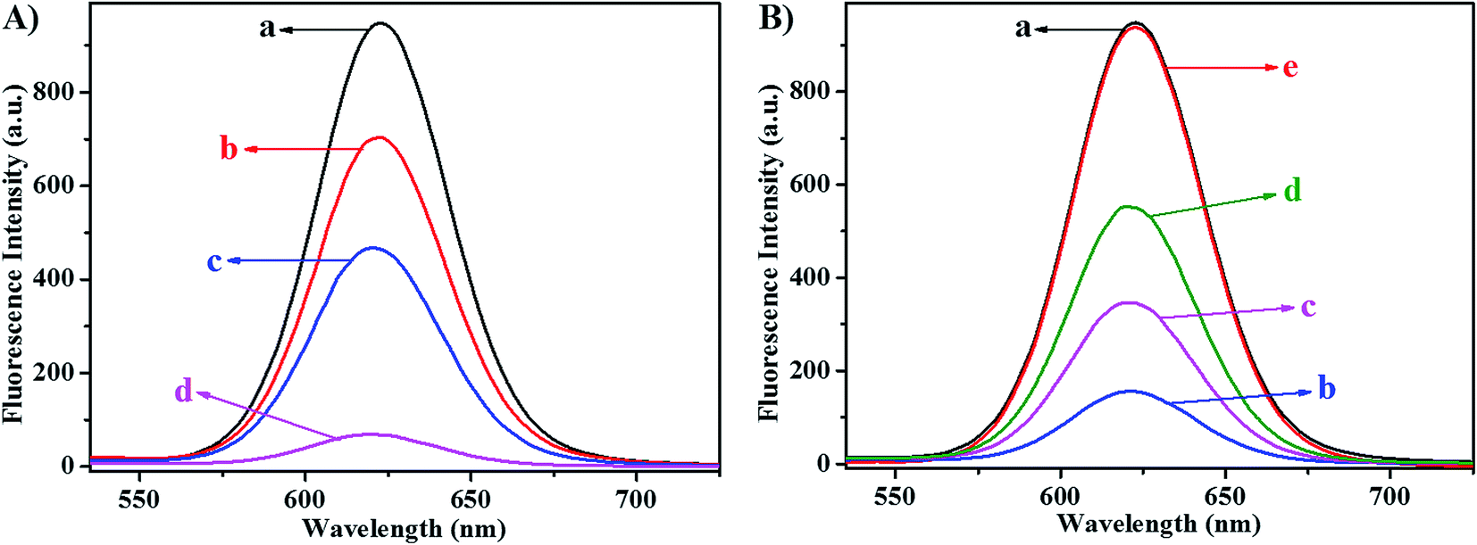 | ||
| Fig. 2 (A) Fluorescence spectra of NAC-capped CdTe/CdS/ZnS core/shell/shell QDs at different concentrations of KMnO4 in Tris–HCl buffer (1.5 mL, pH 7.4). NAC-capped CdTe/CdS/ZnS core/shell/shell QDs: 1.7 × 10⁻⁷ M. KMnO4: (a) 0; (b) 1.7 × 10⁻⁶ M; (c) 2.3 × 10⁻⁶ M; (d) 3.0 × 10⁻⁶ M. (B) Fluorescence spectra of NAC-capped CdTe/CdS/ZnS core/shell/shell QDs alone (a), NAC-capped CdTe/CdS/ZnS core/shell/shell QDs with 1.0 × 10⁻⁶ M L-ascorbic acid (e), and NAC-capped CdTe/CdS/ZnS core/shell/shell QDs–KMnO4 system with different concentrations of L-ascorbic acid. NAC-capped CdTe/CdS/ZnS core/shell/shell QDs: 1.7 × 10⁻⁷ M; KMnO4: 3.0 × 10⁻⁶ M; L-ascorbic acid: (b) 8.0 × 10⁻⁸ M; (c) 5.0 × 10⁻⁷ M; and (d) 1.0 × 10⁻⁶ M. | ||
The fluorescence of NAC-capped CdTe/CdS/ZnS core/shell/shell QDs was switched “ON” after the addition of L-ascorbic acid into the QDs–KMnO4 system. The fluorescence spectra of NAC-capped CdTe/CdS/ZnS core/shell/shell QDs–KMnO4 system with different concentrations of L-ascorbic acid are shown in Fig. 2B. According to Fig. 2B, the fluorescence intensity of NAC-capped CdTe/CdS/ZnS core/shell/shell QDs was gradually restored with increase in the concentration of L-ascorbic acid. Because the fluorescence restoring effect was mainly dependent on the L-ascorbic acid concentration and the addition of 8.0 × 10⁻⁸ M, 5.0 × 10⁻⁷ M and 1.0 × 10⁻⁶ M of L-ascorbic acid presented a restoring effect of 16%, 36% and 58%, respectively, the fluorescence restoration approach could be used for the sensitive detection of L-ascorbic acid. Furthermore, the fluorescence intensity of NAC-capped CdTe/CdS/ZnS core/shell/shell QDs was not affected by 1.0 × 10⁻⁶ M L-ascorbic acid, validating the specific interaction between KMnO4 and L-ascorbic acid that resulted in the fluorescence “ON” status of QDs.
3.3 Effect of reaction time
The effect of reaction time on the fluorescence quenching of NAC-capped CdTe/CdS/ZnS core/shell/shell QDs by KMnO4 was investigated. Preliminary experiments demonstrated that the fluorescence quenching of NAC-capped CdTe/CdS/ZnS core/shell/shell QDs by KMnO4 was completed within 3 min and the fluorescence signals remained constant for more than 60 min, indicating that the NAC-capped CdTe/CdS/ZnS core/shell/shell QDs–KMnO4 system exhibited good stability. Thus, the fluorescence intensity of NAC-capped CdTe/CdS/ZnS core/shell/shell QDs–KMnO4 system was recorded after the addition of KMnO4 for 5 min.The effect of reaction time on the fluorescence of NAC-capped CdTe/CdS/ZnS core/shell/shell QDs–KMnO4 system by L-ascorbic acid was also studied. The results indicated that the fluorescence restoration of NAC-capped CdTe/CdS/ZnS core/shell/shell QDs by L-ascorbic acid was completed within 10 min and lasted for more than 90 min, which indicated that the NAC-capped CdTe/CdS/ZnS core/shell/shell QDs–KMnO4–L-ascorbic acid system exhibited higher stability. Therefore, the fluorescence intensity of NAC-capped CdTe/CdS/ZnS core/shell/shell QDs–KMnO4–L-ascorbic acid system was measured after adding L-ascorbic acid for 15 min.
3.4 Influence of pH value and buffer volume
Because the pH value of the solution played an important role in the interaction of QDs with other molecules,6,42 the influence of different pH values on the fluorescence intensity reflecting the interaction of both NAC-capped CdTe/CdS/ZnS core/shell/shell QDs with KMnO4 and NAC-capped CdTe/CdS/ZnS core/shell/shell QDs–KMnO4 system with L-ascorbic acid was investigated from pH 6.5 to 10.0. The variation in fluorescence intensities between the QDs–KMnO4 system and the QDs–KMnO4–L-ascorbic acid system is shown in Fig. 3A. It was found that the change in fluorescence intensity gradually increased with the increase in pH value from 6.5 to 7.4. When pH value was higher than 7.4, the change in fluorescence intensity decreased dramatically. The maximum change in fluorescence intensities was observed when pH value was 7.4. Therefore, Tris–HCl buffer with pH 7.4 was chosen for further experiments. | ||
| Fig. 3 The influences of pH value (A), buffer volume (B) and QDs volume (C) on the fluorescence intensity response of the system. (A) NAC-capped CdTe/CdS/ZnS core/shell/shell QDs: 1.7 × 10⁻⁷ M; KMnO4: 3.0 × 10⁻⁶ M; L-ascorbic acid: 1.0 × 10⁻⁷ M; buffer: 1.5 mL Tris–HCl buffer. (B) NAC-capped CdTe/CdS/ZnS core/shell/shell QDs: 1.7 × 10⁻⁷ M; KMnO4: 3.0 × 10⁻⁶ M; L-ascorbic acid: 1.0 × 10⁻⁷ M; buffer: pH 7.4 Tris–HCl buffer. (C) KMnO4: 3.0 × 10⁻⁶ M; buffer: 1.5 mL Tris–HCl buffer (pH 7.4). | ||
Simultaneously, the impact of the volume of Tris–HCl buffer on the fluorescence intensities between NAC-capped CdTe/CdS/ZnS core/shell/shell QDs–KMnO4 system and NAC-capped CdTe/CdS/ZnS core/shell/shell QDs–KMnO4–L-ascorbic acid system was also investigated. As shown in Fig. 3B, the change in fluorescence intensity gradually increased with the increase in Tris–HCl buffer volume from 0 to 1.5 mL. When the volume of Tris–HCl buffer was higher than 1.5 mL, the change of fluorescence intensity gradually decreased. The results indicated that the maximum change in fluorescence intensities occurred when the volume of Tris–HCl buffer was 1.5 mL; thus, 1.5 mL Tris–HCl buffer was selected as the reaction medium.
3.5 Influence of QDs volume
The influence of NAC-capped CdTe/CdS/ZnS core/shell/shell QDs volume on the fluorescence “OFF–ON” systems was also tested. The results of the previous experiment indicated that the quench efficiency of the fluorescence intensity of QDs by KMnO4 obviously increased when the volume of QDs was in the range of 10–50 μL. The quenching efficiency gradually decreased when the volume of QDs was higher than 50 μL, because higher QDs volume would disturb the electrostatic balance of QDs themselves and affect the electron transfer process from QDs to KMnO4. Because the maximum change in the fluorescence intensities between NAC-capped CdTe/CdS/ZnS core/shell/shell QDs and NAC-capped CdTe/CdS/ZnS core/shell/shell QDs–KMnO4 system appeared when the volume of QDs was 50 μL (Fig. 3C), 50 μL of NAC-capped CdTe/CdS/ZnS core/shell/shell QDs was chosen in this study.3.6 Influence of KMnO4 concentration
In order to improve the sensitivity of this NAC-capped CdTe/CdS/ZnS core/shell/shell QDs-based “OFF–ON” fluorescent biosensor, the concentration of the quencher was considerably important and should be investigated. As shown in Fig. 4, KMnO4 efficiently quenched the fluorescence of QDs and the fluorescence quenching effect of the NAC-capped CdTe/CdS/ZnS core/shell/shell CdTe QDs–KMnO4 system was mainly concentration dependent. The fluorescence intensity quenching of NAC-capped CdTe/CdS/ZnS core/shell/shell CdTe QDs was proportional to the concentration of KMnO4 in the range of 2.0 × 10⁻⁸ M to 1.7 × 10⁻⁶ M, which could be described by the Stern–Volmer equation:43herein, I0 and I are the fluorescence intensities of QDs in the absence and presence of KMnO4, respectively. KSV represents the Stern–Volmer quenching constant and [Q] represents the concentration of KMnO4, respectively. As shown in the inset in Fig. 4, the linear regression equation was I0/I − 1 = 0.19 × [KMnO4] and the Ksv value of KMnO4 was 1.9 × 10⁵ M−1.

 | ||
| Fig. 4 The influences of KMnO4 concentration on the fluorescence intensity response of the system in Tris–HCl buffer (1.5 mL, pH 7.4). NAC-capped CdTe/CdS/ZnS core/shell/shell QDs: 1.7 × 10⁻⁷ M. KMnO4: (a) 0; (b) 2.0 × 10⁻⁸ M; (c) 8.0 × 10⁻⁸ M; (d) 1.7 × 10⁻⁷ M; (e) 3.3 × 10⁻⁷ M; (f) 6.7 × 10⁻⁷ M; (g) 1.0 × 10⁻⁶ M; (h) 1.3 × 10⁻⁶ M; (i) 1.7 × 10⁻⁶ M; (j) 2.0 × 10⁻⁶ M; (k) 2.3 × 10⁻⁶ M; (l) 2.7 × 10⁻⁶ M; and (m) 3.0 × 10⁻⁶ M. The insert was the linear relationship between I0/I and KMnO4 concentration in the range of 2.0 × 10⁻⁸ M to 1.7 × 10⁻⁶ M with a correlation coefficient of 0.9918. | ||
In order to provide an optimal “OFF” state of the biosensor, a higher concentration of quencher should be applied to completely turn off the fluorescence of QDs. However, a significant amount of quencher in the system will hamper the response sensitivity of subsequent L-ascorbic acid detection. According to the preliminary experimental results, when the concentration of KMnO4 was 3.0 × 10⁻⁶ M, a tiny concentration of L-ascorbic acid (8.0 × 10⁻⁹ M) could effectively restore the fluorescence intensity of NAC-capped CdTe/CdS/ZnS core/shell/shell QDs. Thus, 3.0 × 10⁻⁶ M KMnO4 was employed to be the quencher of the NAC-capped CdTe/CdS/ZnS core/shell/shell QDs in the biosensor preparation.
3.7 Detection of L-ascorbic acid
The fluorescence of NAC-capped CdTe/CdS/ZnS core/shell/shell QDs was switched “ON” after the addition of L-ascorbic acid into the NAC-capped CdTe/CdS/ZnS core/shell/shell QDs–KMnO4 system. The fluorescence spectra of NAC-capped CdTe/CdS/ZnS core/shell/shell QDs–KMnO4 system with different concentrations of L-ascorbic acid are shown in Fig. 5. Under the optimum conditions, the fluorescence intensity restoration of NAC-capped CdTe/CdS/ZnS core/shell/shell QDs–KMnO4 system was proportional to the concentration of L-ascorbic acid in the range of 8.0 × 10⁻⁹ M to 1.0 × 10⁻⁷ M with a correlation coefficient of 0.9971. The linear relationship between the change in fluorescence intensity of NAC-capped CdTe/CdS/ZnS core/shell/shell QDs and L-ascorbic acid concentration in that range is also shown in the inset of Fig. 5. The linear regression equation was I − I0 = 0.91 × [L-ascorbic acid]. The relative standard deviation for 6.0 × 10⁻⁸ M L-ascorbic acid was 2.1% (n = 5). On the basis of the three times standard deviation of 12 measurements of NAC-capped CdTe/CdS/ZnS core/shell/shell QDs–KMnO4 system alone, the limit of detection for L-ascorbic acid was up to 1.8 × 10⁻⁹ M, which could be comparable to the most sensitive methods reported for L-ascorbic acid detection (Table 1).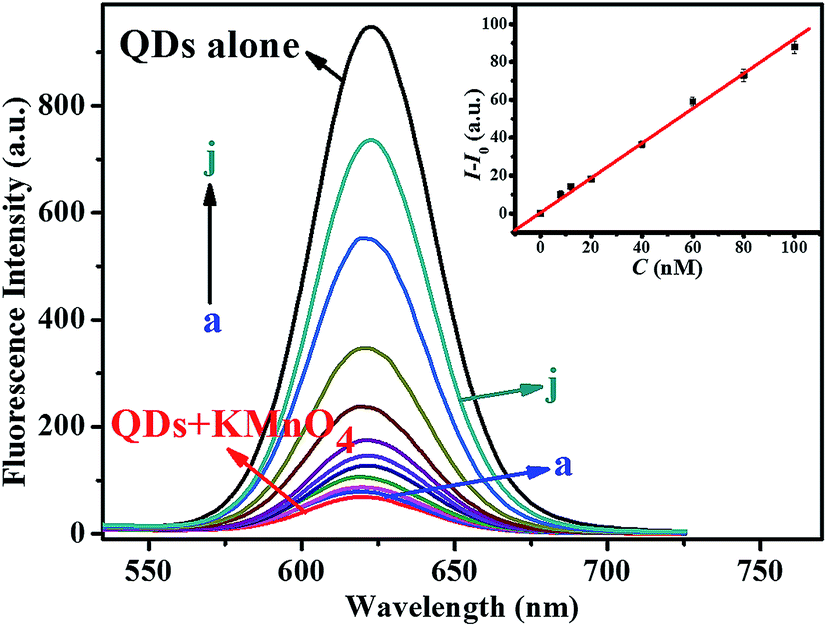 | ||
| Fig. 5 Fluorescence spectra of NAC-capped CdTe/CdS/ZnS core/shell/shell QDs and NAC-capped CdTe/CdS/ZnS core/shell/shell QDs–KMnO4 system with different concentrations of L-ascorbic acid: (a) 8.0 × 10⁻⁹ M; (b) 1.2 × 10⁻⁸ M; (c) 2.0 × 10⁻⁸ M; (d) 4.0 × 10⁻⁸ M; (e) 6.0 × 10⁻⁸ M; (f) 1.0 × 10⁻⁷ M; (g) 3.0 × 10⁻⁷ M; (h) 7.5 × 10⁻⁷ M; (i) 1.0 × 10⁻⁶ M; and (j) 1.5 × 10⁻⁶ M. The inset is the linear relationship between I − I0 and L-ascorbic acid concentration in the range of 8.0 × 10⁻⁹ M to 1.0 × 10⁻⁷ M with a correlation coefficient of 0.9971. NAC-capped CdTe/CdS/ZnS core/shell/shell QDs: 1.7 × 10⁻⁷ M; KMnO4: 3.0 × 10⁻⁶ M. | ||
| Detection method | Linear range (×10−6 M) | Detection limit (×10−6 M) | Samples detection | Reference |
|---|---|---|---|---|
| Spectrophotometry | 0.1–1000000 |
0.1 | Fruit juices, urine, serum, vitamin C tablets | 28 |
| Enzymatic analysis | 0.1–10 | 0.1 | Milk and sour-milk products | 29 |
| Electroanalysis | 1–150 | 0.76 | Blood serum and pharmaceutical samples | 30 |
| Chemiluminescence | 0.1–100 | 0.0067 | Human serum | 31 |
| Colorimetry | 0.044–0.3 | 0.003 | Orange juice and grapefruit juice | 32 |
| Colorimetry | 0.1–2.5 | 0.049 | Not given | 33 |
| Colorimetry | 0.11–85 | 0.019 | Human urine | 34 |
| Phosphorimetry | 2.5–37.5 | 0.72 | Human urine | 35 |
| Liquid chromatography | Not given | 1.1 | Citrus fruits | 36 |
| Fluorometry | 0.3–10 | 0.074 | Human urine and plasma | 37 |
| Fluorometry | 0.008–0.1 | 0.0018 | Human urine and vitamin C tablets | This method |
3.8 Effect of foreign substances and sample determination
The influence of some common ions, carbohydrates, nucleotides and amino acids was investigated to verify the applicability of this NAC-capped CdTe/CdS/ZnS core/shell/shell QDs-based “OFF–ON” fluorescent biosensor for the determination of L-ascorbic acid in biological samples, and the results are shown in Table 2. The coexisting compounds are considered to have no interference with the detection if they cause a relative error of less than ±5% in the fluorescence intensity of the system. As shown in Table 2, most common ions, carbohydrates, four nucleotides and twenty amino acids with higher concentration had almost no distinct influence on the determination of 3.0 × 10⁻⁷ M L-ascorbic acid in the given conditions. The data revealed that the proposed method might be applied to the detection of L-ascorbic acid in biological samples.| Foreign substances | Concentration coexisting (M) | Change of FL Intensity (%) | R.S.D. (%) | Foreign substances | Concentration coexisting (M) | Change of FL Intensity (%) | R.S.D. (%) |
|---|---|---|---|---|---|---|---|
| a NAC-capped CdTe/CdS/ZnS core/shell/shell QDs: 1.7 × 10⁻⁷ M; KMnO4: 3.0 × 10⁻⁶ M; L-ascorbic acid: 3.0 × 10⁻⁷ M; buffer: 1.5 mL Tris–HCl buffer (pH 7.4). | |||||||
| K+ | 5 × 10⁻⁴ | −2.8 | 1.4 | L-Arg | 2 × 10⁻⁵ | −2.4 | 1.1 |
| Na+ | 1 × 10⁻³ | −2.1 | 0.9 | L-Cys | 1 × 10⁻⁵ | −2.8 | 0.7 |
| Ca2+ | 2 × 10⁻⁴ | +1.6 | 0.5 | L-Val | 2 × 10⁻⁵ | +1.4 | 0.6 |
| Mg2+ | 5 × 10⁻⁴ | −2.1 | 2.7 | L-Ala | 2 × 10⁻⁵ | +2.6 | 2.1 |
| Cu2+ | 1 × 10⁻⁴ | −1.2 | 0.8 | L-Gly | 2 × 10⁻⁵ | −3.3 | 0.9 |
| Al3+ | 3 × 10⁻⁴ | +1.6 | 1.9 | L-Lys | 2 × 10⁻⁵ | +4.2 | 1.3 |
| C2O42− | 2 × 10⁻⁴ | +1.1 | 0.9 | L-Trp | 2 × 10⁻⁵ | +3.6 | 1.8 |
| NO3− | 5 × 10⁻⁴ | −2.9 | 1.6 | L-Asp | 2 × 10⁻⁵ | −2.6 | 1.2 |
| SO42− | 1 × 10⁻⁴ | −2.8 | 1.8 | L-Pro | 2 × 10⁻⁵ | +1.8 | 1.5 |
| Cl− | 1 × 10⁻³ | +1.1 | 2.5 | L-Leu | 2 × 10⁻⁵ | −2.2 | 2.7 |
| Glucose | 1 × 10⁻³ | −2.2 | 1.1 | L-Glu | 2 × 10⁻⁵ | −4.4 | 1.5 |
| Sucrose | 5 × 10⁻⁴ | −3.1 | 2.3 | L-Tyr | 2 × 10⁻⁵ | +4.9 | 2.1 |
| Lactose | 2 × 10⁻⁴ | +2.7 | 0.9 | L-Met | 2 × 10⁻⁵ | −3.6 | 0.7 |
| Urea | 5 × 10⁻⁴ | +2.3 | 1.3 | L-Ser | 2 × 10⁻⁵ | +1.7 | 1.7 |
| Adenine | 2 × 10⁻⁵ | −3.9 | 1.2 | L-Phe | 2 × 10⁻⁵ | −3.5 | 1.5 |
| Cytosine | 2 × 10⁻⁵ | −2.7 | 1.5 | L-Thr | 2 × 10⁻⁵ | −3.1 | 1.9 |
| Thymine | 1 × 10⁻⁵ | −3.9 | 2.2 | L-His | 2 × 10⁻⁵ | −2.4 | 2.1 |
| Guanine | 1 × 10⁻⁵ | −1.5 | 1.0 | L-Ile | 2 × 10⁻⁵ | +1.8 | 1.7 |
| L-Asn | 2 × 10⁻⁵ | +1.8 | 2.8 | L-Gln | 2 × 10⁻⁵ | −2.6 | 2.2 |
To confirm the feasibility, this present method was first applied to the detection of L-ascorbic acid in three synthetic samples, which contained four common chemical substances, three carbohydrates, four nucleotides and twenty amino acids. As indicated in Table 3, the values found for the three synthetic samples were identical to the expected values and the recoveries were from 98.9% to 101.3%, which indicated the suitability for the determination of L-ascorbic acid in the presence of these substances.
| Synthetic samples | Taken (×10−8 M) | Found (×10−8 M) | Recovery (%) | R.S.D. (%) |
|---|---|---|---|---|
| a NAC-capped CdTe/CdS/ZnS core/shell/shell QDs: 1.7 × 10⁻⁷ M; KMnO4: 3.0 × 10⁻⁶ M; buffer: 1.5 mL Tris–HCl buffer (pH 7.4).b The concentrations of twenty amino acids were all 1.0 × 10⁻⁵ M.c The concentrations of glucose, sucrose, lactose and urea were all 2.0 × 10⁻⁴ M. The concentrations of adenine, cytosine, thymine and guanine were all 1.0 × 10⁻⁵ M.d The concentrations of KNO3, NaNO3, Ca(C2O4)2, Mg(NO3)2, Al(NO3)3, CuSO4 were all 1.0 × 10⁻⁴ M. | ||||
| Sample 1b | 1.5 | 1.52 ± 0.03 | 101.3 | 1.4 |
| Sample 2c | 5.0 | 5.01 ± 0.02 | 100.2 | 1.8 |
| Sample 3d | 8.0 | 7.91 ± 0.05 | 98.9 | 1.2 |
To further confirm the feasibility for L-ascorbic acid assay in biological conditions, this method was applied to the determination of L-ascorbic acid in human urine samples. The urine of individuals was treated as described in Section 2.4 and their L-ascorbic acid concentrations were measured according to the proposed method. The recovery of L-ascorbic acid was determined by comparing the results obtained before and after the addition of standard L-ascorbic acid to the diluted urine samples. As listed in Table 4, the recoveries of different known amounts of L-ascorbic acid were obtained from 95.6% to 104.5% with a satisfying analytical precision (R.S.D. ≤ 3.6%) validating the reliability and practicality of this method. In addition, this present approach was applied to L-ascorbic acid detection in vitamin C tablets. As indicated in Table 5, the value obtained for the sample was highly consistent with the expected values, and the recoveries of spiked L-ascorbic acid were obtained from 95.5% to 103.1% with a satisfying analytical precision (R.S.D. ≤ 3.0%), which validated the reliability and practicality of this strategy.
| Samples | Taken (×10−8 M) | Found (×10−8 M) | Recovery (%) | R.S.D. (%) |
|---|---|---|---|---|
| a NAC-capped CdTe/CdS/ZnS core/shell/shell QDs: 1.7 × 10⁻⁷ M; KMnO4: 3.0 × 10⁻⁶ M; buffer: 1.5 mL Tris–HCl buffer (pH 7.4). | ||||
| Human urine sample 1 | 0.0 | 5.12 ± 0.05 | — | — |
| 2.0 | 7.21 ± 0.04 | 104.5 | 2.0 | |
| 5.0 | 10.10 ± 0.12 | 99.6 | 2.4 | |
| 8.0 | 12.81 ± 0.08 | 96.1 | 1.0 | |
| Human urine sample 2 | 0.0 | 6.91 ± 0.01 | — | — |
| 2.0 | 8.94 ± 0.05 | 102.0 | 2.5 | |
| 5.0 | 12.05 ± 0.09 | 103.0 | 1.8 | |
| 8.0 | 14.82 ± 0.15 | 99.0 | 1.9 | |
| Human urine sample 3 | 0.0 | 3.12 ± 0.04 | — | — |
| 2.0 | 5.11 ± 0.05 | 100.5 | 2.5 | |
| 5.0 | 7.92 ± 0.10 | 96.4 | 2.0 | |
| 8.0 | 10.82 ± 0.18 | 96.5 | 2.3 | |
| Human urine sample 4 | 0.0 | 9.81 ± 0.03 | — | — |
| 2.0 | 11.85 ± 0.06 | 102.5 | 3.0 | |
| 5.0 | 14.71 ± 0.12 | 98.2 | 2.4 | |
| 8.0 | 17.93 ± 0.21 | 101.6 | 2.6 | |
| Human urine sample 5 | 0.0 | 2.44 ± 0.12 | — | — |
| 2.0 | 4.47 ± 0.05 | 103.5 | 2.5 | |
| 5.0 | 7.18 ± 0.18 | 95.6 | 3.6 | |
| 8.0 | 10.75 ± 0.19 | 104.4 | 2.4 | |
| Sample | Taken (×10−8 M) | Found (×10−8 M) | Recovery (%) | R.S.D. (%) |
|---|---|---|---|---|
| a NAC-capped CdTe/CdS/ZnS core/shell/shell QDs: 1.7 × 10⁻⁷ M; KMnO4: 3.0 × 10⁻⁶ M; buffer: 1.5 mL Tris–HCl buffer (pH 7.4). | ||||
| Vitamin C tablets | 0.0 | 24.10 ± 0.82 | — | — |
| 1.0 | 25.12 ± 0.03 | 102.0 | 3.0 | |
| 4.0 | 27.92 ± 0.11 | 95.5 | 2.7 | |
| 8.0 | 32.35 ± 0.21 | 103.1 | 2.6 | |
3.9 Possible fluorescence quenching mechanism
It is widely reported that the fluorescence quenching mechanism usually includes the inner filter effect (IFE), non-radiative ground state complex formation (static quenching) and electron transfer processes (dynamic quenching).44 The IFE quenching mechanism could be proved through the UV-vis absorption spectrum of KMnO4 and the emission spectrum of NAC-capped CdTe/CdS/ZnS core/shell/shell QDs. As shown in Fig. 6A, the absorption spectrum of KMnO4 had three typical absorption bands at 506 nm, 525 nm and 545 nm, respectively. However, the emission peak of NAC-capped CdTe/CdS/ZnS core/shell/shell QDs was centered at 622 nm under the excitation of 388 nm. Thus, almost no spectral overlap took place between the absorption spectrum of KMnO4 and the emission spectrum of NAC-capped CdTe/CdS/ZnS core/shell/shell QDs. Furthermore, the absorbance of KMnO4 at 388 nm was also very weak. Therefore, KMnO4 could not efficiently shield the excitation light for QDs nor effectively absorb the emission light from QDs, suggesting that the quenching mechanism between KMnO4 and NAC-capped CdTe/CdS/ZnS core/shell/shell QDs was not IFE.45,46 | ||
| Fig. 6 (A) UV-vis absorption spectrum of KMnO4 (a) and the emission spectrum of NAC-capped CdTe/CdS/ZnS core/shell/shell QDs at 388 nm excitation wavelength (b). (B) UV-vis absorption spectra of NAC-capped CdTe/CdS/ZnS core/shell/shell QDs, KMnO4 and NAC-capped CdTe/CdS/ZnS core/shell/shell QDs–KMnO4 system; the difference absorption spectra between NAC-capped CdTe/CdS/ZnS core/shell/shell QDs–KMnO4 system and NAC-capped CdTe/CdS/ZnS core/shell/shell QDs, and the summed absorption spectrum between NAC-capped CdTe/CdS/ZnS core/shell/shell QDs and KMnO4, respectively. NAC-capped CdTe/CdS/ZnS core/shell/shell QDs: 8.5 × 10⁻⁷ M; KMnO4: 6.0 × 10⁻⁶ M. | ||
Charge transfer often occurs in the dynamic quenching and fluorescence is subsequently quenched when the electron acceptor collides with the excited fluorophore; thus, no variations in the absorption spectra of the fluorophore will be expected. However, the ground state complex formation during the state quenching can perturb the absorption spectra of the fluorophore, which results in the variation of the absorption spectra of the fluorophore.47 In order to precisely reveal the fluorescence quenching mechanism, the UV-vis absorption spectra of NAC-capped CdTe/CdS/ZnS core/shell/shell QDs, KMnO4 and NAC-capped CdTe/CdS/ZnS core/shell/shell QDs–KMnO4 system were recorded. As shown in Fig. 6B, the UV-vis absorption spectrum of NAC-capped CdTe/CdS/ZnS core/shell/shell QDs–KMnO4 system and the summed absorption spectrum between NAC-capped CdTe/CdS/ZnS core/shell/shell QDs and KMnO4 could not be superposed within the experimental error, indicating the ground state recombination between NAC-capped CdTe/CdS/ZnS core/shell/shell QDs and KMnO4. Furthermore, in comparing with the absorption spectra of NAC-capped CdTe/CdS/ZnS core/shell/shell QDs, the difference absorption spectrum between NAC-capped CdTe/CdS/ZnS core/shell/shell QDs–KMnO4 system and KMnO4 exhibited an obvious hypochromic effect. These results reconfirmed the static quenching mechanism attributed to the ground state recombination between NAC-capped CdTe/CdS/ZnS core/shell/shell QDs and KMnO4.
4. Conclusions
A novel NAC-capped CdTe/CdS/ZnS core/shell/shell QDs-based “OFF–ON” fluorescent biosensor was established to detect L-ascorbic acid. The primary advantage of this method is its sensitivity and specificity. The fluorescence of NAC-capped CdTe/CdS/ZnS core/shell/shell QDs was quenched by KMnO4 through the electron transfer process and correspondingly the fluorescence of the QDs into the “OFF” status. Then, the fluorescence of QDs was switched “ON” after the addition of L-ascorbic acid that could bind with KMnO4 and remove it from the surface of the QDs. Under the optimum conditions, the present method had a linear range of 8.0 × 10⁻⁹ M to 1.0 × 10⁻⁷ M, with a correlation coefficient of 0.9971. The limit of detection of L-ascorbic acid was 1.8 × 10⁻⁹ M. Some common ions, carbohydrates, nucleotides and amino acids did not affect the determination of L-ascorbic acid. The presented method has been successfully applied to the determination of L-ascorbic acid in three synthetic samples, human urine samples and vitamin C tablets. The possible fluorescence quenching mechanism of this NAC-capped CdTe/CdS/ZnS core/shell/shell QDs-based “OFF–ON” fluorescent sensor was the static quenching mechanism.Acknowledgements
This work was financially supported by the National Natural Science Foundation of China (21203035, 21403039), the Guangxi Natural Science Foundation (2013GXNSFCA019005, 2013GXNSFBA019029), the Scientific Research Foundation of Guangxi Provincial Education Department (2013YB138, ZD2014081), the Scientific Research Foundation for the Returned Overseas Chinese Scholars, State Education Ministry, the Open Research Fund Program of the State Key Laboratory of Virology of China (2014KF006) and the Innovation Project of Guangxi Graduate Education (YCSZ2014186).References
- M. F. Frasco and N. Chaniotakis, Semiconductor quantum dots in chemical sensors and biosensors, Sensors, 2009, 9, 7266–7286 CrossRef CAS PubMed.
- I. L. Medintz, H. T. Uyeda, E. R. Goldman and H. Mattoussi, Quantum dot bioconjugates for imaging, labelling and sensing, Nat. Mater., 2005, 4, 435–446 CrossRef CAS PubMed.
- R. Freeman, R. Gill, I. Shweky, M. Kotler, U. Banin and I. Willner, Biosensing and probing of intracellular metabolic pathways by NADH-sensitive quantum dots, Angew. Chem., Int. Ed., 2009, 48, 309–313 CrossRef CAS PubMed.
- R. Z. Liang, R. Tian, W. Y. Shi, Z. H. Liu, D. P. Yan, M. Wei, D. G. Evans and X. Duan, A temperature sensor based on CdTe quantum dots-layered double hydroxide ultrathin films via layer-by-layer assembly, Chem. Commun., 2013, 49, 969–971 RSC.
- G. W. Platt, F. Damin, M. J. Swann, I. Metton, G. Skorski, M. Cretich and M. Chiari, Allergen immobilisation and signal amplification by quantum dots for use in a biosensor assay of IgE in serum, Biosens. Bioelectron., 2014, 52, 82–88 CrossRef CAS PubMed.
- W. J. Liang, Z. Q. Liu, S. P. Liu, J. D. Yang and Y. Q. He, A novel surface modification strategy of CdTe/CdS QDs and its application for sensitive detection of ctDNA, Sens. Actuators, B, 2014, 196, 336–344 CrossRef CAS PubMed.
- T. Yu, T. Y. Ying, Y. Y. Song, Y. J. Li, F. H. Wu, X. Q. Dong and J. S. Shen, A highly sensitive sensing system based on photoluminescent quantum dots for highly toxic organophosphorus compounds, RSC Adv., 2014, 4, 8321–8327 RSC.
- X. Y. Cao, F. Shen, M. W. Zhang, J. X. Bie, X. Liu, Y. L. Luo, J. J. Guo and C. Y. Sun, Facile synthesis of chitosan-capped ZnS quantum dots as an eco-friendly fluorescence sensor for rapid determination of bisphenol A in water and plastic samples, RSC Adv., 2014, 4, 16597–16606 RSC.
- S. Huang, Q. Xiao, R. Li, H. L. Guan, J. Liu, X. R. Liu, Z. K. He and Y. Liu, A simple and sensitive method for L-cysteine detection based on the fluorescence intensity increment of quantum dots, Anal. Chim. Acta, 2009, 645, 73–78 CrossRef CAS PubMed.
- M. H. Wang, W. F. Niu, X. Wu, L. X. Li, J. Yang, S. M. Shuang and C. Dong, Fluorescence enhancement detection of uric acid based on water-soluble 3-mercaptopropionic acid-capped core/shell ZnS: Cu/ZnS, RSC Adv., 2014, 4, 25183–25188 RSC.
- D. W. Huang, C. G. Niu, X. Y. Wang, X. X. Lv and G. M. Zeng, “Turn-on” fluorescent sensor for Hg2+ based on single-stranded DNA functionalized Mn: CdS/ZnS quantum dots and gold nanoparticles by time-gated mode, Anal. Chem., 2013, 85, 1164–1170 CrossRef CAS PubMed.
- F. X. Wang, Z. Y. Gu, W. Lei, W. J. Wang, X. F. Xia and Q. L. Hao, Graphene quantum dots as a fluorescent sensing platform for highly efficient detection of copper(II) ions, Sens. Actuators, B, 2014, 190, 516–522 CrossRef CAS PubMed.
- X. Y. Hu, K. Zhu, Q. S. Guo, Y. Q. Liu, M. F. Ye and Q. J. Sun, Ligand displacement-induced fluorescence switch of quantum dots for ultrasensitive detection of cadmium ions, Anal. Chim. Acta, 2014, 812, 191–198 CrossRef CAS PubMed.
- J. Zong, X. L. Yang, A. Trinchi, S. Hardin, I. Cole, Y. H. Zhu, C. Z. Li, T. Muster and G. Wei, Carbon dots as fluorescent probes for “off–on” detection of Cu2+ and L-cysteine in aqueous solution, Biosens. Bioelectron., 2014, 51, 330–335 CrossRef CAS PubMed.
- Y. P. Shi, Y. Pan, H. Zhang, Z. M. Zhang, M. J. Li, C. Q. Yi and M. S. Yang, A dual-mode nanosensor based on carbon quantum dots and gold nanoparticles for discriminative detection of glutathione in human plasma, Biosens. Bioelectron., 2014, 56, 39–45 CrossRef CAS PubMed.
- Z. P. Liu, Q. Ma, X. Y. Wang, Z. H. Lin, H. Zhang, L. L. Liu and X. G. Su, A novel fluorescent nanosensor for detection of heparin and heparinase based on CuInS2 quantum dots, Biosens. Bioelectron., 2014, 54, 617–622 CrossRef CAS PubMed.
- E. Vaishnavi and R. Renganathan, “Turn-on-off-on” fluorescence switching of quantum dots-cationic porphyrin nanohybrid: a sensor for DNA, Analyst, 2014, 139, 225–234 RSC.
- R. Zhang, D. X. Zhao, H. G. Ding, Y. X. Huang, H. Z. Zhong and H. Y. Xie, Sensitive single-color fluorescence “off–on” switch system for dsDNA detection based on quantum dots-ruthenium assembling dyads, Biosens. Bioelectron., 2014, 56, 51–57 CrossRef CAS PubMed.
- S. Huang, H. N. Qiu, Q. Xiao, C. S. Huang, W. Su and B. Q. Hu, A simple QD-FRET bioprobe for sensitive and specific detection of hepatitis B virus DNA, J. Fluoresc., 2013, 23, 1089–1098 CrossRef CAS PubMed.
- S. Huang, Q. Xiao, Z. K. He, Y. Liu, P. Tinnefeld, X. R. Su and X. N. Peng, A high sensitive and specific QDs FRET bioprobe for MNase, Chem. Commun., 2008, 44, 5990–5992 RSC.
- P. P. Li, S. P. Liu, S. G. Yan, X. Q. Fan and Y. Q. He, A sensitive sensor for anthraquinone anticancer drugs and hsDNA based on CdTe/CdS quantum dots fluorescence reversible control, Colloids Surf., A, 2011, 392, 7–15 CrossRef CAS PubMed.
- Z. Q. Liu, S. P. Liu, X. D. Wang, P. P. Li and Y. Q. He, A novel quantum dots-based OFF–ON fluorescent biosensor for highly selective and sensitive detection of double-strand DNA, Sens. Actuators, B, 2013, 176, 1147–1153 CrossRef CAS PubMed.
- R. F. Cathcart, A unique function for ascorbate, Med. Hypotheses., 1991, 35, 32–37 CrossRef CAS.
- S. J. Padayatty, A. Katz, Y. Wang, P. Eck, O. Kwon, J. H. Lee and S. Chen, Vitamin C as an antioxidant: evaluation of its role in disease prevention, J. Am. Coll. Nutr., 2003, 22, 18–35 CrossRef CAS.
- P. Janda, J. Weber, L. Dunsch and A. B. P. Lever, Detection of ascorbic acid using a carbon fiber microelectrode coated with cobalt tetramethylpyridoporphyrazine, Anal. Chem., 1996, 68, 960–965 CrossRef CAS.
- O. Arrigori and C. D. Tullio, Ascorbic acid: much more than just an antioxidant, Biochim. Biophys. Acta, 2002, 1569, 1–3 CrossRef.
- L. K. Massey, M. Liebman and S. A. Kynast-Gales, Ascorbate increases human oxaluria and kidney stone risk, J. Nutr., 2005, 135, 1673–1677 CAS.
- B. Mudabuka, D. Ondigo, S. Degni, S. Vilakazi and N. Torto, A colorimetric probe for ascorbic acid based on copper–gold nanoparticles in electrospun nylon, Microchim. Acta, 2014, 181, 395–401 CrossRef CAS.
- T. N. Shekhovtsova, S. V. Muginova, J. A. Luchinina and A. Z. Galimova, Enzymatic methods in food analysis: determination of ascorbic acid, Anal. Chim. Acta, 2006, 573–574, 125–132 CrossRef PubMed.
- T. Madrakian, E. Haghshenas and A. Afkhami, Simultaneous determination of tyrosine, acetaminophen and ascorbic acid using gold nanoparticles/multiwalled carbon nanotube/glassy carbon electrode by differential pulse voltammetric method, Sens. Actuators, B, 2014, 193, 451–460 CrossRef CAS PubMed.
- H. Chen, R. B. Li, L. Lin, G. S. Guo and J. M. Lin, Determination of L-ascorbic acid in human serum by chemiluminescence based on hydrogen peroxide–sodium hydrogen carbonate–CdSe/CdS quantum dots system, Talanta, 2010, 81, 1688–1696 CrossRef CAS PubMed.
- Y. F. Zhang, B. X. Li and C. L. Xu, Visual detection of ascorbic acid via alkyne–azide click reaction using gold nanoparticles as a colorimetric probe, Analyst, 2010, 135, 1579–1584 RSC.
- G. Q. Wang, Z. P. Chen and L. X. Chen, Mesoporous silica-coated gold nanorods: towards sensitive colorimetric sensing of ascorbic acid via target-induced silver overcoating, Nanoscale, 2011, 3, 1756–1759 RSC.
- X. X. Wang, J. M. Liu, S. L. Jiang, L. Jiao, L. P. Lin, M. L. Cui, X. Y. Zhang, L. H. Zhang and Z. Y. Zheng, Non-aggregation colorimetric sensor for detecting vitamin C based on surface plasmon resonance of gold nanorods, Sens. Actuators, B, 2013, 182, 205–210 CrossRef CAS PubMed.
- W. Bian, J. Ma, W. R. Guo, D. T. Lu, M. Fan, Y. L. Wei, Y. F. Li, S. M. Shuang and M. M. F. Choi, Phosphorescence detection of L-ascorbic acid with surface-attached N-acetyl-L-cysteine and L-cysteine Mn doped ZnS quantum dots, Talanta, 2013, 116, 794–800 CrossRef CAS PubMed.
- F. Khosravi and H. Asadollahzadeh, Determination of ascorbic acid in different citrus fruits under reversed phase conditions with UPLC, Euro, J. Exp. Biol., 2014, 4, 91–94 CAS.
- Y. J. Chen and X. P. Yan, Chemical redox modulation of the surface chemistry of CdTe quantum dots for probing ascorbic acid in biological fluids, Small, 2009, 5, 2012–2018 CrossRef CAS PubMed.
- Q. Xiao, S. Huang, W. Su, W. H. Chan and Y. Liu, Facile synthesis and characterization of highly fluorescent and biocompatible N-acetyl-L-cysteine capped CdTe/CdS/ZnS core/shell/shell quantum dots in aqueous phase, Nanotechnology, 2012, 23, 495717 CrossRef PubMed.
- A. M. Pisoschi, A. Pop, A. I. Serban and C. Fafaneata, Electrochemical methods for ascorbic acid determination, Electrochim. Acta, 2014, 121, 443–460 CrossRef CAS PubMed.
- W. W. Yu, L. H. Qu, W. Z. Guo and X. G. Peng, Experimental determination of the extinction coefficient of CdTe, CdSe, and CdS nanocrystals, Chem. Mater., 2003, 15, 2854–2860 CrossRef CAS.
- J. N. Demas and G. A. Crosby, The measurement of photoluminescence quantum yields, J. Phys. Chem., 1971, 75, 991–1024 CrossRef.
- S. Huang, Q. Xiao, W. Su, P. Y. Li, J. Q. Ma and Z. K. He, Simple and sensitive determination of papain by resonance light-scattering with CdSe quantum dots, Colloids Surf., B, 2013, 102, 146–151 CrossRef CAS PubMed.
- J. R. Lakowicz, Principles of Fluorescence Spectroscopy, Plenum Press, New York, 3rd edn, 2006, p. 277 Search PubMed.
- Y. Chen and Z. Rosenzweig, Luminescent CdS quantum dots as selective ion probes, Anal. Chem., 2002, 74, 5132–5138 CrossRef CAS.
- N. Shao, Y. Zhang, S. M. Cheung, R. H. Yang, W. H. Chan, T. Mo, K. A. Li and F. Liu, Copper ion-selective fluorescent sensor based on the inner filter effect using a spiropyran derivative, Anal. Chem., 2005, 77, 7294–7303 CrossRef CAS PubMed.
- M. Zheng, Z. G. Xie, D. Qu, D. Li, P. Du, X. B. Jing and Z. C. Sun, On–Off–On fluorescent carbon dot nanosensor for recognition of chromium(VI) and ascorbic acid based on the inner filter effect, ACS Appl. Mater. Interfaces, 2013, 5, 13242–13247 CAS.
- Q. Xiao, S. Huang, Z. D. Qi, B. Zhou, Z. K. He and Y. Liu, Conformation, thermodynamics and stoichiometry of HSA adsorbed to colloidal CdSe/ZnS quantum dots, Biochim. Biophys. Acta, Proteins Proteomics, 2008, 1784, 1020–1027 CrossRef CAS PubMed.
| This journal is © The Royal Society of Chemistry 2014 |