The convenient aqueous synthesis and biological evaluation of ortho-(3,4,5-trimethoxybenzoyl)-acetanilides as novel anti-cancer agents

Jianfei Sheng, Fei Mao, Jun Yan, Ling Huang* and Xingshu Li*
School of Pharmaceutical Sciences, Sun Yat-sen University, Guangzhou, 510006, China. E-mail: huangl72@mail.sysu.edu.cn; lixsh@mail.sysu.edu.cn; Fax: +86-20-3994-3051; Fax: +86-20-3994-3050; Tel: +86-20-3994-3051 Tel: +86-20-3994-3050

Received 2nd August 2014 , Accepted 27th August 2014

First published on 28th August 2014


Abstract

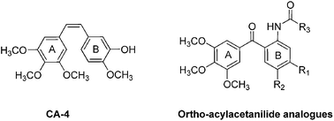
A series of new ortho-(3,4,5-trimethoxybenzoyl)-acetanilides were synthesised by the cross-coupling reaction catalyzed with Pd catalyst in aqueous medium, with polyethylene glycol as additive under very mild conditions. The evaluation of these compounds as tubulin polymerization inhibitors indicated that most of these compounds were potential anti-cancer agents. Among them, compound 13, 2-hydroxy-N-(5-methoxy-2-(3,4,5-trimethoxy benzoyl)phenyl) acetamide exhibited excellent antiproliferative activity against various human cancer cell lines (GI50 = 71 nM for human HeLa cell line), and good activity for inhibiting tubulin polymerization (IC50 = 2.94 μM). The mechanism study indicated that compound 13 could arrest cell-cycle progression at the mitosis phase, block the formation of cdc2/cyclin B1 complex, down-regulate the p-Cdc 25C and the anti-apoptotic proteins Bcl-2 and Bcl-XL, and up-regulate the pro-apoptotic proteins Bax and Bad.


Introduction

Microtubules are cytoskeletal filaments consisting of α, β-tubulin heterodimers and are essential in a wide range of cellular processes including cell shape maintenance, intracellular transport, cell signaling, separation of duplicated chromosomes during cell division, and are required for cell life cycle.1–3 The crucial involvement of microtubules in mitosis makes them an attractive target for anticancer drugs.4,5

There are many chemically diverse compounds targeting the tubulin-microtubule system.6,7 Among them, combretastatin A-4 (CA-4, 1), a natural cis-stilbene derivative first isolated from the bark of the African willow tree Combretum caffrum in 1982,8 is a typical example which strongly inhibits tubulin polymerization (with about 1.9 μM of the IC50) by binding to the colchicine binding site, and then, the disruption of tubulin assembly results in mitotic arrest, and subsequent cancer cell death.9 However, the cis double bond in CA-4 is prone to isomerize to the more thermodynamic stable, but less bioactive trans isomers (with the IC50 above 50 μM against tubulin polymerization).10 For overcoming this disadvantage, numerous modifications to stabilize the conformation by replacement of the olefinic bridge with a ring or carbonyl have been developed in recent years.11–13 Generally, several reaction steps were needed for the synthesis of these compounds, therefore, from both academically and practical standpoints, it would be very interested to develop a highly effective strategy for the preparation of the new effective antimitotic agents by simple, especially one-step procedure.

In view of direct synthesis being able to minimize waste/side product formation, transition-metal-catalyzed direct C–H bond functionalization/cross-coupling as a valuable tool for the modular and facile synthesis of structurally similar, yet diversified organic molecules have attracted increasing attention. A lot of papers have been published on transition metals (e.g., Pd, Ir, Rh, Ru, Cu, Fe, etc.) facilitating the cross-coupling reaction in recent years.14 Recently, we and other groups reported the palladium-catalyzed cross-dehydrogenative coupling between anilides and aromatic aldehydes in toluene.15–17 Subsequently, Novák described that the reaction could be performed mildly in aqueous with the addition of detergent, accompanying the reaction rate acceleration.18,19

Although a lot of progress have been made in transition-metal-catalyzed carbon–carbon coupling reactions, there were very limited reports on the application of this methodology to the preparation of complex molecules and useful medical compounds. Herein, we disclose the synthesis of a series of new ortho-(3,4,5-trimethoxybenzoyl)-acetanilides catalyzed with Pd catalyst in aqueous with polyethyleneglycol as additive, especially the evaluation of them as tubulin polymerization inhibitors, and the study of their anticancer mechanism of these compounds.

The design strategy of these target compounds were showed in Fig. 1. As 3,4,5-trimethoxy groups on the A-ring and the cis-orientation of the two aryl rings on olefinic bond were responsible for efficient binding to tubulin,20,21 we focused on the modification of B-ring and the bridging double bond, using the simple coupling reaction of commercially available 3,4,5-trimethoxybenzaldehyde and different anilides to obtaine these bioactive ortho-(3,4,5-trimethoxybenzoyl)-acetanilides.


image file: c4ra07998a-f1.tif
Fig. 1 Structures of CA-4 and the ortho-acylacetanilide analogues.

Results and discussion

Chemistry

The study of one-step synthesis of the new ortho-acylacetanilides 1–12 is shown in Table 1 and Scheme 1. Inspired by Novák's method with the addition of detergent to accelerate the coupling reaction, and considering polyethylene glycol (PEG), which is soluble in both water and organic solvents, has been successfully used as reaction medium and additive in a variety of organic reactions,22–27 we studied the reaction with 3-methoxyacetanilide and 3,4,5-trimethoxybenzaldehyde as the prototypical substrates, 5% Pd(OAc)2 as the catalyst and polyethylene glycol as the additive. A screening of optimal conditions indicated that the use of PEG was rather significant (Table 1, entries 1–4: 5% mol of PEG-2000, 63% yield; 10% mol of PEG, 71% yield; 20% mol of PEG, 91% yield; 50% mol of PEG, trace product). The temperature effect was also important for the reaction; it could proceed smoothly at 40 °C or 60 °C, but only gave 38% yield at room temperature (Table 1, entries 3, 6–7). We also attempted to reduce the amount of Pd(OAc)2, however, low yields were obtained (Table 1, entry 8–9).
Table 1 Optimization of reaction conditionsa

image file: c4ra07998a-u1.tif

a Reactions was carried out using amide (1.0 mmol), 3,4,5-trimethoxybenzaldehyde (1.0 mmol), Pd(OAc)2, PEG-2000, tert-butylhydroperoxide TBHP (4.0 mmol), trifluoroacetic acid TFA (0.26 mmol, 26% mol), water (1.0 mL) under air for 12 h.b Isolated yields.c PEG-2000 was also as solvent.
Entry Pd(OAc)2 (equiv.) PEG-2000 (equiv.) H2O (mL) T (°C) TFA (equiv.) TBHP (equiv.) Yieldb (%)
1 5% 5% 1 40 26% 4.0 63
2 5% 10% 1 40 26% 4.0 71
3 5% 20% 1 40 26% 4.0 91
4 5% 50% 1 40 26% 4.0 Trace
5c 5% 100% 0 40 26% 4.0 Trace
6 5% 20% 1 rt 26% 4.0 38
7 5% 20% 1 60 26% 4.0 89
8 1.5% 20% 1 40 26% 4.0 Trace
9 3% 20% 1 40 26% 4.0 47


image file: c4ra07998a-s1.tif
Scheme 1 Reaction conditions: corresponding amide (1.0 mmol), 3,4,5-trimethoxybenzaldehyde (1.0 mmol), Pd(OAc)2 (0.05 mmol, 5 mol%), PEG-2000 (0.2 mmol, 20 mol%), tert-butylhydroperoxide TBHP (4.0 mmol), trifluoroacetic acid TFA (0.26 mmol, 26 mol%), water (1.0 mL) at 40 °C under air for 12 h. Isolated yields (1–12) were reported (reaction times for each substrate were not optimized).

With this optimal reaction conditions, compounds 1–12 were prepared by the reaction of substituted anilides and 3,4,5-trimethoxybenzaldehyde (Scheme 1).

Compounds 13–17 were prepared with 12 as the intermediate as the route of Scheme 2. The reaction of 12 with CH3OH in presence of CH3ONa gave compound 13 in good yield.28 Similarly, 12 reacted with methylamine, ethylamine and N,N-dimethylamine in the presence of potassium carbohydrate to provide compounds 14, 15 and 16 in 86–90% yields, respectively.29 Finally, compound 17 was obtained by the reaction of 16 with CH3I in CH2Cl2.30


image file: c4ra07998a-s2.tif
Scheme 2 Conditions: (a) CH3ONa, CH3OH, reflux; (b) NH2CH3, K2CO3, 50 °C; (c) NH2CH2CH3, K2CO3, 50 °C; (d) NH(CH3)2, K2CO3, 50 °C; (e) CH3I, rt.

In vitro cell growth inhibition

To evaluate the antiproliferative activities of all the synthesized compounds, human cancer cell lines HeLa (human epithelial cervical cancer cell line), A549 (non-small cell lung carcinoma), MCF-7 (human breast cancer cell line), and HCT-8 (human colon cancer cell line) were tested using MTT (3-(4,5-dimethylthiazol-2-yl)-2,5-diphenyltetrazolium bromide) assay with CA-4 as the reference compound.

The results shown in Table 2 demonstrated that most of the synthesized compounds displayed good anti-proliferative activity with GI50 values in micromol to sub-micromol range. Among them, compounds 13 and 1 gave the best results (13, with 71 nM of GI50 value for HeLa cell line; 1, GI50 = 113 nM for HeLa). A simple structure–activity relationship analysis showed that the anti-proliferative activity closely related to the substituted groups in B ring, and especially, the methoxy group in R1 (C-3) position was necessary. Compounds 2–7, with H, CH3 and Cl in this position exhibited relatively poor activities with GI50 values from 0.90 to 36.8 μM respectively for the four cell lines. On the other hand, compounds 9, 10, 11, 13, 14, and 15, which possess OCH3 in R1 position, showed moderate to very good activities (e.g., with 692 nM to 71 nM of GI50 values for HeLa, and 750 nM to 71 nM of GI50 values for A549). When moving the methoxy substitution from C-3 to the C-4 position of the B ring (compound 1 vs. 2), almost 10-fold decrease in antiproliferative activity was observed (from 113 nM to 1420 nM). Using the naphthalene ring to replace the benzene ring (compound 3 vs. 4), the antiproliferative activity (GI50 from 20.1 μM to 4.88 μM) was significantly increased.

Table 2 Antiproliferative activity of compounds 1–11, 13–17 and CA-4 against four human cancer cell linesa

image file: c4ra07998a-u2.tif

Compd R1 R2 R3 GI50 (μM)b
HeLa A549 MCF-7 HCT-8
a Cell lines were treated with different concentrations of the compounds for 48 h. Cell viability was measured by MTT assay as described in the Experimental section.b GI50 values are indicated as the mean ± SD (standard error) of three independent experiments.
1 OCH3 H CH3 0.113 ± 0.11 0.18 ± 0.01 0.88 ± 0.02 0.34 ± 0.06
2 H OCH3 CH3 1.40 ± 0.014 1.29 ± 0.19 0.90 ± 0.03 1.59 ± 0.02
3 4.88 ± 0.31 2.65 ± 0.03 1.71 ± 0.02 0.676 ± 0.05
4 H H CH3 20.1 ± 2.15 10.4 ± 0.16 14.2 ± 1.05 16.8 ± 1.10
5 CH3 H CH3 23.5 ± 1.16 13.4 ± 0.62 16.1 ± 3.17 20.5 ± 0.56
6 H CH3 CH3 2.15 ± 0.15 2.00 ± 0.35 3.56 ± 0.22 2.59 ± 0.69
7 Cl H CH3 36.8 ± 4.13 7.60 ± 1.93 12.5 ± 1.2 21.5 ± 1.57
8 H H Ph 0.54 ± 0.03 0.50 ± 0.035 4.34 ± 1.28 0.89 ± 0.05
9 OCH3 H CH2CH3 0.32 ± 0.01 0.36 ± 0.064 2.34 ± 0.03 1.68 ± 0.86
10 OCH3 H n-Propyl 0.23 ± 0.02 0.50 ± 0.03 2.60 ± 0.4 0.84 ± 0.06
11 OCH3 H Isopropyl 0.38 ± 0.05 0.50 ± 0.102 0.91 ± 0.01 1.23 ± 0.75
13 OCH3 H CH2OH 0.071 ± 0.013 0.071 ± 0.004 0.23 ± 0.01 0.12 ± 0.02
14 OCH3 H CH2NHCH3 0.692 ± 0.158 0.613 ± 0.059 0.55 ± 0.13 0.78 ± 0.01
15 OCH3 H CH2NCH2CH3 0.47 ± 0.11 0.590 ± 0.004 0.79 ± 0.02 0.79 ± 0.01
16 OCH3 H CH2N(CH3)2 2.1 ± 0.006 0.75 ± 0.03 1.05 ± 0.05 1.56 ± 0.39
17 OCH3 H CH2N(CH3)3 >10 21.4 ± 3.39 31.5 ± 2.59 35.6 ± 0.45
CA-4 0.0028 ± 0.06 0.018 ± 0.05 0.009 ± 0.01 0.025 ± 0.02


Inhibition of tubulin polymerization

To investigate whether the antiproliferative activities of these compounds were related to their interaction with tubulin, compounds 1, 8, 13, and 15, which showed relatively better activity, were choosed to study for their ability to block the assembly of tubulin (Table 3) by the method originally described by Bonne, D. et al. with moderate modification,31,32 and CA-4 was examined as reference compound. We firstly use 10 μM of the tested compounds for the assays. At this concentration, compound 13 was found to inhibit tubulin polymerization the most (97.8%), while compounds 1, 8, 15 gave 59.3%, 32.9%, and 31.9% inhibitions, respectively. Then, the complete dose-response curves were obtained by assaying these compounds with inhibition rates and the results were summarized in Table 3. The representative raw data for the polymerization assay of compound 13 is shown in Fig. 2. It can be seen that compounds 1 and 13 were effective tubulin polymerization inhibitors with IC50 values of 7.99 and 2.94 μM, respectively. Compounds, 8 and 15, which had relatively poor anti-proliferative activities, exhibited weaker ability to inhibit tubulin polymerization (with IC50 values more than 10 μM) than that of 1 and 13, which suggest that the anti-proliferative activity of these compounds was correlated with their tubulin polymerization inhibition. Moreover, the chemical stability of the target compound 13 and CA-4 were evaluated by HPLC (see ESI). The results show that there is no degradation of compound 13 up to 5 day at room temperature. While three impurities (should be the isomerize or decomposed products) of CA-4 in acetonitrile appeared in 24 h, which is the same as those reported in literature.33 These results indicated that replacement of the bridging double bond of CA-4 with carbonyl group could enhance the configurational stability.
Table 3 Effects of the selected compounds on tublin polymerization inhibitiona
Compd IC50b (μM)
a Inhibition of tubulin polymerization. Tublin was at 20 μM.b IC50 values are indicated as the mean ± SD (standard error) of three independent experiments.
1 7.99 ± 0.05
8 >10
13 2.94 ± 0.02
15 >10
CA-4 1.2 ± 0.2


image file: c4ra07998a-f2.tif
Fig. 2 Effects of compound 13 on mircrotubule dynamics.

The morphological alterations of HeLa cells caused by 13

Base on the assay of antiproliferative activity and tubulin polymerization inhibition, we choose compound 13 for further biological study. Optical microscope was applied to identify the morphological alterations of HeLa cells after treated with 13. As shown in Fig. 3, when exposed to serial concentrations of 13 (0.01, 0.05, 0.1, and 0.5 μM) for 24 h and 48 h, the cells exhibited morphological alterations such as shrinkage, membrane blebbing and chromatin condensation that are normally associated with the occurrence of apoptotic cell death.
image file: c4ra07998a-f3.tif
Fig. 3 The micrographs of HeLa cells exposed to compound 13 (0.01, 0.05, 0.1, and 0.5 μM) or 0.01% DMSO (control) for 24 h (A) and 48 h (B), (magnification ×200).

Analysis of cell cycle arrest

It is well-known that CA-4 blocks the cell cycle in the G2/M phase due to microtubule depolymerization and cytoskeleton disruption. The cell growth inhibitory potency of compound 13 prompted us to evaluate its effects on the cell cycle (Fig. 3). After HeLa cells were treated with DMSO (0.01%) or compound 13 at different concentrations (0.01, 0.05, 0.1, and 0.5 μM) for 24 h and 48 h, the analysis (Fig. 4) of harvested cells by flow cytometry indicated that 13 resulted in significant cell-cycle arrest at G2/M phase, showing a time and dose-dependent manner. The lowest concentration of 13 induced no significant distribution of HeLa cells through the cell cycle (22.0% of cells at G2/M phase for 24 h, and 23.0% for 48 h). Whereas, when the concentration of 13 was increased to 0.05, 0.1, and 0.5 μM for 24 h, the percentage of cells at G2/M phase correspondingly increased to 39.51%, 52.75%, and 56.06%, respectively (Fig. 4A), and the percentage of cells at G2/M phase were 58.70%, 60.07%, and 63.86% (Fig. 4B) after treatment of compound 13 at the same concentration for 48 h. Futhermore, when the cells were exposed to the high concentration, the obvious typical apoptosis sub-peak was observed in DNA histogram. These results revealed that compound 13 could arrest cell-cycle progression at the mitotic phase, which is in agreement with the reported properties for most of the antimitotic agents.
image file: c4ra07998a-f4.tif
Fig. 4 Effect of compound 13 on cell-cycle arrest of HeLa cells. Cells were treated with compound 13 (0.01, 0.05, 0.1, and 0.5 μM) or DMSO (0.01%) for 24 h (A) and 48 h (B). The DNA contents of each cell phase were analyzed by flow cytometry. The experiments were performed for three times, and the results of representative experiments are shown.

Effect of compound 13 on cell-cycle-related-protein expression

In view of the significant ability of compound 13 to induce a G2/M arrest, we further studied the correlation between 13-induced G2/M arrest and alterations of proteins expression that regulate cell division. The formation of a cdc2/cyclin B1 complex is an important event for cell mitosis. Phosphorylation of cdc2 on Tyr15 and phosphorylation of cdc25c phosphatase on Ser216 negatively regulate the activation of the cdc2/cyclin B1 complex. Thus, dephosphorylation of these proteins is necessary to activate the cdc2/cyclin B1 complex, and is mediated by the phosphatase cdc25c, which dephosphorylates cdc2 and autodephosphorylates itself. Phosphorylation of cdc25c directly stimulates both its phosphatase and autophosphatase activities, a condition required to trigger cdc2/cyclin B1 induction of entry of cells into mitosis.34,35 Thus, the protein level of cyclin B1 and the status of both p-cdc2 and cdc25c were examined by Western blot analysis. As shown in Fig. 5, treatment of HeLa cells with 13 at the lowest concentration (0.01 μM) caused no significantly variations in cyclin B1, p-Cdc2, and Cdc25C. However, with the concentration increasing to 0.05, 0.1, and 0.5 μM, a dramatic decrease in the cyclin B1 expression, the phosphorylated form of Cdc2 (p-Cdc2) and Cdc25C, was observed. These results, which were perfectly consistent with the cell cycle arrest analysis, further illustrated the mechanism of the cell-cycle arrest effect of compound 13.
image file: c4ra07998a-f5.tif
Fig. 5 Effects of 13 on G2/M regulatory proteins. HeLa cells were treated with 13 for 24 h at the concentrations of 0.01, 0.05, 0.1, and 0.5 μM. The cells were harvested and lysed for the detection of cyclin B1, p-Cdc2, Cdc25C expression by Western blot analysis, and β-actin was used as a control. The experiments were performed for three times, and the results of representative experiments are shown.

Apoptosis assay

To evaluate the mode of cell death induced by 13, a flow cytometry was performed using propidium iodide (PI), which only stains DNA and enters only dead cells, and the protein annexin-V, which selectively binds to the apoptotic cells that expose on their surface the phospholipid phosphatidylserine. Thus, this analysis is able to distinguish between live cells (annexin-V/PI), early apoptotic cells (annexin-V+/PI), late apoptotic cells (annexin-V+/PI+), and necrotic cells (annexin-V/PI+). As shown in Fig. 6A, when HeLa cells were treated with 13 at different concentrations (0.01, 0.05, 0.1, and 0.5 μM) or DMSO (0.01%) for 24 h, comparing with the control cells, an accumulation of annexin-V positive cells was observed, which meaning the apoptotic cells were increased. And this tendency was also obvious after the cells treated with 13 for 48 h. These data showed 13 induce cell apoptosis in a concentration-and time-dependent manner.
image file: c4ra07998a-f6.tif
Fig. 6 Effect of compound 13 on HeLa cell apoptosis. Cells were treated with compound 13 (0.01, 0.05, 0.1, and 0.5 μM) or DMSO (0.01%) for 24 h (A) and 48 h (B). The percentages of cells in each stage of cell apoptosis were quantitated by flow cytometry. (upper left quadrant) necrotic cells; (upper right quadrant) late apoptotic cells; (bottom left quadrant) live cells; and (bottom right quadrant) early apoptotic cells. The experiments were performed three times, and representative experiments are shown.

Effect of 13 on apoptosis-related-protein expression

It is well-known that tubulin polymerization inhibitors affect signaling pathways that involve the regulation of the Bcl-2 family of proteins.36 Several pro-apoptotic family proteins (e.g., Bax, Bid, Bim, and Bad) promote the release of cytochrome C and further induce cell apoptosis, whereas anti-apoptotic members (Bcl-2, Bcl-XL) are capable of antagonizing the pro-apoptotic proteins and preventing the cells from apoptosis.37–39 To evaluate the effects of 13 on apoptosis-related proteins, HeLa cells were treated with 13 at different concertrations (0.01, 0.05, 0.1, and 0.5 μM) for 24 h. As shown in Fig. 7, the expression of the pro-apoptotic protein Bax and Bad was strongly up-regulated after the treatment, whereas the expression of the anti-apoptotic protein Bcl-2 and Bcl-XL was obviously down-regulated. These results illustrated that the anti-proliferative activity of 13 was due to the regulation of apoptosis-related protein and eventually led to cell death.
image file: c4ra07998a-f7.tif
Fig. 7 Effects of 13 on apoptosis-related proteins. HeLa cells were treated with 13 for 24 h at the concentrations of 0.01, 0.05, 0.1, and 0.5 μM. The cells were harvested and lysed for the detection of Bad, Bax, Bcl-xl, Bcl-2 expression by Western blot analysis, and β-actin was used as a control. The experiments were performed for three times, and the results of representative experiments are shown.

Conclusion

In conclusion, we have developed a new series of ortho-(3,4,5-trimethoxybenzoyl)-acetanilides by the simple cross-coupling reaction in aqueous solution with polyethylene glycol as additive at a very mild conditions. The evaluation of these compounds as anti-cancer agents exhibited excellent antiproliferative activity against various human cancer cell lines and good activity for inhibiting tubulin polymerization. The study of the relation of the structure and activity indicated that the methoxyl group on C-3 position of B ring is crucial for the activity, and at the same time the acetyl group was helpful to improve their activity. It is worth noting that compound 13, which bears a hydroxyl in acetyl group, showed the best antiproliferative activity (GI50 = 71 nM, for human HeLa cell line) and inhibited tubulin polymerization with IC50 value of 2.94 μM. The mechanism study indicated that compound 13 could arrest cell-cycle progression at the mitosis phase, block the formation of cdc2/cyclin B1 complex and down-regulated the p-Cdc 25C. The western blot analysis revealed the pro-apoptotic protein Bax and Bad was strongly up-regulated and the anti-apoptotic protein Bcl-2 and Bcl-XL was obviously down-regulated. The further study of compound 13 as anti-cancer agent in vitro and in vivo is in progress.

Experimental

Materials and methods

All reagents used in the synthesis were obtained commercially and used without further purification, unless otherwise specified. The 1H NMR and 13C NMR spectra were recorded using TMS as the internal standard on a Bruker BioSpin GmbH spectrometer at 400 and 101 MHz, respectively, and the coupling constants are reported in hertz. The reactions were monitored by thinlayer chromatography (TLC) on glass-packed precoated silica gel plates and visualized in an iodine chamber or with a UV lamp. Flash column chromatography was performed using silica gel (200−300 mesh) purchased from Qingdao Haiyang Chemical Co. Ltd. The high-resolution mass spectra were obtained using a Shimadzu LCMS-ITTOF mass spectrometer. The term “dried” refers to the use of anhydrous sodium sulfate. The purity (≥95%) of the samples was determined by high-performance liquid chromatography (HPLC), conducted on a Shimadzu LC-20AT series system, a TC-C18 column (4.6 mm × 250 mm, 5 μm), eluted with a 40[thin space (1/6-em)]:[thin space (1/6-em)]60 acetonitrile–1% Ammonium acetate, at a flow rate of 1 mL min−1.

Preparation of substituted acetanilide substrates40–42

All the substituted anilides were prepared from their corresponding precursors with their corresponding acyl chlorides in CH2Cl2 according to the literature method without modifications.

General procedures for the coupling reactions15,18,19

Substituted anilide (1.0 mmol), 3,4,5-trimethoxybenzaldehyde (1.0 mmol), Pd(OAc)2 (12 mg, 0.05 mmol) and PEG-2000 (400 mg, 0.2 mmol) were loaded into a Schlenk tube, and then water (1.0 mL) was added. After the mixture was stirred for 2 minutes, TBHP (4.0 mmol) and trifluoroacetic acid (0.26 mmol) were added at the room temperature. The mixture was stirred at 40 °C for 12 hours and then cooled to room temperature. Saturated solution of K2CO3 was added to the mixture and diluted with ethyl acetate. The organic layer was separated, washed with brine and concentrated under reduced pressure. The crude products were purified by flash column chromatography on silica gel (200–300 mesh) to afford the desired product.
N-(5-Methoxy-2-(3,4,5-trimethoxybenzoyl)phenyl)acetamide (1). White solid; 91% yield; mp: 156.7–157.9 °C; 1H NMR (400 MHz, CDCl3) δ 11.30 (s, 1H), 8.35 (d, J = 2.4 Hz, 1H), 7.56 (d, J = 8.9 Hz, 1H), 6.89 (s, 2H), 6.59 (d, J = 8.9 Hz, 1H), 3.93 (s, 3H), 3.91 (s, 3H), 3.87 (s, 6H), 2.24 (s, 3H). 13C NMR (101 MHz, CDCl3) δ 197.82, 169.41, 164.45, 152.83, 143.47, 141.55, 135.65, 134.40, 115.82, 108.81, 107.22, 105.12, 60.90, 56.28, 55.60, 25.37. HRMS found (ESI) (M + H)+ 360.1440 C19H22NO6, requires 360.1442. Purity: 98.3% (by HPLC).
N-(4-Methoxy-2-(3,4,5-trimethoxybenzoyl)phenyl)acetamide (2). White solid; 71% yield; mp: 144.4–145.6 °C; 1H NMR (400 MHz, CDCl3) δ 9.98 (s, 1H), 8.39 (d, J = 9.1 Hz, 1H), 7.13 (dd, J = 9.1, 2.9 Hz, 1H), 7.07 (d, J = 2.9 Hz, 1H), 7.01 (s, 2H), 3.95 (s, 3H), 3.88 (s, 6H), 3.77 (s, 3H), 2.17 (s, 3H). 13C NMR (101 MHz, CDCl3) δ 197.48, 168.72, 154.33, 152.86, 142.44, 132.93, 132.78, 125.99, 123.95, 119.02, 117.31, 107.79, 60.96, 56.32, 55.67, 24.81. HRMS found (ESI) (M + H)+ 360.1442 C19H22NO6, requires 360.1442. Purity: 98.1% (by HPLC).
N-(2-(3,4,5-Trimethoxybenzoyl)naphthalen-1-yl)acetamide (3). White solid; 53% yield; mp: 209.7–211.1 °C;1H NMR (400 MHz, CDCl3) δ 8.79 (s, 1H), 7.98 (d, J = 7.4 Hz, 1H), 7.86 (d, J = 6.0 Hz, 1H), 7.72 (d, J = 8.4 Hz, 1H), 7.59 (d, J = 3.5 Hz, 2H), 7.44 (d, J = 8.5 Hz, 1H), 7.13 (s, 2H), 3.95 (s, 3H), 3.85 (s, 6H), 2.23 (s, 3H). 13C NMR (101 MHz, CDCl3) δ 197.14, 169.68, 152.86, 142.73, 134.78, 132.44, 132.11, 130.84, 129.00, 128.00, 127.63, 126.74, 125.55, 125.21, 124.51, 108.06, 60.91, 56.25, 23.64. HRMS found (ESI) (M + H)+ 380.1495 C22H22NO5, requires 380.1492. Purity: 99.3% (by HPLC).
N-(2-(3,4,5-Trimethoxybenzoyl)phenyl)acetamide (4). White solid; 66% yield; mp: 171.5–172.8 °C; 1H NMR (400 MHz, CDCl3) δ 10.49 (s, 1H), 8.56 (d, J = 8.7 Hz, 1H), 7.57 (t, J = 6.5 Hz, 2H), 7.11 (t, J = 7.6 Hz, 1H), 6.97 (s, 2H), 3.95 (s, 3H), 3.87 (s, 6H), 2.22 (s, 3H). 13C NMR (101 MHz, CDCl3) δ 198.31, 169.03, 152.86, 142.28, 139.98, 133.91, 133.39, 132.82, 124.01, 122.08, 121.92, 107.75, 60.96, 56.31, 25.10. HRMS found (ESI) (M + H)+ 330.1339 C18H20NO5, requires 330.1336. Purity: 99.5% (by HPLC).
N-(5-Methyl-2-(3,4,5-trimethoxybenzoyl)phenyl)acetamide (5). White solid; 73% yield; mp: 148.5–150.2 °C; 1H NMR (400 MHz, CDCl3) δ 10.71 (s, 1H), 8.44 (s, 1H), 7.49 (d, J = 7.9 Hz, 1H), 6.96 (s, 2H), 6.92 (d, J = 7.7 Hz, 1H), 3.95 (s, 3H), 3.79 (d, J = 73.4 Hz, 6H), 2.45 (s, 3H), 2.23 (s, 3H). 13C NMR (101 MHz, CDCl3) δ 198.27, 169.08, 152.82, 145.42, 141.98, 140.32, 133.79, 133.22, 122.92, 122.06, 121.16, 107.57, 60.94, 56.28, 25.20, 22.07. HRMS found (ESI) (M + H)+ 344.1494 C19H22NO5, requires 344.1492. Purity: 98.7% (by HPLC).
N-(4-Methyl-2-(3,4,5-trimethoxybenzoyl)phenyl)acetamide (6). White solid; 67% yield; mp: 128.7–131.0 °C; 1H NMR (400 MHz, CDCl3) δ 10.34 (s, 1H), 8.44 (d, J = 8.3 Hz, 1H), 7.40 (d, J = 10.6 Hz, 2H), 7.00 (s, 2H), 3.98 (s, 3H), 3.90 (s, 6H), 2.34 (s, 3H), 2.21 (s, 3H). 13C NMR (101 MHz, CDCl3) δ 198.30, 168.93, 152.86, 142.25, 137.46, 134.53, 133.45, 132.94, 131.76, 124.22, 122.02, 107.76, 60.99, 56.34, 25.05, 20.71. HRMS found (ESI) (M + H)+ 344.1490 C19H22NO5, requires 344.1492. Purity: 96.1% (by HPLC).
N-(5-Chloro-2-(3,4,5-trimethoxybenzoyl)phenyl)acetamide (7). White solid; 48% yield; mp: 158.6–160.1 °C; 1H NMR (400 MHz, CDCl3) δ 10.66 (s, 1H), 8.71 (s, 1H), 7.54 (d, J = 8.4 Hz, 1H), 7.09 (d, J = 8.4 Hz, 1H), 6.94 (s, 2H), 3.96 (s, 3H), 3.89 (s, 6H), 2.24 (s, 3H). 13C NMR (101 MHz, CDCl3) δ 197.68, 169.13, 152.97, 142.43, 141.27, 140.45, 133.99, 133.20, 122.22, 121.70, 121.62, 107.59, 61.01, 56.35, 25.19. HRMS found (ESI) (M + H)+ 364.0946 C18H19ClNO5, requires 364.0946. Purity: 99.8% (by HPLC).
N-(5-Methoxy-2-(3,4,5-trimethoxybenzoyl)phenyl)benzamide (8). White solid; 86% yield; mp: 156.4–158.2 °C; 1H NMR (400 MHz, CDCl3) δ 12.43 (s, 1H), 8.62 (d, J = 2.6 Hz, 1H), 8.17–8.03 (m, 2H), 7.64 (d, J = 8.9 Hz, 1H), 7.60–7.49 (m, 3H), 6.92 (s, 2H), 6.64 (dd, J = 8.9, 2.6 Hz, 1H), 3.97 (s, 3H), 3.94 (s, 3H), 3.89 (s, 6H). 13C NMR (101 MHz, CDCl3) δ 198.48, 166.19, 164.77, 152.89, 144.12, 141.42, 136.13, 134.63, 134.53, 132.13, 128.87, 127.44, 115.98, 109.38, 107.09, 104.91, 60.98, 56.32, 55.74. HRMS found (ESI) (M + H)+ 422.1600 C24H24NO6, requires 422.1598. Purity: 97.4% (by HPLC).
N-(5-Methoxy-2-(3,4,5-trimethoxybenzoyl)phenyl)propionamide (9). White solid; 81% yield; mp: 141.7–143.1 °C; 1H NMR (400 MHz, CDCl3) δ 11.37 (s, 1H), 8.41 (d, J = 2.6 Hz, 1H), 7.57 (d, J = 8.9 Hz, 1H), 6.89 (s, 2H), 6.59 (dd, J = 8.9, 2.6 Hz, 1H), 3.92 (d, J = 7.0 Hz, 6H), 3.87 (s, 6H), 2.50 (q, J = 7.6 Hz, 2H), 1.29 (t, J = 7.6 Hz, 3H). 13C NMR (101 MHz, CDCl3) δ 197.93, 173.35, 164.52, 152.83, 143.69, 141.41, 135.74, 134.52, 115.72, 109.00, 107.12, 104.85, 60.94, 56.27, 55.62, 31.59, 9.45. HRMS found (ESI) (M + H)+ 374.1599 C20H24NO6 requires 374.1598. Purity: 99.9% (by HPLC).
N-(5-Methoxy-2-(3,4,5-trimethoxybenzoyl)phenyl) butyramide (10). White solid; 76% yield; mp: 90.7–92.1 °C; 1H NMR (400 MHz, CDCl3) δ 11.37 (s, 1H), 8.41 (d, J = 2.6 Hz, 1H), 7.57 (d, J = 8.9 Hz, 1H), 6.89 (s, 2H), 6.59 (dd, J = 8.9, 2.6 Hz, 1H), 3.92 (d, J = 7.3 Hz, 6H), 3.88 (s, 6H), 2.44 (t, J = 7.5 Hz, 2H), 1.88–1.74 (m, 2H), 1.03 (t, J = 7.4 Hz, 3H). 13C NMR (101 MHz, CDCl3) δ 197.95, 172.63, 164.55, 152.85, 143.70, 141.48, 135.76, 134.52, 115.70, 109.01, 107.18, 104.86, 60.95, 56.29, 55.64, 40.56, 18.89, 13.73. HRMS found (ESI) (M + H)+ 388.1754 C21H26NO6, requires 388.1755. Purity: 99.8% (by HPLC).
N-(5-Methoxy-2-(3,4,5-trimethoxybenzoyl)phenyl)isobutyramide (11). White solid; 69% yield; mp: 117.1–118.8 °C; 1H NMR (400 MHz, CDCl3) δ 11.48 (s, 1H), 8.44 (d, J = 2.6 Hz, 1H), 7.58 (d, J = 8.9 Hz, 1H), 6.86 (d, J = 19.9 Hz, 2H), 6.59 (dd, J = 8.9, 2.6 Hz, 1H), 3.93 (s, 3H), 3.92 (s, 3H), 3.88 (s, 6H), 2.73–2.54 (m, 1H), 1.32 (s, 3H), 1.30 (s, 3H). 13C NMR (101 MHz, CDCl3) δ 198.00, 176.72, 164.58, 152.84, 143.95, 141.38, 135.83, 134.58, 115.68, 109.14, 107.11, 104.68, 60.94, 56.28, 55.63, 37.46, 19.46. HRMS found (ESI) (M + H)+ 388.1755 C2H26NO6, requires 388.1755. Purity: 99.0% (by HPLC).
2-Bromo-N-(5-methoxy-2-(3,4,5-trimethoxybenzoyl)phenyl) acetamide (12). White solid; 76% yield; mp: 127.1–128.8 °C; 1H NMR (400 MHz, CDCl3) δ 11.93 (s, 1H), 8.30 (d, J = 2.5 Hz, 1H), 7.61 (d, J = 8.9 Hz, 1H), 6.91 (s, 2H), 6.66 (dd, J = 8.9, 2.6 Hz, 1H), 4.03 (s, 2H), 3.93 (s, 3H), 3.92 (s, 3H), 3.88 (s, 6H). 13C NMR (101 MHz, CDCl3) δ 197.56, 165.35, 164.30, 152.86, 142.45, 141.64, 135.73, 134.14, 116.64, 109.49, 107.30, 105.57, 60.97, 56.32, 55.70, 29.50. LCMS found (ESI) 437.0 (M + H)+.
Synthesis of 2-hydroxy-N-(5-methoxy-2-(3,4,5-trimethoxy benzoyl)phenyl) acetamide (13)18. To a stirred solution of 2-bromo-N-(5-methoxy-2-(3,4,5-trimethoxybenzoyl)phenyl) acetamide (12, 60 mg) in CH3OH (dry) at room temperature, CH3ONa (30 mg) was added and then refluxed for 6 h. The mixture was evaporated under reduced pressure and the residue was purified by column chromatography (petroleum ether–ethyl acetate, 5[thin space (1/6-em)]:[thin space (1/6-em)]1) to give the product as a white solid. 85% yield; mp: 146.8–148.7 °C; 1H NMR (400 MHz, CDCl3) δ 11.84 (s, 1H), 8.37 (s, 1H), 7.60 (s, 1H), 6.91 (s, 2H), 6.64 (d, J = 9.0 Hz, 1H), 5.36 (s, 1H), 4.31 (s, 2H), 4.19–3.68 (m, 12H). 13C NMR (101 MHz, CDCl3) δ 197.54, 171.38, 164.33, 152.80, 142.29, 141.60, 135.62, 134.22, 116.56, 109.17, 107.31, 105.53, 62.97, 60.97, 56.29, 55.66. HRMS found (ESI) (M + H)+ 376.1393 C19 H22NO7, requires 376.1391. Purity: 99.8% (by HPLC).

General procedure for the synthesis of 14–16 (ref. 29)

To a stirred solution of 2-bromo-N-(5-methoxy-2-(3,4,5-trimethoxybenzoyl)phenyl) acetamide (12, 50 mg) in acetone, K2CO3 (30 mg) and the corresponding amine were added. After stirred at 50 °C for 5 h, the mixture was extracted with ethyl acetate (3 × 10 mL), washed with brine (twice), dried over Na2SO4 and concentrated in vacue to give the crude product which was purified by column chromatography (petroleum ether–ethyl acetate, 5[thin space (1/6-em)]:[thin space (1/6-em)]1) to give a yellow solid.
N-(5-Methoxy-2-(3,4,5-trimethoxybenzoyl)phenyl)-2-(methylamino)acetamide (14). Yellow solid; 90% yield; mp: 123.6–125.4 °C; 1H NMR (400 MHz, CDCl3) δ 12.04 (s, 1H), 8.42 (d, J = 2.5 Hz, 1H), 7.55 (d, J = 8.8 Hz, 1H), 6.93 (s, 2H), 6.61 (dd, J = 8.8, 2.6 Hz, 1H), 3.92 (d, J = 3.9 Hz, 6H), 3.87 (s, 6H), 3.43 (s, 2H), 2.55 (s, 3H). 13C NMR (101 MHz, CDCl3) δ 196.95, 171.76, 164.09, 152.77, 142.40, 141.48, 135.31, 134.48, 117.04, 108.91, 107.34, 105.45, 60.91, 56.27, 55.84, 55.59, 36.84. HRMS found (ESI) (M + H)+ 389.1710 C20 H25N2O6, requires 389.1707. Purity: 99.5% (by HPLC).
2-(Ethylamino)-N-(5-methoxy-2-(3,4,5-trimethoxybenzoyl) phenyl)acetamide (15). Yellow solid; 86% yield; mp: 99.1–100.3 °C; 1H NMR (400 MHz, CDCl3) δ 12.04 (s, 1H), 8.41 (d, J = 2.5 Hz, 1H), 7.53 (d, J = 8.8 Hz, 1H), 6.93 (s, 2H), 6.61 (dd, J = 8.8, 2.6 Hz, 1H), 3.92 (d, J = 5.9 Hz, 6H), 3.87 (s, 6H), 3.46 (s, 2H), 2.75 (q, J = 7.1 Hz, 2H), 1.20 (t, J = 7.1 Hz, 3H). 13CNMR (101 MHz, CDCl3) δ 196.70, 172.20, 163.97, 152.76, 142.29, 141.45, 135.15, 134.49, 117.20, 108.86, 107.33, 105.44, 60.89, 56.26, 55.56, 53.54, 44.53, 15.19. HRMS found (ESI) (M + H)+ 403.1866 C21H27N2O6, requires 403.1864. Purity: 98.8% (by HPLC).
2-(Dimethylamino)-N-(5-methoxy-2-(3,4,5-trimethoxy benzoyl)phenyl) acetamide (16). Yellow solid; 88%yield; mp: 138.4–140.3 °C; 1H NMR (400 MHz, CDCl3) δ 11.94 (s, 1H), 8.39 (d, J = 2.5 Hz, 1H), 7.53 (d, J = 8.8 Hz, 1H), 6.92 (s, 2H), 6.61 (dd, J = 8.8, 2.6 Hz, 1H), 3.92 (d, J = 5.2 Hz, 7H), 3.87 (s, 6H), 3.14 (s, 2H), 2.42 (s, 6H). 13C NMR (101 MHz, CDCl3) δ 196.80, 170.90, 164.00, 152.80, 142.28, 141.33, 135.30, 134.64, 117.21, 108.93, 107.17, 105.52, 64.34, 60.90, 56.28, 55.58, 46.07. HRMS found (ESI) (M + H)+ 403.1866 C21H27N2O6, requires 403.1864. Purity: 97.6% (by HPLC).
Synthesis of 2-(5-methoxy-2-(3,4,5-trimethoxybenzoyl) phenylamino)-N,N,N-trimethyl-2-oxoethanaminium (17)30. To a stirred solution of 2-(dimethylamino)-N-(5-methoxy-2-(3,4,5-trimethoxybenzoyl)phenyl)acetamide (50 mg) in CH2Cl2, CH3I (85 mg) and triethylamine (35 mg) were added and then the mixture was stirred at room temperature overnight. The solid was filtered and dried to give a white solid. 91% yield; mp: 127.6–128.9 °C; 1H NMR (400 MHz, CDCl3) δ 11.48 (s, 1H), 7.86 (d, J = 2.5 Hz, 1H), 7.48 (d, J = 8.8 Hz, 1H), 6.84 (s, 2H), 6.67 (dd, J = 8.8, 2.5 Hz, 1H), 5.10 (s, 2H), 3.92 (s, 3H), 3.89 (s, 3H), 3.83 (s, 6H), 3.68 (s, 9H). 13C NMR (101 MHz, CDCl3) δ 196.73, 163.76, 161.69, 152.74, 141.63, 140.00, 134.97, 133.55, 118.64, 110.11, 107.35, 65.20, 60.94, 56.38, 55.76, 55.10. HRMS found (ESI) (M+) 417.2018 C22H29N2O6, requires 417.2020. Purity: 99.1% (by HPLC).

Cell culture and maintenance

Four cancer cell lines, HeLa (human epithelial cervical cancer), A549 (non-small cell lung carcinoma), MCF-7 (human breast cancer cell line), and HCT-8 (human colon cancer cell line) were obtained from the laboratory animal center of Sun Yat-sen University, which were cultivated in the Dulbecco's modified Eagle medium (DMEM, GIBCO), containing 10% (v/v) heat inactivated fetal bovine serum (FBS, GIBCO), 100 IU mL−1 penicillin and 100 μg mL−1 streptomycin (GIBCO) in culture dish in a humidified atmosphere of 5% CO2 at 37 °C.

Antiproliferative activity assays in vitro

The antiproliferative activity of the target compounds were examined in four human cancer cell lines (HeLa, HCT-8, A549 and MCF-7) by MTT assay. When the cells grew in the logarithmic phase, 5 × 10³ cells per well cells were harvested and plated into the 96-well plates for 24 h, and then the cells were exposed to different concentrations of the test compounds for 48 h in three replicates. Afterward, 20 μL of MTT (5 mg mL−1, Sigma) was added and incubated for another 4 h. Then, the suspension was discarded and 150 μL of DMSO was added to each well. After shooked the plateds to dissolve the dark blue crystals (formazan) for 10 min, the absorbance at 570 nm was measured using a multifunction mircoplate reader (Moleculardevices, Flex Station 3). All experiments were repeated at least three times. The data were calculated using Grap Pad Prism version 5.0. The IC50 values were fitted using a nonlinear regression model with a sigmodial dose response.

Flow cytometric analysis of cell-cycle

For flow cytometric analysis of DNA content, HeLa cells were plated in 6-well plates (3 × 10⁵ cells per well) and incubated in the presence or absence of compound 13 at various concentrations (0.01, 0.05, 0.1 and 0.5 μM) for 24 or 48 h. The cells were harvested with 0.25% trypsin and fixed with ice-cold 70% ethanol overnight. Ethanol was removed by centrifugation, and the cells were washed with cold 10% phosphate buffer solution (PBS), then, treated with RNAse A (100 μg mL−1, Beyotime) at 37 °C for 30 min and DNA staining solution (PI, Beyotime) at 4 °C for 15 min. The DNA contents of 10[thin space (1/6-em)]000 events were measured by flow cytometer (Beckman Coulter, Epics XL) at 488 nm. The data regarding the number of cells in different phases of the cell cycle were analyzed by EXPO32 ADC Analysis software.

Flow cytometric analysis of apoptotic cells

HeLa cells were plated in 6-well plates (3 × 10⁵ cells per well) and incubated in the presence or absence of compound 13 at various concentrations (0.01, 0.05, 0.1, and 0.5 μM) for 24 h or 48 h to induce cell apoptosis; 0.1% DMSO was used as a vehicle control. The percentages of apoptotic cells were estimated by staining with Annexin-V-FITC and PI (Beyotime Annexin-V-FITC Apoptosis Detection Kit) according to the manufacturer's instructions with moderate modification. After the cells were induced for 24 h or 48 h, both treated and untreated cells were harvested and incubated with 5 μL of Annexin-V-FITC in binding buffer (10 mM HEPES, 140 mM NaCl, and 2.5 mM CaCl2 at Ph 7.4) at room temperature for 15 min. PI was then dropwise added to the medium at cold temperature, and the mixture was incubated for 10 min. Almost 10[thin space (1/6-em)]000 events were collected for each sample and analyzed by flow cytometer (Beckman Coulter, Epics XL). The percentage of cells undergoing apoptosis was calculated using EXPO32 ADC Analysis software.

Tublin polymerization assay in vitro

Tublin polymerization is followed by fluorescence enhancement due to the incorporation of a fluorescent reporter into microtubules as polymerization occurs. The tublin polymerization assay was monitored by an increase in fluorescence emission at 410 nm over a 60 minute period at 37 °C (excitation wavelength is 340 nm) using a modification of methods described by Bonne, D. et al. Purified brain tubulin polymerization kit was purchased from Cytoskeleton (BK110P, Denver, CO). The final buffer concentrations for tubulin polymerization contained 80.0 mM piperazine-N,N′-bis(2-ethanesulfonic acid)sequisodium salt (pH 6.9), 2.0 mM MgCl2, 0.5 mM ethylene glycol bis(β-aminoethyl ether)-N,N,N′,N′-tetraacetic acid (EGTA), 1 mM GTP, and 10.2% glycerol. Firstly, 5 μL of test compounds were added in different concentrations, and then were warmed to 37 °C for 1 min. The reaction was initiated by the addition of 55 μL of the tubulin reaction mix.

Western blot analysis

HeLa cells (8.0 × 10⁵ cells per dish) were incubated with or without at various concentrations (0.01, 0.05, 0.1, and 0.5 μM) for 24 h. After incubation, the cells were collected by centrifugation and washed twice with phosphate-buffered saline chilled to 0 °C. The cells were then suspended in a lysis buffer at 0 °C for 30 min, and then centrifuged at 15[thin space (1/6-em)]000 g for 30 min at 4 °C. Total protein content was determined with the BCA protein kit (Pierce, Italy), and Bovine Serum Albumin (BSA) as a standard. The protein extracts were reconstituted in sample loading buffer (62 mM Tris–HCl, 2% SDS, 10% glycerol, and 5% β-mercaptoethanol), and the mixture was boiled at 100 °C for 5 min. Equal amounts (50 μg) of denatured protein samples were loaded into each lane, separated by sodium dodecyl sulfate-polyacrylamide gel electrophoresis (SDS-PAGE) on 8−12% polyacrylamide gradient, and then transferred onto a polyvinylidene difluoride (PVDF) membrane and then were blocked with 5% non-fat dried milk in PBS containing 1% Tween-20 for 2 h at room temperature followed by incubation with primary antibodies overnight and either horseradish peroxidase-conjugated goat anti-rabbit or anti-mouse antibodies for 2 h. The blots were detected by ImageQuant LAS 4000 mini (Fujifilm) with the Super-Signal West Pico chemiluminescence substrate (Thermo Scientific, IL). Western blot analyses were performed using antibodies against cyclin B1 (Cell Signaling, Boston, U.S.A.), Cdc2 (Cell Signaling, Boston, U.S.A.). Cdc25C (Cell Signaling, Boston, U.S.A.), Bcl-2 (Cell Signaling, Boston, U.S.A.), Bax (Cell Signaling, Boston, U.S.A.), Bad (Santa Cruz, Texas, U.S.A.), Bcl-xl (Santa Cruz, Texas, U.S.A.). Densitometry analyses were performed using quantitative software (AlphaEase, Genetic Technology Inc. Miami, FL) with the control representing 1-fold as shown just below the data.

Acknowledgements

We thank the Natural Science Foundation of China (no. 21302235, 20972198), the Ph.D. Programs Foundation of the Ministry of Education of China (20120171120045) and the Opening Project of Guangdong Provincial Key Laboratory of New Drug Design and Evaluation (2011A060901014) for financial support of this study.

Notes and references

  1. M. Kavallaris, Nat. Rev. Cancer, 2010, 10, 194 CrossRef CAS PubMed.
  2. F. Pellegrini and D. R. Budman, Cancer Invest., 2005, 23, 264 CrossRef CAS PubMed.
  3. C. E. Walczak, Curr. Opin. Cell Biol., 2000, 12, 52 CrossRef CAS.
  4. M. A. Jordan and L. Wilson, Nat. Rev. Cancer, 2004, 4, 253 CrossRef CAS PubMed.
  5. C. Dumontet and M. A. Jordan, Nat. Rev. Drug Discovery, 2010, 9, 790 CrossRef CAS PubMed.
  6. G. R. Pettit, J. R. C. Temple, V. Narayanan, R. Varma, M. J. Simpson, M. R. Boyd, G. A. Rener and N. Bansal, Anti-Cancer Drug Des., 1995, 10, 299 CAS.
  7. R. O. Carlson, Expert Opin. Invest. Drugs, 2008, 17, 707 CrossRef CAS PubMed.
  8. G. R. Pettit, G. M. Cragg, D. L. Herald, J. M. Schmidt and P. Lohavanijaya, Can. J. Chem., 1982, 60, 1374 CrossRef CAS.
  9. G. R. Pettit, E. Singh Sb Fau-Hamel, C. M. Hamel E Fau-Lin, D. S. Lin Cm Fau-Alberts, D. Alberts Ds Fau-Garcia-Kendall and D. Garcia-Kendall, Cell. Mol. Life Sci., 1989, 45, 209 CrossRef CAS.
  10. M. Cushman, D. Nagarathnam, D. Gopal, A. K. Chakraborti, C. M. Lin and E. Hamel, J. Med. Chem., 1991, 34, 2579 CrossRef CAS.
  11. G. C. Tron, T. Pirali, G. Sorba, F. Pagliai, S. Busacca and A. A. Genazzani, J. Med. Chem., 2006, 49, 3033 CrossRef CAS PubMed.
  12. Y. Shan, J. Zhang, Z. Liu, M. Wang and Y. Dong, Curr. Med. Chem., 2011, 18, 523 CrossRef CAS.
  13. N.-H. Nam, Curr. Med. Chem., 2003, 10, 1697 CrossRef CAS.
  14. E. Negishi, Angew. Chem., Int. Ed., 2011, 50, 6738 CrossRef CAS PubMed.
  15. Y. Wu, B. Li, F. Mao, X. Li and F. Y. Kwong, Org. Lett., 2011, 13, 3258 CrossRef CAS PubMed.
  16. C. Li, L. Wang, P. Li and W. Zhou, Chem.–Eur. J., 2011, 17, 10208 CrossRef CAS PubMed.
  17. C.-W. Chan, Z. Zhou and W.-Y. Yu, Adv. Synth. Catal., 2011, 353, 2999 CrossRef CAS PubMed.
  18. F. Szabó, J. Daru, T. Z. Nagy, A. Stirling and Z. Novák, Adv. Synth. Catal., 2013, 355, 685 CrossRef PubMed.
  19. F. Szabó, D. Simkó and Z. Novák, RSC Adv., 2014, 4, 3883 RSC.
  20. R. Romagnoli, P. G. Baraldi, C. Lopez-Cara, D. Preti, M. Aghazadeh Tabrizi, J. Balzarini, M. Bassetto, A. Brancale, X.-H. Fu, Y. Gao, J. Li, S.-Z. Zhang, E. Hamel, R. Bortolozzi, G. Basso and G. Viola, J. Med. Chem., 2013, 56, 9296 CrossRef CAS PubMed.
  21. J.-P. Liou, C.-W. Chang, J.-S. Song, Y.-N. Yang, C.-F. Yeh, H.-Y. Tseng, Y.-K. Lo, Y.-L. Chang, C.-M. Chang and H.-P. Hsieh, J. Med. Chem., 2002, 45, 2556 CrossRef CAS PubMed.
  22. A. Haimov and R. Neumann, Chem. Commun., 2002, 876 RSC.
  23. S. Chandrasekhar, C. Narsihmulu, S. S. Sultana and N. R. Reddy, Org. Lett., 2002, 4, 4399 CrossRef CAS PubMed.
  24. S. Chandrasekhar, C. Narsihmulu, S. S. Sultana and N. R. Reddy, Chem. Commun., 2003, 1716 RSC.
  25. K. Tanemura, T. Suzuki, Y. Nishida and T. Horaguchi, Chem. Lett., 2005, 34, 576 CrossRef CAS.
  26. G. Pozzi, M. Cavazzini, S. Quici, M. Benaglia and G. Dell'Anna, Org. Lett., 2004, 6, 441 CrossRef CAS PubMed.
  27. M. Benaglia, G. Celentano, M. Cinquini, A. Puglisi and F. Cozzi, Adv. Synth. Catal., 2002, 344, 149 CrossRef CAS.
  28. F. F. Wong, P.-W. Chang, H.-C. Lin, B.-J. You, J.-J. Huang and S.-K. Lin, J. Med. Chem., 2009, 694, 3452 CAS.
  29. C. C. Sheng, Y. Xiao, W. Wang, S. Wang, Y. Cao, Z. Mao, J. Yao and W. Zhang, Chem. Biol. Drug Des., 2011, 78, 309 CAS.
  30. O. V. e. a. Gashkova, Pharm. Chem. J., 2008, 42, 665 CrossRef CAS.
  31. D. Bonne, C. Heuséle, C. Simon and D. Pantaloni, J. Biol. Chem., 1985, 260, 2819 CAS.
  32. J. F. Diaz and J. M. Andreu, Biochemistry, 1993, 32, 2747 CrossRef CAS.
  33. M. Nakamura, D. Kajita, Y. Matsumoto and Y. Hashimoto, Bioorg. Med. Chem., 2013, 21, 7381 CrossRef CAS PubMed.
  34. P. R. Clarke and L. A. Allan, Trends Cell Biol., 2009, 19, 89 CrossRef CAS PubMed.
  35. H. K. a. D. Ray, Anti-Cancer Agents Med. Chem., 2008, 8, 832 CrossRef.
  36. F. Mollinedo and C. Gajate, Apoptosis, 2003, 8, 413 CrossRef CAS.
  37. S. Haldar, C. M. Basu A Fau-Croce and C. M. Croce, Cancer Res., 1997, 57, 229 CAS.
  38. M. S. Poruchynsky, C. M. Wang Ee Fau-Rudin, M. V. Rudin Cm Fau-Blagosklonny, T. Blagosklonny Mv Fau-Fojo and T. Fojo, Cancer Res., 1998, 58, 3331 CAS.
  39. A. Gross, IUBMB Life, 2001, 52, 231 CrossRef CAS PubMed.
  40. F. Zhou, X. Han and X. Lu, Tetrahedron Lett., 2011, 52, 4681 CrossRef CAS PubMed.
  41. S. Kirubakaran, S. K. Gorla, L. Sharling, M. Zhang, X. Liu, S. S. Ray, I. S. MacPherson, B. Striepen, L. Hedstrom and G. D. Cuny, Bioorg. Med. Chem. Lett., 2012, 22, 1985 CrossRef CAS PubMed.
  42. V. R. Choudhary and D. K. Dumbre, Catal. Commun., 2011, 12, 1351 CrossRef CAS PubMed.

Footnote

Electronic supplementary information (ESI) available: NMR spectrums of target compounds. See DOI: 10.1039/c4ra07998a

This journal is © The Royal Society of Chemistry 2014
Click here to see how this site uses Cookies. View our privacy policy here.