Jinxiu Guo,
Shenghong Yang,
Xianglu Peng,
Fengyun Li,
Lei Zhou and
Qiaosheng Pu*
State Key Laboratory of Applied Organic Chemistry, Key Laboratory of Nonferrous Metals Chemistry and Resources Utilization of Gansu Province, Department of Chemistry, Lanzhou University, Lanzhou, Gansu 730000, China. E-mail: puqs@lzu.edu.cn; Fax: +86 931 891 2582; Tel: +86 931 8913813
First published on 23rd September 2014
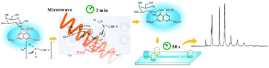
For rapid and low-cost determination of saccharides with microchip electrophoresis, a domestic microwave oven was used to perform the derivatization of saccharides with 8-aminonaphthalene-1,3,6-trisulfonate (ANTS) for laser-induced fluorescence detection with a 405 nm laser diode. The influence of the reaction conditions was systematically examined. Derivatization of xylose, glucose and lactose was fulfilled within 3 min under the optimized condition. With appropriate selection of the separation medium, these model analytes could be resolved within 30 s in borate solutions containing hydroxypropyl cellulose using disposable cyclic olefin copolymer microchips. Theoretical plate numbers of 1.0 × 10⁶ m−1 and limits of detection of 0.15–0.23 μM were achieved. Peak areas were linear up to 1000 μM for all analytes. The applicability of the proposed method was verified by the determination of glucose in human urine and serum. Separation of oligosaccharides from konjac powder was also demonstrated. The domestic microwave oven, the cheap laser diode and disposable microchips used here make the method an economical way for rapid analysis of saccharides.
However, the analysis of saccharides is still a challenge due to their structural complexity with microheterogeneity, and the lack of charged groups or chromophoric moieties.4 Currently, the preferred techniques of saccharide analysis are the chromatography and electrophoresis with different detection routes.4–6 Tedious chemical modifications are always needed in these techniques for certain purposes, such as improving thermal stability and volatility for gas-phase analyses, attaching fluorophores for optical detection and introducing charge for mass spectral analyses.6 Fast sample preparation, efficient separation and sensitive detection are hardly achievable concurrently, the development of rapid, efficient and sensitive methods for analysis of saccharides is still necessary.
As a newly emerging analytical technique, microchip electrophoresis (MCE) has been successfully used in the analysis of DNA fragments,7,8 amino acids,9 proteins,10,11 neurotransmitters,12 saccharides13,14 and other biological molecules with its outstanding advantages, such as high speed, low sample/reagent consumption and easy automation. MCE has shown capability of miniaturization and high sensitivity when laser-induced fluorescence detection (LIF) and electrochemical detection are used. Baba's group has proved that MCE-LIF was suitable for rapid separation of saccharides.14,15 Recently, we have developed economical electrophoresis devices including disposable cyclic olefin copolymer (COC) microchips,16 power supplies and LIF detectors that assembled with cheap optical and electronic parts.17 These home-made MCE devices are portable due to the relatively small dimensions (30.0 cm × 18.0 cm × 31.2 cm). They have been successfully employed for the rapid analysis of samples with complicated matrices.16–19 It should be possible to develop economical ways of saccharides analysis with these devices.
Derivatization is indispensable for introduction of fluorophores when MCE-LIF is used for saccharides. Some aromatic primary amines have been used as fluorescent tags based on reductive amination, such as 2-aminopyridine (AP),20 5-aminonaphthalene-2-sulfonic acid (5-ANSA),21 8-aminopyrene-1,3,6-trisulfate (APTS),14,15 2-aminoacrydone (AMAC)22 and 8-aminonaphthalene-1,3,6-trisulfonate (ANTS).23 ANTS was first demonstrated by Jackson24 for use in polyacrylamide-gel electrophoresis. The optimum excitation wavelength of ANTS is around 356 nm,25 but it can be readily excited with widely available low-cost diode lasers of 405 nm which are mainly used in DVD laser heads. The use of 405 nm laser diodes can ensure the low-cost nature of the apparatus. However, the derivatization time needed for ANTS is quite long,23,26 which significantly compromise the speed of MCE separation. Therefore, it is vital to develop a rapid and effective derivatization protocol for saccharides that matches the high-speed analysis of MCE.
The application of microwave in chemical reactions has been widely recognized including synthetic,27 environmental28 and analytical chemistry. Microwave-assisted sample extraction29 and digestion30 are typical examples in analytical area. We have previously demonstrated a rapid analysis method for histidine, 1-methylhistidine and 3-methylhistidine in human urine by using microwave-accelerated derivatization.31 Momenbeik and Khorasani32 reported a protocol for the pre-column derivatization of sugar samples with p-nitroaniline within 5 min under microwave irradiation before reversed-phase high-performance liquid chromatographic analysis with UV detection. However, microwave-assisted derivatization of saccharides before fluorescence detection was rarely seen.
In the present work, we incorporated microwave-assisted derivatization and MCE-LIF to realize rapid and efficient analysis of saccharides labeled with ANTS. Effects of experimental conditions were systematically investigated with xylose, glucose and lactose as the model analytes. The applicability of the proposed method was evaluated through the determination of glucose in human urine and serum samples. Its capability for analysis of oligosaccharides was evaluated by the separation of extracts of konjac powder, which is a kind of food supplement that was believed to be beneficial to control glycemia, lipids, and systolic blood pressure.33
A domestic microwave oven (P70D20P-TD(W0), 700 W, Galanz, Guangzhou, China) was used for the derivatization reaction. To avoid damage to the magnetron caused by low load, continuous pumping of water was maintained through a PTFE tubing (2.0 mm i.d.) that was introduced into the microwave chamber through the vent holes (Fig. 1). The length of the tubing inside the chamber was approximately 30 cm, which contains 0.94 mL of water. Fluorescence spectra of ANTS and its sugar derivatives were recorded on a fluorescence spectrophotometer (F97Pro, Lengguang Technology Co., Ltd., Shanghai, China).
![]() | ||
Fig. 1 Schematic illustration of the chamber of the microwave oven and the position of the ‘cooling coil’. |
Stock standard solutions of xylose, glucose and lactose with concentrations of 0.1 M were prepared in distilled water and stored in a refrigerator (4 °C). The running buffers for MCE were prepared by mixing given amounts of hydroxypropyl cellulose to sodium tetraborate with appropriate concentrations. The pH of the running buffer was adjusted using 1.0 M hydrochloric acid or 1.0 M sodium hydroxide. All solutions were filtered through 0.45 μm membranes (Shanghai Xinya Purification Materials Factory, Shanghai, China) before use.
Glucomanno-oligosaccharides were extracted with the following procedure: 1.00 g konjac powder was slowly added into 100 mL distilled water and then mixed with 0.49 g neutral β-mannanase to start the reaction in a water bath at 41 °C. The pH of the mixture was adjusted to 7.0. After 3.4 h of incubation, the mixture was boiled for 15 min and centrifuged at 3900 rpm for 10 min.34 The supernatant was freeze-dried (DZF-6020, Shanghai Yiheng Technology Co., Ltd., Shanghai, China) to get glucomanno-oligosaccharides.
The effect of buffer acidity on saccharides separation was studied in a pH range of 3.0 to 8.0, while concentration of borate was maintained at 80 mM and HPC concentration was fixed at 1.0% (m/v). The results suggested that all analytes migrated to the anode and pH had profound effect on the resolution. Xylose and glucose could not fully resolved when pH > 7.0. At pH 4.0–6.0, all these compounds and ANTS were well separated. Hence, pH 5.0 was used for the following experiments based on the compromise of resolution and theoretical plate numbers (Fig. 2a and b).
The effect of borate concentration was examined in the range of 20–100 mM with 1.0% HPC at pH 5.0. The resolution between xylose and glucose was dependent on the borate concentration and reached the highest at 80 mM (Fig. S1a†). The migration time of all derivatives of saccharides decreased with increase of the borate concentration, most probably due to the further suppression of EOF toward the cathode.18 The electrophoretic behavior became unstable and tiny bubbles appeared in the separation channel when the concentration of borate increased to 100 mM. Therefore, 80 mM borate solution was selected for the further experiments.
The influence of HPC concentration on separation was studied from 0.5% to 3.0% (w/v) when borate solution was maintained at 80 mM and pH was adjusted to 5.0. With a small amount of HPC present in the running buffer, satisfactory resolution of three analytes could be achieved. When the concentration of HPC increased from 0.5% to 3.0%, migration time could increase by 79.8% for all analytes. The change of resolution between xylose and glucose was shown in Fig. S1b.† Based on the resolution, 1.0% HPC was selected.
When 80 mM of borate containing 1.0% HPC at pH 5.0 was used, xylose, glucose and lactose could be baseline separated within 30 s, which was pretty match the requirement of rapid and economical separation of saccharides. However, the derivatization needed at least one hour if the conventional procedure was adopted,35 which is approximately 100 times longer than separation. Exploring the efficient derivatization of saccharides that was compatible with the established MCE separation is critical for this work.
Traditional derivatization with ANTS or APTS takes much longer time than that for microchip electrophoretic separation. A domestic microwave oven was employed to perform the derivatization. As shown in Scheme S1,† the formation of CN bond between a saccharide and ANTS is somewhat reversible. Further reduction of C
N bond to C–N leads to a more stable product. Microwave could improve both steps of the reaction due to the polar groups involved.36 To get the optimum condition, various factors were systematically investigated, including the amounts of reagents (ANTS and NaBH3CN), reaction time, and the location of the reaction vial in the microwave chamber.
To run a domestic microwave oven without load may damage the oven. To avoid this type of damage to the magnetron caused by small load, a PTFE ‘cooling coil’ that was filled with flowing water was used. The volume of water in the PTFE tubing was only 0.94 mL, but it was sufficient to avoid the damage of the microwave oven even without any other load in the chamber.
Because the distribution of microwave inside the chamber is not uniform, the best location of the reaction vial should be defined. To find the best location, we put 27 vials (with exactly same reaction mixture) evenly distributed in 3 layers, 9 points in each layer (as shown in Fig. 3), and checked the derivatization performance in each vial. The peak areas of xylose obtained from the mixtures in every vial were plotted in Fig. 3. The best was the center of the bottom layer, which is the center of the rotary tray of the oven.
![]() | ||
Fig. 3 Contours of peak area of the ANTS-derivatized xylose placed at 27 positions (circled in white) in the microwave chamber of the oven. Derivatization conditions: 0.2 M ANTS, reaction under microwave irradiation of 700 W for 3 min. X-, Y- and Z-directions were shown by axis in Fig. 1. Other conditions are same as in Fig. 2b. |
ANTS was commonly used for saccharides labeling via acid catalyzed reductive amination. ANTS provides both fluorescent tag and negative charge from three sulfonic groups, which enables neutral sugar molecules to migrate under the action of electric field.24,35 Polar solvents, especially water, can facilitate the absorption of microwave36 and are therefore favorable to the reaction. As shown in Fig. 4, the fluorescence intensities of ANTS labeled analytes increased evidently with the concentration of ANTS from 0.05 to 0.125 M and plateaued at higher concentrations. Therefore 0.125 M ANTS was used for the derivatization, which corresponds to 25 μL of 0.2 M ANTS added to the mixture. It was found that the concentration of acetic acid used to dissolve the ANTS affected the fluorescence intensity, too. We studied its influence in a concentration range of 0–25% (v/v). The results are shown in Fig. S3.† The maximum fluorescence intensities were achieved when 15% acetic acid was used in ANTS solution. Lower concentration of acetic acid gave rise to the apparent decrease of the signal, because acetic acid is a catalyst of the formation of the Schiff base,35 while too high concentration of acetic acid might protonate –NH2 of ANTS and lead to decrease of the signal. The effect of NaBH3CN was also examined, 0.25 M NaBH3CN gave the optimum results, as shown in Fig. 5.
![]() | ||
Fig. 4 Effect of the concentration of ANTS. Conditions are same as in Fig. 3 except the microcentrifuge vial containing sample was placed at the center of the rotary tray. |
![]() | ||
Fig. 5 Effect of the amount of NaBH3CN. Conditions are same as in Fig. 4. |
The output power of the domestic microwave oven can normally be adjusted by the ‘firepower’, but this adjustment is actually realized through switching the magnetron on and off intermittently.37 For the derivatization, we need microwave to be applied constantly, so 100% fire power (700 W) was used in all of our experiments. The fluorescence of all analytes increased with the increase of the time of microwave treatment. To evaluate the efficiency of the derivatization, the samples obtained with microwave was compared with that derivatized in an 80 °C boiling bath for 2.5 h, which was used in previous works.23 The experimental results indicated that 3 min was enough to reach the consequence of traditional derivatization, so 3 min was used for the next steps. The peak heights of xylose, glucose and lactose obtained with microwave were 126.1%, 97.7%, and 77.0% of that with water bath, respectively. The difference in the derivatization efficiencies between normal heating and microwave-assisted derivatization might be related to the molecular sizes that affect the motion and reorientation of the molecules under electromagnetic fields provided by the microwave.
Sugars | Regression equation | LOD (μM) | LOQ (μM) | Linear range (μM) | RSD (%) | |||
---|---|---|---|---|---|---|---|---|
Slope (×10−4) | Intercept (×10−3) | Correl. coeff. | Migration time (n = 5) | Peak area (n = 5) | ||||
Xylose | 8.51 | 4.29 | 0.9971 | 0.23 | 0.76 | 0.8–1000 | 0.88 | 2.7 |
Glucose | 4.54 | 7.88 | 0.9995 | 0.16 | 0.55 | 0.6–1000 | 1.9 | 3.1 |
Xylose | 4.42 | 2.69 | 0.9990 | 0.15 | 0.49 | 0.5–1000 | 0.40 | 2.9 |
![]() | ||
Fig. 6 Electropherograms of (a) human urine sample (Samples 1) (A), and one spiked with 1 mM of saccharides (B); and (b) human serum sample (A), and one spiked with 8 mM of saccharides (B). Conditions and peak identification are same as in Fig. 2b. The urine and serum samples were diluted by factors of 10 and 100, respectively. |
Analyte | Content (mM) | Added (mM) | Totally found (mM) | Recovery (%) | Within-day RSD (%) (n = 5) | Between-day RSDb (%) (n = 3) |
---|---|---|---|---|---|---|
a Represents not detected.b Represents results detected in three days. | ||||||
Xylose | n.d.a | 1.00 | 1.10 | 109.8 | 0.5 | 1.6 |
2.00 | 2.12 | 106.2 | 1.8 | 3.4 | ||
3.00 | 2.95 | 98.3 | 0.7 | 1.5 | ||
Glucose | 0.62 | 1.00 | 1.64 | 102.5 | 1.3 | 2.9 |
2.00 | 2.64 | 101.3 | 2.8 | 3.8 | ||
3.00 | 3.82 | 106.7 | 1.7 | 2.6 | ||
Lactose | n.d. | 1.00 | 1.05 | 105.3 | 0.9 | 0.6 |
2.00 | 1.95 | 97.6 | 2.4 | 2.1 | ||
3.00 | 3.34 | 111.4 | 1.4 | 1.9 |
For the method validation, three addition levels, 1.00, 2.00 and 3.00 mM sugar standards were spiked into urine samples, 5.00, 8.00 and 10.00 mM were spiked into serum samples to check the standard recoveries. These spiked saccharides in both types of real samples were determined successfully (Tables 2, 3 and S1†). The recoveries for all three addition levels were in a range of 91.0–118.3%. There was no significant difference in RSDs of within-day and between-day values and the maximum was below 5%.
Analyte | Content (mM) | Added (mM) | Totally found (mM) | Recovery (%) | Within-day RSD (%) (n = 5) | Between-day RSD (%) (n = 3) |
---|---|---|---|---|---|---|
Xylose | n.d. | 5.00 | 5.60 | 112.1 | 3.1 | 3.7 |
8.00 | 7.42 | 92.8 | 1.8 | 2.9 | ||
10.00 | 10.24 | 102.4 | 0.6 | 4.7 | ||
Glucose | 6.44 | 5.00 | 12.36 | 118.3 | 3.7 | 4.3 |
8.00 | 14.94 | 106.3 | 3.3 | 2.1 | ||
10.00 | 17.23 | 107.8 | 2.9 | 2.7 | ||
Lactose | n.d. | 5.00 | 4.81 | 96.1 | 4.7 | 4.0 |
8.00 | 7.81 | 97.6 | 1.9 | 0.9 | ||
10.00 | 9.66 | 96.6 | 3.1 | 1.5 |
After microwave-assisted derivatization with ANTS, GMOs were separated on COC disposable microchips. Parameters including borate concentration (20–120 mM), HPC concentration (0.5–2.5%, m/v) and buffer pH (5.0–10.0) of running buffer were screened for improving the separation efficiency. The results suggested that the concentration of borate and HPC could significantly influence the migration time of GMOs, similar to that for xylose etc. Effect of acidity on the separation of oligosaccharides was negligible in a pH range of 5.0–10.0. The electropherogram of GMOs from konjac powder at pH 8.0 were shown in Fig. 7. Under this condition, at least seven components in GMO could be well resolved.
![]() | ||
Fig. 7 Electropherogram of glucomanno-oligosaccharides (GMOs) from konjac powder. Conditions are same as in Fig. 2b, except buffer pH was 8.0. |
Footnote |
† Electronic supplementary information (ESI) available: Fig. S1–S3, Scheme S1. See DOI: 10.1039/c4ra07934e |
This journal is © The Royal Society of Chemistry 2014 |