SERS performance of graphene oxide decorated silver nanoparticle/titania nanotube array

Yibing Xie*ab and Yujie Mengab
aSchool of Chemistry and Chemical Engineering, Southeast University, Nanjing 211189, China. E-mail: ybxie@seu.edu.cn
bSuzhou Research Institute of Southeast University, Suzhou 215123, China

Received 31st July 2014 , Accepted 27th August 2014

First published on 27th August 2014


Abstract

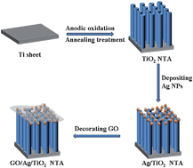
A graphene oxide (GO) decorated silver nanoparticle/titania nanotube array (Ag/TiO2 NTA) has been designed as a surface-enhanced Raman scattering (SERS) substrate for the sensitive detection of organic molecules. Anatase TiO2 NTA was synthesized by controlled anodic oxidation and annealing treatment processes. Ag/TiO2 NTA was formed by depositing silver nanoparticles (Ag NPs) on the surface of anatase TiO2 NTA through the polyol process. GO/Ag/TiO2 NTA was prepared by decorating GO on the surface of Ag/TiO2 NTA through an impregnation process. Methyl blue (MB) was used as the probe molecule to evaluate the adsorption capability and SERS activity of the as-prepared SERS substrates. The adsorption ratio of MB was increased from 25.2% for Ag/TiO2 NTA up to 38.0% for GO/Ag/TiO2 NTA, presenting an obvious improvement of the adsorption capability. The analytical enhancement factor was increased from 1.06 × 10⁴ for Ag/TiO2 NTA up to 3.67 × 10⁴ for GO/Ag/TiO2 NTA, presenting an obvious improvement of SERS detection performance. The GO/Ag/TiO2 NTA substrate accordingly achieved a very low detection limit of 1.0 × 10⁻⁹ M MB. GO/Ag/TiO2 NTA also exhibited good adsorption capability to bisphenol A (BPA) with a weak affinity, presenting a SERS detection limit of 5 × 10⁻⁷ M BPA. Moreover, GO/Ag/TiO2 NTA was able to achieve self-cleaning functionality through photocatalytic degradation of organic molecules adsorbed on the SERS substrate under solar light irradiation. GO/Ag/TiO2 NTA having good adsorption capability, self-cleaning properties, reproducibility and cycleability contributed to a promising application for sensitive and recyclable SERS detection.


1. Introduction

Surface-enhanced Raman scattering (SERS) spectroscopy, as a quite powerful tool, has been applied in environmental monitoring, chemical analysis and biomedical research due to its supersensitivity, rapid response and fingerprint effect.1–3 Two main mechanisms generally contribute to the Raman scattering enhancement observed in SERS, which involve the electromagnetic mechanism and the chemical enhancement mechanism. The former is based on the enhancement of the local electromagnetic field, whereas the latter is based on charge transfer between absorbed molecules and metal surface.4 Since the surface morphologies and structures of the substrates determine the generation and intensity of Raman signals, the substrates play a vital role in SERS detection.5 An efficient SERS substrate can not only provide strong surface enhancement of Raman scattering, but also stable, reproducible and recyclable SERS detection.6,7 It is well known that the nanostructured noble metals (such as silver and gold) can be well used as SERS substrates to generate strong SERS effect.8,9 Such noble metals with well-controlled nanoarray structure can further improve the sensitivity and reproducibility of SERS detection.10,11 TiO2 or TiN nanotube or nanopore arrays with well ordered and aligned nanostructure have been well fabricated as feasible supporting materials for versatile applications.12–20 It has been found that TiO2 can also generate weak SERS activity.21 In addition, anatase TiO2 has been extensively used for photocatalytic degradation of organic compounds because of its high photocatalytic activity, nontoxicity and stability.22 Therefore, it is reasonable to fabricate noble metal decorated TiO2 for both SERS detection and photocatalysis application. After SERS measurements, the substrates can achieve a self-cleaning function by a photocatalytic process and recycled for subsequent SERS detection.23,24

In addition to the nanostructured noble metals, the sensitivity of SERS detection also relies on the amount of analyte molecules in the enhancement region.25 In other words, SERS effect is observed when organic molecules are adsorbed on the SERS substrate. The insufficient molecule adsorption usually causes low electromagnetic enhancement and chemical enhancement effect. Therefore, SERS substrate with high adsorption capacity is essential for sensitive SERS detection. Grapheme oxide (GO) is a one-atom-thick and two dimensional carbon network containing carboxylic acid groups at the sheet edges, as well as hydroxyl and epoxy groups on the graphite plane.26 Owing to the presence of aromatic domains and high specific surface area, GO is an ideal host material to adsorb a wide variety of organic molecules. Moreover, GO can chemically enhance the Raman signals of adsorbed molecules because the graphene has been recently reported to be used as a substrate with Raman enhancement effect. The observed SERS phenomenon has been ascribed to chemical enhancement mechanism.27 Recently, it is reported that a noble metal/graphene oxide as SERS substrate is capable of sensitive detection of a variety of organic molecules, especially aromatic compounds keeping high affinity toward GO.25 Ag NPs/GO hybrid material can be applied for SERS detection, showing 2–3 times of Raman signal enhancement when compared to pure Ag NPs.28 Ag NPs/reduced graphene oxide on silicon surface as the substrate can be used as an efficient SERS substrate to detect aromatic molecules with a low detection limit at scale of 10−9 M.29

In this study, GO decorated Ag/TiO2 NTA (GO/Ag/TiO2 NTA) was design as a stable and recyclable SERS substrate for sensitive SERS detection of organic compounds and photocatalytic self-cleaning.30 Fig. 1 shows the schematic displaying the fabrication process of GO/Ag/TiO2 NTA substrate. Briefly, highly ordered anatase TiO2 NTA was synthesized by a controlled anodic oxidation and annealing treatment process. The well-dispersed Ag NPs were deposited on the surface of anatase TiO2 NTA to form Ag/TiO2 NTA via the polyol process.31 GO was then decorated on the surface of Ag/TiO2 NTA to form GO/Ag/TiO2 NTA through an impregnation process.32 The adsorption capability and SERS activity of Ag/TiO2 NTA and GO/Ag/TiO2 NTA were investigated. The sensitivity and recyclability of GO/Ag/TiO2 NTA were fully evaluated using methyl blue as the probe molecule. GO/Ag/TiO2 NTA was also used for SERS detection of bisphenol A with a weak adsorption affinity.


image file: c4ra07865a-f1.tif
Fig. 1 Schematic displaying the fabrication process of GO/Ag/TiO2 NTA substrate.

2. Experimental

2.1 Material

Titanium sheet (Ti, thickness of 0.2 mm, purity >99.6%) was purchased from Good fellow Cambridge Ltd. Ethylene glycol (EG), ammonium fluoride (NH4F), phosphoric acid (H3PO4) silver nitrate (AgNO3), sodium borohydride (NaBH4), polyvinylpyrrolidone (PVP) and methyl blue (MB) bisphenol A (BPA) and ethanol purchased from Sinopharm chemical company are analytical grade and used without further purification. Distilled water was fully used in all experiments.

2.2 Preparation of GO/Ag/TiO2 NTA

TiO2 NTA was fabricated by anodic oxidation of titanium sheet. The anodization process was conducted at 30 V for 2 h in water and ethylene glycol mixture solution (volume ratio, 1[thin space (1/6-em)]:[thin space (1/6-em)]1) containing 0.5 M H3PO4 and 0.2 M NH4F in a two-electrode cell using Ti foil as the working electrode and a Pt sheet as the counter electrode. The obtained amorphous TiO2 was annealed at 450 °C for 2 h to form anatase TiO2 NTA with photocatalytic activity. Ag/TiO2 NTA was fabricated by depositing Ag NPs on the surface of TiO2 NTA through the polyol process. Briefly, the as-prepared TiO2 NTA was immersed in water and ethylene glycol mixture solution (volume ratio, 1[thin space (1/6-em)]:[thin space (1/6-em)]1) containing 0.001 M PVP, 0.035 M NaBH4, 0.055 M AgNO3 at ambient temperature for 7 h, and subsequently rinsed with DI water and dried in vacuum. GO was prepared through chemically oxidizing graphite powders by a modified Hummers method.33 The GO/Ag/TiO2 NTA was obtained by decorating GO on Ag/TiO2 through an impregnation method. Typically, the as-prepared Ag/TiO2 NTA was immersed in a 0.5 mg mL−1 GO aqueous solution for 3 h, and subsequently rinsed with DI water and dried in vacuum. For a comparison, GO/TiO2 NTA was also fabricated by coating GO on the surface of TiO2 NTA using the same above method.

2.3 Characterization

The surface morphologies of TiO2, Ag/TiO2, and GO/Ag/TiO2 NTA were characterized by the Field-Emission Scanning Electron Microscope (FE-SEM, Zeiss Ultra-Plus). UV-Vis spectrum was measured to analyze MB concentration by UV/Vis Spectrometer (Shimadzu UV-2100 UV/Vis Spectrometer). Raman spectrum was measured to investigate SERS substrates using a Renishaw Invia Reflex System Raman spectrometer. The single layer structure of graphene oxide was investigated using a transmission electron microscope (TEM, JEOJ-2010) with an acceleration voltage of 200 kV. The excitation wavelength was 785 nm. The 50× objective lens was used to focus the laser light on the samples. The laser power was 1%, and integration time for each sample was 10 s.

2.4 Adsorption measurement

TiO2, Ag/TiO2, GO/TiO2, and GO/Ag/TiO2 NTA were respectively immersed in freshly prepared 1.0 × 10⁻⁵ M MB and 2.2 × 10⁻⁴ M BPA solution for 2 h in the dark at ambient temperature to achieve the adsorption equilibrium. The concentration change of MB and BPA solution before and after the adsorption on different substrates was analyzed according to the of characteristic peak intensity of UV-Vis spectrum.

2.5 SERS measurement

All SERS substrates were immersed in freshly prepared MB or BPA solution with controlled concentration for 2 h in the dark, then rinsed with solvent and dried in vacuum for the Raman spectrum measurements. The Ag/TiO2, GO/TiO2 and GO/Ag/TiO2 NTA were used for SERS detection of 10−5 M MB solution. The GO/Ag/TiO2 NTA was used for SERS detection of MB solution with different concentration from 10−9 to 10−4 M. The Ag/TiO2 and GO/Ag/TiO2 NTA were used for SERS detection of 5 × 10⁻⁴ M BPA solution. The GO/Ag/TiO2 NTA was used for SERS detection of BPA ethanol solution with different concentrations from 5 × 10⁻⁷ M to 5 × 10⁻⁴ M.

3. Results and discussion

3.1 Characterization of Ag/TiO2 NTA and GO/Ag/TiO2 NTA

Fig. 2(A–E) shows the SEM images of TiO2, Ag/TiO2 NTA and GO/Ag/TiO2 NTA. TiO2 NTA exhibited highly ordered, vertically aligned nanotube microstructure with tube diameter of 100–120 nm, tube length of 850–950 nm and tube wall thicknesses of 15–20 nm. The interspace between the neighboring tubes is 40–60 nm (see Fig. 2(A and B)). Concerning Ag/TiO2 NTA, Ag NPs with average diameter of 10–15 nm were evenly distributed on the pore mouth of TiO2 NTA. Such Ag NPs were hardly observed on the tube walls of TiO2 NTA (see Fig. 2(C and D)). Concerning GO/Ag/TiO2 NTA, a continuous and transparent GO monolayer film was well coated on the top surface of Ag/TiO2 NTA (see Fig. 2(E and F)). Fig. 2(G) shows the TEM image of GO. Obviously, GO kept a transparent and well expanding monolayer structure. Such a GO was very suitable for functional modification of Ag NPs to achieve good adsorption capability and SERS activity as well.
image file: c4ra07865a-f2.tif
Fig. 2 SEM images of (A and B) TiO2 NTA; (C and D) Ag/TiO2 NTA and (E and F) GO/Ag/TiO2 NTA; TEM image of GO.

Raman spectroscopy was employed to further confirm the transparent GO coating layer on the surface of Ag/TiO2 NTA. Fig. 3 shows the Raman spectra of Ag/TiO2 NTA and GO/Ag/TiO2 NTA. The Raman characteristic peaks at 155, 396, 517, 638 cm−1 were observed in both samples, which were assigned to the Eg, B1g, B1g or A1g Eg modes of anatase TiO2, respectively.34 For the GO/Ag/TiO2 NTA, the two additional prominent peaks at 1320 cm−1 and 1597 cm−1 were regarded as the characteristic D band and G band of GO. The G-band appearing at around 1597 cm−1 was a significant characteristic of sp2 hybridized carbon, which could provide the information on the in-plane vibration of sp2-bonded carbon domains.35 The D-band appearing at around 1320 cm−1 revealed the presence of sp3 defects within the hexagonal graphitic structure,36 which was associated with the amorphous carbon, or edges that break the symmetry and selection rule.37 Such characteristics demonstrate that the expanding GO sheets were really decorated on the Ag/TiO2 NTA.


image file: c4ra07865a-f3.tif
Fig. 3 Raman spectra of (a) Ag/TiO2 NTA and (b) GO/Ag/TiO2 NTA.

3.2 Adsorption capability of GO/Ag/TiO2 NTA

The adsorption capability of Ag/TiO2 NTA and GO/Ag/TiO2 NTA were investigated by immersing these substrates in 1.0 × 10⁻⁵ M MB solution for 2 h in the dark. MB concentration was evaluated before and after adsorption according to the characteristic peak intensity at 664 nm using UV-Vis spectrophotometer. Fig. 4(A) shows UV-Vis spectra of MB solution before and after adsorption on different SERS substrates. They demonstrated different adsorption effects. Fig. 4(B) shows the adsorption ratio of MB on different SERS substrates. The adsorption ratio of MB followed the decreasing order of GO/Ag/TiO2 > GO/TiO2 > Ag/TiO2. Obviously, both Ag/TiO2 NTA and GO/TiO2 NTA exhibited a similar adsorption capacity, but much higher than TiO2 NTA. The covalent bonding between Ag NPs and MB molecule contributed to improving the adsorption capability of Ag/TiO2 NTA.38 Moreover, GO/Ag/TiO2 NTA showed the highest adsorption capacity. This is owing to the unique chemical structure of GO. The highly negative charged GO possesses the oxygen-containing functional groups, which can interact with the positive charged MB molecule through electrostatic-interaction.39 In addition, GO with aromatic domains, can also form strong π–π stacking interactions with π-electron-rich MB molecule.40 GO/Ag/TiO2 NTA exhibited higher adsorption capability than GO/TiO2 NTA, indicating that the introduction of Ag NPs between TiO2 NTA and GO enlarged the specific surface area of the composite.41
image file: c4ra07865a-f4.tif
Fig. 4 UV-Vis spectra and adsorption ratio of (A and B) 10−5 M MB solution and (C and D) 2.2 × 10⁻⁴ M BPA solution on the substrate of TiO2 NTA, Ag/TiO2 NTA, GO/TiO2 NTA and GO/Ag/TiO2 NTA.

Fig. 4(C) shows UV-Vis spectra of 2.2 × 10⁻⁴ M BPA solution before and after adsorption on SERS substrates. The similar trend was obtained for the adsorption ratio of MB and BPA on SERS substrates. Comparatively, due to the weak adsorption affinity to the substrate, BPA exhibited weaker adsorption capability than MB. Fig. 4(D) shows the adsorption ratio of BPA on different SERS substrates. Both TiO2 NTA and Ag/TiO2 NTA had similar and comparable adsorption ratio of BPA, presenting low adsorption capability. This was owing to the weak affinity between Ag NPs and BPA. However, the adsorption capability of both GO/TiO2 NTA and GO/Ag/TiO2 NTA was much higher than that of Ag/TiO2 NTA. This indicates that GO played an important role on the adsorption of BPA. The benzene rings of BPA could interact with GO through π–π interaction. The hydroxyl groups of GO could form hydrogen bonds with hydroxyl groups of BPA.42 These results suggest that GO/Ag/TiO2 NTA indeed exhibited higher adsorption capability.

3.3 SERS performance of Ag/TiO2 NTA and GO/Ag/TiO2 NTA

The SERS activity of the Ag/TiO2 NTA and GO/Ag/TiO2 NTA was evaluated using MB as probe molecule. Fig. 5(A) shows the SERS spectra of 10−5 M MB aqueous solution adsorbed on Ag/TiO2, GO/TiO2 and GO/Ag/TiO2 NTA. Obviously, SERS signals of MB molecule on all SERS substrates were observed and the characteristic bands could be clearly distinguished. The Raman peaks at 1622 cm−1 and 1397 cm−1 were corresponding to the C–C ring stretching vibration and C–N–C skeletal bending vibration of MB molecule, respectively.43 Concerning GO/Ag/TiO2 NTA and GO/TiO2 NTA, the additional prominent peak at 1320 cm−1 was the characteristic D band of GO. On the other hand, the G band overlapped significantly with the C–C ring stretching vibration of MB molecule at 1622 cm−1, making it difficult to distinguish above two vibration modes from each other. The SERS peak intensity obtained on the GO/Ag/TiO2 NTA and Ag/TiO2 NTA were far higher than that on GO/TiO2 NTA. This indicates that the SERS chemical enhancement effect of GO was much weaker than SERS electromagnetic field enhancement effect of Ag NPs. The SERS peak intensity at 1622 cm−1 obtained on the GO/TiO2 NTA was only 3.6% of that on GO/Ag/TiO2 NTA. The contribution of GO layer to the Raman intensity of 1622 cm−1 was so small that it could be ignored in the calculation of SERS enhancement factor. Moreover, GO/Ag/TiO2 NTA had better SERS performance than Ag/TiO2 NTA. Firstly, it is reported that graphene coating layer did not alter the plasmonic properties of Ag NPs, causing less influence on its electromagnetic SERS enhancement.44 Therefore, the ultrathin GO layer coating on the Ag NPs did not impede SERS enhancement of Ag NPs.45 Secondly, GO was regarded as a favorable SERS platform. When MB was adsorbed on GO, the charge transfer process occurred between the oxygen-functional groups and MB molecule, which was the dominant mechanism contributing to the additional SERS enhancement. It was reported that the Raman peak intensity of the probe molecules descended along with the increase of the reduction degree of GO when GO was converted to reduced graphene oxide (RGO).46 The chemical enhancement of SERS using GO would arise from π–π stacking and charge transfer from the oxygen-functional groups to the probe molecules. When GO was fully converted to RGO, the number of the oxygen-functional groups highly decreased, resulting in a corresponding decrease of the Raman peak intensity of the probe molecules28 Thirdly, GO had a double-sided polyaromatic scaffold with an ultrahigh specific surface area and therefore was capable of adsorbing a larger number of MB molecule. The improved adsorption of MB molecule on GO occurred in the SERS enhancement region, leading to better SERS enhancement effect.25
image file: c4ra07865a-f5.tif
Fig. 5 (A) SERS spectra of 10−5 M MB aqueous solution adsorbed on (a) GO/Ag/TiO2 NTA, (b) Ag/TiO2 NTA and (c) GO/TiO2 NTA; (B) normal Raman spectra of 10−2 M MB aqueous solution.

The enhancement effect of SERS substrates was estimated by calculating enhancement factor (EF) and analytical enhancement factor (AEF) using the equations of EF = (ISERS/Nsurface)/(IRS/Nbulk) and AEF = (ISERS/CSERS)/(IRS/CRS).47 Herein, ISERS and IRS are the intensities of SERS signal and normal Raman signal at 1622 cm−1; CRS and CSERS are MB concentration under normal Raman and SERS condition; Nbulk and Nsurface are MB molecule number illuminated by the laser focus spot under normal Raman and SERS condition. The adsorption quantity per unit area can be calculated using the equation of Qe = [(C0Ce)V]/S, where C0 and Ce are MB concentration before and after adsorption by Ag/TiO2 NTA or GO/Ag/TiO2 NTA; V is the volume of MB solution; S is the surface area of Ag/TiO2 NTA or GO/Ag/TiO2 NTA. The C0 was determined to be 10−5 M. Ce was calculated according to the absorbance change before and after adsorption and Lambert–Beer Law. The laser spot has a diameter of 1.0 μm and surface area of about 7.9 × 10⁻⁹ cm−2. The volume of laser focus spot is 7.9 × 10⁻¹⁰ L. Fig. 5 shows SERS spectra of 10−5 M MB on Ag/TiO2 NTA, GO/Ag/TiO2 NTA and normal Raman spectra of 10−2 M MB. The MB concentration was determined to be 10−5 M for CSERS and 10−2 M for CRS. The adsorption quantity per unit area was estimated to be 1.0 nmol cm−2 for Ag/TiO2 NTA and 1.8 nmol cm−2 for GO/Ag/TiO2 NTA. Nsurface was accordingly calculated to be 4.8 × 10⁶ and 8.5 × 10⁶. Nbulk was calculated to be 4.8 × 10¹². As a result, the EF values of Ag/TiO2 NTA and GO/Ag/TiO2 NTA were estimated to be 1.0 × 10⁷ and 2.1 × 10⁷. The corresponding AEF values were estimated to be 1.06 × 10⁴ and 3.67 × 10⁴. For a comparison, the Raman peak at 1397 cm−1 was also used for the enhancement factor calculation because it was free from potential signal interference that may arise from GO. On the base of Raman peak at 1397 cm−1, the EF values of Ag/TiO2 NTA and GO/Ag/TiO2 NTA were estimated to be 2.2 × 10⁷ and 3.6 × 10⁷. The corresponding AEF values were estimated to be 2.2 × 10⁴ and 6.4 × 10⁴. Similar results of the enhancement factor were obtained according to the different Raman peaks at 1622 and 1397 cm−1. The Raman signal of MB was enhanced about 2–3 times using GO/Ag/TiO2 NTA instead of Ag/TiO2 NTA, indicating an improved SERS enhancement effect. It was also reported that GO coated on Ag octahedron was used as SERS substrate for 4-aminothiophenol detection and the EF value of GO/Ag substrate was about 106 (ref. 28). The SERS activity of GO/Ag/TiO2 NTA substrate was comparable with or even superior to as-reported GO/Ag substrate.

3.4 SERS performance of GO/Ag/TiO2 NTA

The Raman enhancement effect of GO/Ag/TiO2 NTA was further investigated through SERS detection of MB solution with different concentration. Fig. 6(A) shows the Raman spectra of 10−9 M to 10−4 M MB solution adsorbed on GO/Ag/TiO2 NTA substrate. The Raman characteristic peak intensity at 1622 cm−1 was quite sensitive to MB concentration. The corresponding Raman peak intensity descended along with the decrease of MB concentration. Fig. 6(B) shows the Raman characteristic peak intensity at 1622 cm−1 in terms of MB concentration. A quasi-linear relationship was determined at a low concentration range from 10−9 to 10−6 M. It indicates that the SERS active sites were enough for the full adsorption of MB in this concentration range. However, such an increase of Raman peak intensity would become insignificant at the higher concentration above 10−5 M due to the saturated coverage of SERS active sites. The Raman signals of MB could still clearly be observed at a concentration as low as 10−9 M. The low detection limit at a scale of 10−9 M demonstrated the high sensitivity of SERS detection performance using GO/Ag/TiO2 NTA substrate. The similar result with a detection limit of 10−9 M was also reported when using Ag NPs-decorated single-layer reduced graphene oxide film on silicon surface as the substrate for Raman spectrum detection of MB.29
image file: c4ra07865a-f6.tif
Fig. 6 (A) Raman spectra of 10−9 M to 10−4 M MB adsorbed on GO/Ag/TiO2 NTA; (B) the intensity of Raman characteristic peaks at 1622 cm−1 in terms of MB concentration.

3.5 Stability of GO/Ag/TiO2 NTA

The stability of Ag/TiO2 NTA and GO/Ag/TiO2 NTA substrates were investigated through exposure treatment under ambient condition. Fig. 7 shows Raman spectra of 10−4 M MB solution adsorbed on Ag/TiO2 NTA and GO/Ag/TiO2 NTA with exposure treatment for 0 day and 60 days. Concerning freshly prepared Ag/TiO2 NTA and GO/Ag/TiO2 NTA substrates, the Raman characteristic peaks of MB at 1622 cm−1 and 1397 cm−1 could be clearly detected. In view of the same substrates with exposure treatment for 60 days, The Raman characteristic peak intensity at 1622 cm−1 decreased 55% for Ag/TiO2 NTA and only 5% for GO/Ag/TiO2 NTA. It indicates that the GO coating on the Ag NPs surface indeed could enhance the stability of Ag/TiO2 NTA substrate and maintained comparable detection sensitivity. It is well known that Ag NPs are easily oxidized when exposed to ambient condition. The oxidation of the Ag NPs results in significant changes of plasmonic properties and thus will greatly affect their SERS performance.48 Besides, the photobleaching phenomenon raised from the decomposition or carbonization of the organic molecules can cause a significant decrease of the SERS activity.49 Concerning GO/Ag/TiO2 NTA substrate, the highly impermeable GO serves as a barrier isolates Ag NPs from the oxygen in ambient environment. The GO coating layer on Ag NPs is able to prevent Ag NPs from oxidation. Meanwhile, the GO also stabilizes the probe molecule via π–π interaction. This interaction enables the charge transfer between GO and MB molecule. MB molecule can be excited from ground state to excitation state when absorbing suitable photon energy. The GO in the GO/Ag/TiO2 NTA provides additional path for the MB molecule to relax from excitation state to ground state, avoiding the probability of photobleaching phenomenon.50
image file: c4ra07865a-f7.tif
Fig. 7 Raman spectra of 10−4 M MB solution adsorbed on (A) Ag/TiO2 NTA and (B) GO/Ag/TiO2 NTA with exposure treatment for (a) 0 day and (b) 60 days.

3.6 Self-cleaning performance of GO/Ag/TiO2 NTA

The recyclability of SERS substrate is very important for practical SERS spectrum detection application. The GO/Ag/TiO2 NTA showed the SERS detection limit of 1.0 × 10⁻⁹ M MB. The SERS activity of GO/Ag/TiO2 NTA substrate was comparable and even superior to some reported Ag-based substrates.28,51 Furthermore, GO/Ag/TiO2 NTA substrate could achieve self-cleaning function through photocatalytic degradation of organic molecules absorbed on the surface of SERS substrate. The self-cleaning performance of GO/Ag/TiO2 NTA was investigated through photocatalytic decomposition of MB molecule under solar light irradiation. A 150 W Xenon arc lamp served as a simulated solar light source. Prior to solar light irradiation, both GO/Ag/TiO2 NTA and Ag/TiO2 NTA were immersed in 1.2 × 10⁻⁵ M MB solution for 2 h in the dark to reach adsorption equilibrium. The solution samples were collected at regular intervals to analyze MB concentration during photocatalytic degradation of MB. Fig. 8(A–C) shows the UV-Vis spectra of MB during direct photolysis, Ag/TiO2 NTA and GO/Ag/TiO2 NTA photocatalysis processes. The Raman characteristic peak intensity at 664 nm was slightly decreased during direct photolysis process. The degradation ratio was about 10% after 150 min. Comparatively, the Raman characteristic peak almost completely disappeared after 150 min during Ag/TiO2 NTA and GO/Ag/TiO2 NTA photocatalysis processes. Fig. 8(D) shows the photocatalytic degradation curves of MB on the base of Ag/TiO2 NTA and GO/Ag/TiO2 NTA under solar light irradiation. The degradation ratio of MB was 85.3% for the Ag/TiO2 NTA and 92.3% for GO/Ag/TiO2 NTA. So, the GO/Ag/TiO2 NTA exhibited the higher photocatalytic degradation efficiency than Ag/TiO2 NTA. In general, TiO2 photocatalysis degradation reaction often follows the pseudo first-order kinetic law.52 The photocatalysis degradation curves is generally fitted using a Langmuir–Hinshelwood kinetic equation of ln[thin space (1/6-em)]C/C0 = kt, where C/C0 is the degradation ratio of MB; t is reaction time; and k is the apparent reaction-rate constant.53 Accordingly, the reaction rate constant was determined to be 0.0132 min−1 for Ag/TiO2 NTA and 0.0165 min−1 for GO/Ag/TiO2 NTA. GO/Ag/TiO2 NTA had higher photocatalysis reactivity than Ag/TiO2 NTA to form a self-cleaning surface of SERS substrate. For Ag/TiO2 NTA, the work function of Ag (4.26 eV) is lying below the conduction band of TiO2 (4.2 eV),54 the Schottky barriers are formed between Ag NPs and TiO2 NTA. As a result, the photogenerated electrons facilely transfer from the conduction band of TiO2 NTA to Ag NPs. For GO/Ag/TiO2 NTA, both Ag NPs and GO serve as acceptors of the photogenerated electrons from TiO2 NTA. Ag NPs get intimate contact with GO at the top surface of TiO2 NTA. The photogenerated electrons are transferred from Ag NPs to GO. The electrons will react with surface adsorbed oxygen molecules or dissolved oxygen in water form various reactive oxidative radical species leading to the degradation of MB. The electron-accepting and transporting properties of GO and the intimate contact are formed between the GO and Ag NPs, which greatly facilitates the transfer of the photoinduced electrons.55 Additionally, GO can well adsorb MB molecule and then concentrate them near the GO/Ag/TiO2 NTA surface to promote the photocatalytic degradation of organic molecule.
image file: c4ra07865a-f8.tif
Fig. 8 UV-Vis spectra of 1.2 × 10⁻⁵ M MB through (A) direct photolysis, (B) Ag/TiO2 NTA photocatalysis, and (C) GO/Ag/TiO2 NTA photocatalysis under solar light irradiation; (D) photocatalytic degradation curves of MB on the base of Ag/TiO2 NTA and GO/Ag/TiO2 NTA.

The recyclability of GO/Ag/TiO2 NTA was further investigated in SERS spectrum detection application. Briefly, the GO/Ag/TiO2 NTA was immersed in 10−6 M MB solution for 2 h and conducted Raman spectrum measurement. This GO/Ag/TiO2 NTA substrate with adequate chemisorption of MB was immersed in distilled water and conducted photocatalysis degradation reaction for 150 min under solar light irradiation. The complete degradation of organic molecule caused to form the recovered GO/Ag/TiO2 NTA substrate, achieving a self-cleaning function. This recovered SERS substrate was applied for Raman spectrum measurement again. The above procedure was denoted as one whole recycle. SERS activity and self-cleaning performance of GO/Ag/TiO2 NTA substrate was conducted for twenty recycles. Fig. 9(A) shows SERS spectra of MB for twenty recycles through photocatalysis self-cleaning treatment of GO/Ag/TiO2 NTA. Obviously, GO/Ag/TiO2 NTA substrate with adequate chemisorption of MB had similar Raman spectrum during twenty recycling applications. Comparatively, all characteristic peaks of MB were completely disappeared in Raman spectrum after complete photocatalysis degradation of the adsorbed MB under solar light irradiation. Herein, MB molecule adsorbed on the substrate was degraded into some small molecules such as CO2 and H2O. So, the Raman peak could be completely neglected on the self-cleaning GO/Ag/TiO2 NTA substrate. Fig. 9(B) shows Raman characteristic peak intensity before and after self-cleaning treatment. In view of the recycling SERS detection application of GO/Ag/TiO2 NTA, the Raman peak intensity of MB decreases along with the increase of recycles. However, the Raman characteristic peaks of MB could be fully observed to keep almost same intensity even after five recycles through photocatalysis self-cleaning process. The Raman spectra of MB on the recovered substrate were very similar to that on a new fresh substrate. The Raman peak intensity almost kept the similar level for the recovered GO/Ag/TiO2 NTA substrate. The Raman peak intensity of MB still kept 55% of the original intensity even after twenty recycles through photocatalysis self-cleaning process. The result revealed the good recyclability of GO/Ag/TiO2 NTA substrate. The GO/Ag/TiO2 NTA was feasible to use as a recyclable SERS substrate in the SERS detection application.


image file: c4ra07865a-f9.tif
Fig. 9 (A) SERS spectra of MB for twenty recycles through photocatalysis self-cleaning treatment of GO/Ag/TiO2 NTA; (B) Raman characteristic peak intensity before and after self-cleaning treatment.

3.7 SERS detection of BPA

Ag/TiO2 NTA and GO/Ag/TiO2 NTA were further applied for SERS detection of BPA, which was regarded as a typical endocrine disruptor in the environment. Fig. 10(A) shows the SERS spectra of 5 × 10⁻⁴ M BPA solution adsorbed on Ag/TiO2 and GO/Ag/TiO2 NTA. Raman characteristic peaks of BPA molecule were observed on GO/Ag/TiO2 NTA. Comparatively, these characteristic peaks of BPA were not observed on Ag/TiO2 NTA. The affinity of BPA molecule to Ag NPs surface was rather weak, resulting in poor SERS signals on Ag/TiO2 NTA. GO was capable of adsorbing BPA molecule through π–π interaction and hydrogen bond. Accordingly, BPA molecule could be accessibly trapped on GO/Ag/TiO2 NTA in the Raman enhancement region and resulted in better SERS activity performance.
image file: c4ra07865a-f10.tif
Fig. 10 (A) SERS spectra of 5 × 10⁻⁴ M BPA solution adsorbed on (a) Ag/TiO2 NTA and (b) GO/Ag/TiO2 NTA; (B) Raman spectra of BPA at different concentration of (a) 5 × 10⁻⁴ M, (b) 5 × 10⁻⁵ M, (c) 5 × 10⁻⁶ M and (d) 5 × 10⁻⁷ M adsorbed on GO/Ag/TiO2 NTA.

GO/Ag/TiO2 NTA substrate was used to SERS detection of BPA with different concentration. Fig. 10(B) shows the Raman spectra of BPA with a concentration from 5 × 10⁻⁷ M to 5 × 10⁻⁴ M. SERS signals of BPA were observed and the characteristic peaks could be clearly distinguished. The Raman peaks at 638 cm−1, 822 cm−1 and 1176 cm−1 were assigned to phenyl ring skeletal vibration. The Raman peaks at 912 cm−1 and 1110 cm−1 were assigned to C–C–C asymmetric stretching vibration and = C–C bending vibration, respectively.56 The Raman peaks at 1320 cm−1 and 1597 cm−1 were considered as the characteristic D band and G band of GO. The intensity of Raman peaks of BPA descended with the decrease of BPA concentration, whereas the intensities of the D and G bands of GO kept nearly unchanged. Such SERS signals of BPA could still be well observed even at a low concentration of 5 × 10⁻⁷ M. This result indicates that GO/Ag/TiO2 NTA could be used for sensitive SERS detection of these organic molecules with weak affinity to Ag/TiO2 NTA substrate.

4. Conclusions

Graphene oxide decorated silver nanoparticles/titania nanotube array had been designed as SERS substrate for SERS detection of organic molecules. Well-aligned TiO2 NTA was fabricated by a controlled anodic oxidation process. Ag/TiO2 NTA was prepared by uniformly depositing Ag NPs on the surface of anatase TiO2 NTA via a polyol process. GO/Ag/TiO2 NTA were obtained by decorating GO on the surface of Ag/TiO2 NTA through an impregnation method. The GO/Ag/TiO2 NTA exhibited adsorption ratio of 38.0% MB, AEF of 3.67 × 10⁴ and a detection limit of 1.0 × 10⁻⁹ M, presenting good adsorption capability and high sensitivity for SERS detection of MB. The GO/Ag/TiO2 NTA was used for SERS detection of BPA with a weak affinity, achieving good adsorption capability of 23.8% BPA and a detection limit as low as 5 × 10⁻⁷ M. Furthermore, the GO/Ag/TiO2 NTA substrate could achieve self-cleaning function through photocatalytic degradation of organic molecules absorbed on the surface of SERS substrate under solar light irradiation. GO/Ag/TiO2 NTA shows higher adsorption capability and better SERS performance than Ag/TiO2 NTA. Therefore, the sensitive and recyclable GO/Ag/TiO2 NTA substrate presented the promising application for SERS detection of diversified organic molecules.

Acknowledgements

The work was supported by National Natural Science Foundation of China (no. 21373047 and 20871029), Program for New Century Excellent Talents in University of the State Ministry of Education (no. NCET-08-0119), Science & Technology Program of Suzhou City (no. ZXG2012026, SYN201208, SYG201017).

Notes and references

  1. M. K. Fan, G. F. S. Andrade and A. G. Brolo, Anal. Chim. Acta, 2011, 693, 7–25 CrossRef CAS PubMed.
  2. R. A. Halvorson and P. J. Vikesland, Environ. Sci. Technol., 2010, 44, 7749–7755 CrossRef CAS PubMed.
  3. S. Shanmukh, L. Jones, J. Driskell, Y. P. Zhao, R. Dluhy and R. A. Tripp, Nano Lett., 2006, 6, 2630–2636 CrossRef CAS PubMed.
  4. J. R. Lombardi and R. L. Birke, J. Phys. Chem. C, 2008, 112, 5605–5617 CAS.
  5. B. L. Sun, X. H. Jiang, S. X. Dai and Z. L. Du, Mater. Lett., 2009, 63, 2570–2573 CrossRef CAS.
  6. Y. D. Wang, N. Lu, W. T. Wang, L. X. Liu, L. Feng, Z. F. Zeng, H. B. Li, W. Q. Xu, Z. J. Wu, W. Hu, Y. Q. Lu and L. F. Chi, Nano Res., 2013, 6, 159–166 CrossRef CAS.
  7. X. H. Li, G. Y. Chen, L. B. Yang, Z. Jin and J. H. Liu, Adv. Funct. Mater., 2010, 20, 2815–2824 CrossRef CAS.
  8. H. Y. Chu, Y. J. Liu, Y. W. Huang and Y. P. Zhao, Opt. Express, 2007, 15, 12230–12239 CrossRef CAS PubMed.
  9. R. Zhang, M. Hummelgård and H. Olin, PLoS One, 2012, 7, e30469 CAS.
  10. X. Liu, B. Choi, N. Gozubenli and P. Jiang, J. Colloid Interface Sci., 2013, 409, 52–58 CrossRef CAS PubMed.
  11. P. Pinkhasova, H. Chen, M. W. G. M. Verhoeven, S. Sukhishvili and H. Du, RSC Adv., 2013, 3, 17954–17961 RSC.
  12. W. Wang, Y. Xie, Y. Wang, H. Du, C. Xia and F. Tian, Microchim. Acta, 2014, 181, 381–387 CrossRef CAS.
  13. Y. Xie, Y. Wang and H. Du, Mater. Sci. Eng., B, 2013, 178, 1443–1451 CrossRef CAS.
  14. Y. Xie and Y. Zhao, Mater. Sci. Eng., C, 2013, 33, 5028–5035 CrossRef CAS.
  15. Y. Xie and X. Fang, Electrochim. Acta, 2014, 120, 273–283 CrossRef CAS.
  16. H. Du, Y. Xie, C. Xia, W. Wang and F. Tian, New J. Chem., 2014, 38, 1284–1293 RSC.
  17. H. Du, Y. Xie, C. Xia, W. Wang, F. Tian and Y. Zhou, Mater. Lett., 2014, 132, 417–420 CrossRef CAS.
  18. W. Wang, Y. Xie, C. Xia, H. Du and F. Tian, Microchim. Acta, 2014, 181, 1325–1331 CrossRef CAS.
  19. C. Xia, Y. Xie, Y. Wang, W. Wang, H. Du and F. Tian, J. Appl. Electrochem., 2013, 43, 1225–1233 CrossRef CAS.
  20. C. Xia, Y. Xie, W. Wang and H. Du, Synth. Met., 2014, 192, 93–100 CrossRef CAS.
  21. A. Musumeci, D. Gosztola, T. Schiller, N. M. Dimitrijevic, V. Mujica, D. Martin and T. Rajh, J. Am. Chem. Soc., 2009, 131, 6040–6041 CrossRef CAS PubMed.
  22. Y. Li, T. Sasaki, Y. Shimizu and N. Koshizaki, J. Am. Chem. Soc., 2008, 130, 14755–14762 CrossRef CAS PubMed.
  23. Y. Xie, Y. Jin, Y. Zhou and Y. Wang, Appl. Surf. Sci., 2014, 133, 549–557 CrossRef.
  24. Y. J. Chen, G. H. Tian, K. Pan, C. G. Tian, J. Zhou, W. Zhou, Z. Y. Ren and H. G. Fu, Dalton Trans., 2012, 41, 1020–1026 RSC.
  25. X. J. Liu, L. Y. Cao, W. Song, K. L. Ai and L. H. Lu, ACS Appl. Mater. Interfaces, 2011, 3, 2944–2952 CAS.
  26. Z. M. Liu, H. Q. Zhong, Z. Y. Guo and B. W. Yang, Chin. Opt. Lett., 2013, 11, 83001–83003 CrossRef.
  27. X. Ling, L. M. Xie, Y. Fang, H. Xu, H. L. Zhang, J. Kong, M. S. Dresselhaus, J. Zhang and Z. F. Liu, Nano Lett., 2010, 10, 553–561 CrossRef CAS PubMed.
  28. W. Fan, Y. H. Lee, S. Pedireddy, Q. Zhang, T. X. Liu and X. Y. Ling, Nanoscale, 2014, 6, 4843–4851 RSC.
  29. G. Lu, H. Li, C. Liusman, Z. Y. Yin, S. X. Wu and H. Zhang, Chem. Sci., 2011, 2, 1817–1821 RSC.
  30. L. Tzounis, R. Contreras-Caceres, L. Schellkopf, D. Jehnichen, D. Fischer, C. Cai, P. Uhlmann and M. Stamm, RSC Adv., 2014, 4, 17846–17855 RSC.
  31. Y. Q. Liang, Z. D. Cui, S. L. Zhu, Y. Liu and X. J. Yang, J. Catal., 2011, 278, 276–287 CrossRef CAS.
  32. P. Song, X. Y. Zhang, M. X. Sun, X. L. Cui and Y. H. Lin, Nanoscale, 2012, 4, 1800–1804 RSC.
  33. W. S. Hummers and R. E. Offeman, J. Am. Chem. Soc., 1958, 80, 1339 CrossRef CAS.
  34. T. Ohsaka, F. Izumi and Y. Fujiki, J. Raman Spectrosc., 1978, 7, 321–324 CrossRef.
  35. C. Chen, W. M. Cai, M. C. Long, B. X. Zhou, Y. H. Wu, D. Y. Wu and Y. J. Feng, ACS Nano, 2010, 4, 6425–6432 CrossRef CAS PubMed.
  36. D. Graf, F. Molitor, K. Ensslin, C. Stampfer, A. Jungen, C. Hierold and L. Wirtz, Nano Lett., 2007, 7, 238–242 CrossRef CAS PubMed.
  37. M. C. Long, Y. L. Qin, C. Chen, X. Y. Guo, B. H. Tan and W. M. Cai, J. Phys. Chem. C, 2013, 117, 16734–16741 CAS.
  38. X. G. M. Shiqing, Chem. Phys. Lett., 2007, 447, 305–309 CrossRef.
  39. G. K. Ramesha, A. V. Kumara, H. B. Muralidhara and S. Sampath, J. Colloid Interface Sci., 2011, 361, 270–277 CrossRef CAS PubMed.
  40. J. Balapanuru, J. X. Yang, S. Xiao, Q. L. Bao, M. Jahan, L. Polavarapu, J. Wei, Q. H. Xu and K. P. Loh, Angew. Chem., Int. Ed., 2010, 49, 6549–6553 CrossRef CAS PubMed.
  41. Y. Wang, Y. H. Tang, Y. Chen, Y. Li, X. N. Liu, S. L. Luo and C. B. Liu, J. Mater. Sci., 2013, 48, 6203–6211 CrossRef CAS.
  42. J. Xu, L. Wang and Y. F. Zhu, Langmuir, 2012, 28, 8418–8425 CrossRef CAS PubMed.
  43. S. Pal, L. E. Depero and I. Alessandri, Nanotechnology, 2010, 21, 425701 CrossRef PubMed.
  44. Q. Z. Hao, B. Wang, J. A. Bossard, B. Kiraly, Y. Zeng, I. K. Chiang, L. Jensen, D. H. Werner and T. J. Huang, J. Phys. Chem. C, 2012, 116, 7249–7254 CAS.
  45. J. F. Li, Y. F. Huang, Y. Ding, Z. L. Yang, S. B. Li, X. S. Zhou, F. R. Fan, W. Zhang, Z. Y. Zhou, D. Y. Wu, B. Ren, Z. L. Wang and Z. Q. Tian, Nature, 2010, 464, 392–395 CrossRef CAS PubMed.
  46. X. X. Yu, H. B. Cai, W. H. Zhang, X. J. Li, N. Pan, Y. Luo, X. P. Wang and J. G. Hou, ACS Nano, 2011, 5, 952–958 CrossRef CAS PubMed.
  47. E. C. Le Ru, E. Blackie, M. Meyer and P. G. Etchegoin, J. Phys. Chem. C, 2007, 111, 13794–13803 CAS.
  48. Y. M. Liu, Y. Hu and J. Zhang, J. Phys. Chem. C, 2014, 118, 8993–8998 CAS.
  49. G. M. H. Goncher and C. B. Harris, J. Chem. Phys., 1982, 77, 3767–3768 CrossRef CAS.
  50. Y. D. Zhao, Y. Z. Xie, Z. Y. Bao, Y. H. Tsang, L. M. Xie and Y. Chai, J. Phys. Chem. C, 2014, 118, 11827–11832 CAS.
  51. Y. Yang, Z. Y. Li, K. Yamaguchi, M. Tanemura, Z. R. Huang, D. L. Jiang, Y. H. Chen, F. Zhou and M. Nogami, Nanoscale, 2012, 4, 2663–2669 RSC.
  52. Y. Xie and D. Fu, Mater. Res. Bull., 2010, 45, 628–635 CrossRef CAS.
  53. S. Y. Kim, T. H. Lim, T. S. Chang and C. H. Shin, Catal. Lett., 2007, 117, 112–118 CrossRef CAS.
  54. H. B. Michaelson, J. Appl. Phys, 1977, 48, 4729–4733 CrossRef CAS.
  55. S. X. Min and G. X. Lu, J. Phys. Chem. C, 2011, 115, 13938–13945 CAS.
  56. S. T. Wang, H. Z. Lu, N. Ma, Y. Bao, H. Y. Wang, Z. G. Liu and W. R. Yao, Spectrosc. Spectral Anal., 2011, 31, 1006–1009 CAS.

This journal is © The Royal Society of Chemistry 2014
Click here to see how this site uses Cookies. View our privacy policy here.