Effect of ceria morphology on the activity of MnOx/CeO2 catalysts for the catalytic combustion of chlorobenzene

Pei Zhao, Chengnan Wang, Fei He and Shantang Liu*
Key Laboratory for Green Chemical Process of Ministry of Education, School of Chemistry and Environmental Engineering, Wuhan Institute of Technology, Xiongchu Avenue 693, Wuhan 430074, China. E-mail: liushantang@mail.wit.edu.cn; Fax: +86 27 87195001; Tel: +86 27 87195001

Received 30th July 2014 , Accepted 15th September 2014

First published on 15th September 2014


Abstract

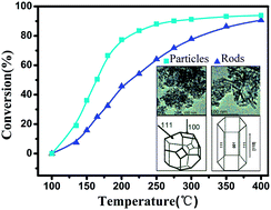
The effect of CeO2 morphology on the performance of a MnOx/CeO2 catalyst was investigated for the catalytic combustion of chlorobenzene (CB), which was used as a model compound for chlorinated volatile organic compounds (CVOCs). The catalytic activity tests revealed that MnOx/CeO2 nanoparticles (NPS) achieved relatively higher CB conversions than MnOx/CeO2 nanorods (NR). The MnOx/CeO2 catalysts were characterized by X-ray diffraction (XRD), Raman spectroscopy, Brunauer–Emmett–Teller (BET) N2 adsorption, transmission electron microscopy (TEM), X-ray photoelectron spectroscopy (XPS) and hydrogen temperature-programmed reduction (H2-TPR). The characterization of the MnOx/CeO2 catalysts indicated that CeO2-NPS had a higher exposure of the (100) crystal plane and possessed more Mn4+ species, oxygen vacancies and surface-adsorbed oxygen. It was suggested that the CeO2-NPS had a stronger interaction with MnOx species, which resulted in greater catalytic activity in the combustion of CB. The catalytic activity of MnOx/CeO2-NPS could be attributed to higher concentrations of Mn4+ species, oxygen vacancies, and surface-adsorbed oxygen, which were associated with the exposed (100) crystal planes. Therefore, these results demonstrated that the catalytic performance of the MnOx/CeO2 catalyst was greatly affected by the CeO2 morphology. Therefore, catalytic materials that have increased activity can be obtained through morphology-controlled synthesis.


1. Introduction

Chlorinated volatile organic compounds (CVOCs) which are mainly generated from industrial processes, and medical waste are recognized as major contributors to air pollutants.1,2 Generally, CVOCs can lead to photochemical smog and are highly toxic to humans and animals. Therefore, it is essential to develop practical and cost-effective methods to eliminate CVOCs in gases. Of the available techniques, catalytic combustion is one of the most effective technologies for the removal of destruction of CVOCs emissions due to its high removal efficiency and low operating temperature.

Among the catalysts used for the catalytic combustion of CVOCs, manganese oxides are known to be active catalysts on account of their high oxygen storage ability and redox properties.3–8 Furthermore, catalysts based on MnOx supported on metal oxides, such as MnOx/CeO2 and MnOx/TiO2 catalysts, have been reported to display higher catalytic performance for the oxidation of CVOCs compared with MnOx catalysts alone.9–11 Accordingly, the nature of the support material is critical to the catalytic performance of MnOx-based catalysts.12–15 In particular, the support material ceria has been widely utilized for the MnOx-based catalyzed oxidation of chlorobenzene (CB) due to its high oxygen storage capacity (OSC) and reducibility in the oxidation of CVOCs. Reports have shown that the capacity for oxygen storage is dependent on the morphology of ceria.10,16,17 Thus far, several studies document the shape effects of ceria in CO oxidation, ethanol steam reforming, and NO reduction with NH3. Huang and co-workers18 investigated the catalytic performance of various Au-supported ceria shapes for CO oxidation. They found that predominantly exposed (100)/(110) surface structures of ceria nanorods are superior for dispersing and stabilizing Au nanoclusters. This leads to a higher redox activity for CO oxidation, while relatively lower reducing the dispersal of Au nanoclusters on the exposed (111) plane for ceria nanoparticles, as determined by its lower activity for CO oxidation. Soykal and co-workers16 also indicated that CeO2 nanocubes were more active for ethanol steam reforming than ceria nanorods due to the presence of a higher exposure of the (110) plane for nanocubes.

Additional studies revealed that MnOx/CeO2 catalysts exhibited excellent catalytic performance in the combustion of CB. However, to the best our knowledge, little attention has been given to the morphology dependence of ceria nanomaterials in Mn species deposited on CeO2 for the catalytic combustion of CB. Additionally, various ceria morphology supports may possess different capacities for oxygen storage,19 which can then influence the catalytic performance of Mn species. Herein, we present the effect of ceria morphology on a Mn-based catalytic system for the catalytic combustion of CB. Ceria nanorods and ceria nanoparticles were synthesized by hydrothermal and co-precipitation methods, respectively. A wet impregnation method was utilized to incorporate manganese oxides over CeO2 nanomaterials. The catalytic performance of MnOx/CeO2 nanomaterials with two different shapes was examined in the catalytic combustion of CB.

2. Experimental section

2.1 Preparation

CeO2 nanorods were prepared by the hydrothermal method as follows.20 Ce(NO3)3·6H2O (3.45 mmol) was dissolved in deionized water (5 mL) and mixed with an aqueous sodium hydroxide aqueous solution (CNaOH = 6 M). The mixture was then stirred at room temperature for 30 min to obtain a milky slurry and subsequently transferred to a 50 mL autoclave, sealed, and heated at 100 °C for 24 h. After the hydrothermal treatment, the resulting precipitate was filtered, washed thoroughly with deionized water, and dried at 60 °C, and finally, calcination occurred at 400 °C for 4 h. The CeO2 nanoparticles were prepared by the precipitation method that has been reported in the literature.21 Ce(NO3)3·6H2O was dissolved in distilled water, and the pH value of the solution was rapidly adjusted to pH 12 with a 10% NaOH solution with stirring. The precipitate was filtered, washed with deionized water, and dried at 60 °C for 12 h, and then, calcination occurred at 400 °C for 4 h. The synthesized CeO2 nanorods and CeO2 nanoparticles were denoted as CeO2-NR and CeO2-NPS, respectively.

MnOx/CeO2 catalysts with different morphologies were prepared by incipient wetness impregnation. Manganese was loaded onto the CeO2 support with 50% Mn(NO3)2. The manganese loading was selected as 11 wt% (Mn/(Ce + Mn) = 0.27, molar ratio). Following impregnation, the catalysts were aged at room temperature for 24 h and dried at 80 °C in air overnight, and calcination took place at 400 °C for 4 h. The MnOx/CeO2 nanorods and MnOx/CeO2 nanoparticles were denoted as MnOx/CeO2-NR and MnOx/CeO2-NPS, respectively.

2.2 Characterization techniques

Transmission electron microscopy (TEM) tests were recorded on a JEM-2100 electron microscope operating at 200 kV. The samples were ultrasonically dispersed in ethanol and deposited on copper grids coated with lacey carbon. Energy dispersion spectra (EDS) were performed on a FALCON 80 X-ray energy-dispersive spectrometer that was used for the elemental analysis of the samples. X-ray diffraction (XRD) patterns of the MnOx/CeO2 and bare CeO2 were collected on a Bruker D8 Advance X-ray diffractometer using Cu Kα radiation (λ = 1.5405 Å) operated at 40 kV and 50 mA. The diffraction patterns were taken in the 2θ angle of 20–70°. The BET surface areas of the catalysts were obtained from N2 adsorption and desorption tests at 77 K using a NOVA2000e analyzer. The samples were degassed at 120 °C for 12 h before measurements were taken. The Raman spectra were recorded on a laser confocal microscopy Raman spectrometer (DXR, American Thermo Electron) operated at a wavelength of 532 nm. XPS measurements were carried out with a VG Multilab 2000 spectrometer using Al Kα ( = 1253.6 eV) radiation. The TPR runs were conducted with a linear heating rate (10 °C min−1) in a flow of 10% H2 in argon at a flow rate of 40 mL min−1. Hydrogen consumption was measured quantitatively by a thermal conductivity detector (TCD), which was calibrated by quantitative reduction of a given quantity of CuO to metallic copper.

2.3 Catalytic activity tests

Catalytic activity tests were performed in a fixed-bed flow reactor made of an 8 mm inner diameter quartz tube under an atmospheric pressure between 100–400 °C. Two-hundred milligrams of the (50–80 mesh) catalysts were placed in the reactor. The feed gas consisted of 5000 ppm CB, 20% O2, and balanced N2. The total flow rate was 100 mL min−1, with the gas hourly space velocity (GHSV) at 20[thin space (1/6-em)]000 h−1. The effluent was analyzed by an on-line gas chromatograph (GC) equipped with a flame ionization detector (FID) for the quantitative analysis of CB.

3. Results and discussion

3.1 XRD and N2 adsorption results

The BET surface areas of the supports and MnOx/CeO2 nanostructures are listed in Table 1. The BET surface area of CeO2-NR and CeO2-NPS were 113.4 and 127.7 m2 g−1, respectively. After loading MnOx, the specific surface areas of the MnOx/CeO2-NR and MnOx/CeO2-NPS decreased to 79.5 and 82.4 m2 g−1. Compared with the BET surface area of CeO2-NR and CeO2-NPS, a decrease in the specific surface areas of MnOx/CeO2-NR and MnOx/CeO2-NPS can be attributed to pore blockage by MnOx.22
Table 1 The properties of the different catalysts
Sample Surface area (m2 g−1) D (CeO2) (nm)/by XRDa D (CeO2) (nm)/by TEM
a From the Scherrer equation, based on the (111) reflection of the CeO2.
CeO2-NR 113.4
CeO2-NPS 127.7
MnOx/CeO2-NR 79.5 15.1 13–90
MnOx/CeO2-NPS 82.4 9.8 10 ± 2


X-ray diffraction patterns of MnOx/CeO2 nanostructures with different morphologies are shown in Fig. 1. All of the diffraction peaks were attributed to the cubic phase of CeO2 (JCPDS 34-0394), but no peaks of manganese oxides were observed, indicating a high dispersion of MnOx on the CeO2 nanostructures. Moreover, diffraction peaks of MnOx/CeO2-NR were broader compared with those of MnOx/CeO2-NPS, suggesting the formation of a smaller size of MnOx/CeO2-NPS. CeO2 crystallite sizes were calculated by the Scherrer equation, and the results (Table 1) indicated that CeO2-NR and CeO2-NPS were 15.1 and 9.8 nm, respectively, which are in agreement with the TEM results. Table 2 shows the peak intensities normalized with respect to the peak intensity of the (111) plane of ceria for the MnOx/CeO2-NPS and MnOx/CeO2-NR. As shown in Table 2, the (220) plane for both catalysts possessed similar relative intensities, while the (200) plane showed significantly higher diffraction for MnOx/CeO2-NPS, with a 45% relative intensity compared to 30% for MnOx/CeO2-NR. This indicates that the CeO2-NPS morphology may expose more of the (100) plane compared with CeO2-NR.


image file: c4ra07843h-f1.tif
Fig. 1 XRD patterns of (a) CeO2-NR, (b) CeO2-NPS, (c) MnOx/CeO2-NR, and (d) MnOx/CeO2-NPS.
Table 2 XRD intensities for MnOx/CeO2 catalysts normalize with respect to CeO2 (111) reflection
2θ 28.5 33.0 47.5 56.3
Respective planes CeO2 (111) CeO2 (200) CeO2 (220) CeO2 (311)
MnOx/CeO2-NR 100% 30% 56% 40%
MnOx/CeO2-NPS 100% 45% 57% 45%


Si and co-workers reported that the formation energies for oxygen vacancies for ceria oxide surface follow the sequence (111) > (100).23 Therefore, the (100) plane more readily form oxygen vacancies. It is also conceivable that the introduction of oxygen vacancies assists in improving catalyst performance significantly.16 Therefore, more (100) plane on the MnOx/CeO2-NPS may help improve the catalytic performance.

3.2 TEM and HRTEM results

The TEM images, structural models, and EDS spectra of synthesized materials are shown in Fig. 2. The two different CeO2 nanomaterials maintain their original crystal shapes after deposition of MnOx. MnOx/CeO2-NR has a uniform diameter of 10 ± 3 nm and a length within 13–90 nm. Fig. 2b shows the HRTEM image of MnOx/CeO2-NR. It can be observed that MnOx/CeO2-NR exposed the (100) and (111) planes, with an interplanar spacing of 0.27 nm and 0.31 nm, respectively. The structural model of the MnOx/CeO2-NR is shown in Fig. 2c. The single crystal nanorod grew along the [110] direction. Fig. 2c shows the MnOx/CeO2-NR hexangular prism enclosed by four (111) and two (100) planes.20 The TEM image of MnOx/CeO2-NPS (Fig. 2e) reveals that the nanoparticles have a size of 10 ± 2 nm. The structural model of the MnOx/CeO2-NPS shows that MnOx/CeO2-NPS is approximately determined to be the truncated octahedra which was surrounded by eight (111) and six (100) planes.24 The EDS spectra in Fig. 2d and h shows that the actual content of Mn element for MnOx/CeO2-NR and MnOx/CeO2-NPS were 11.39 wt% and 16.50 wt%, respectively. In addition, the EDS spectra in Fig. 2d and h confirms the presence of Mn and suggests that the MnOx species are highly dispersed on the surface of the CeO2 nanostructure.
image file: c4ra07843h-f2.tif
Fig. 2 TEM and HRTEM images of (a and b) MnOx/CeO2-NR, (e and f) MnOx/CeO2-NPS; the structural model of (c) MnOx/CeO2-NR, (g) MnOx/CeO2-NPS; EDS patterns of (d) MnOx/CeO2-NR, (h) MnOx/CeO2-NPS.

3.3 H2-TPR results

Fig. 3 displays the H2-TPR profiles of the CeO2 and MnOx/CeO2 nanostructures. For CeO2-NR, the reduction of CeO2 starts at 350 °C and a broad peak centered at 490 °C, which is attributed to the reduction of the surface oxygen of CeO2.25 Meanwhile, CeO2-NPS shows a similar reduction profile at a slightly higher temperature (540 °C), while the MnOx/CeO2 nanostructures showed different profiles of the reduction peaks. MnOx/CeO2-NR showed three distinct reduction peaks, with a shoulder peak at 368 °C. The peak at 227 °C could be attributed to reduction of MnO2 to Mn2O3, and the peak at 302 °C, with a shoulder peak at 368 °C, was a further reduction of Mn2O3 to Mn3O4, while the peak at 475 °C ascribed to the combined reductions of Mn3O4 to MnO and surface oxygen removal of ceria.24,26–30 A similar reduction profile was observed for MnOx/CeO2-NPS, except that the reduction peaks shifted to higher temperature regions, indicating that interaction between MnOx and CeO2-NPS is stronger than that with CeO2-NR. Quantitative evaluation of the reduction peaks (Table 3) revealed that the H2 consumption of the first two peaks on the MnOx/CeO2-NPS is greater than the MnOx/CeO2-NR, demonstrating that MnOx/CeO2-NPS has more surface oxygen and that a larger quantity of strongly interactive MnOx species exist on the CeO2-NPS.26 Therefore, the more surface oxygen on CeO2-NPS and the stronger interaction between MnOx and CeO2-NPS, result in the higher oxidative ability of the MnOx species.31
image file: c4ra07843h-f3.tif
Fig. 3 H2-TPR profiles of (a) CeO2-NR, (b) CeO2-NPS, (c) MnOx/CeO2-NR, and (d) MnOx/CeO2-NPS.
Table 3 Hydrogen consumption of H2-TPR tests
Sample T (°C) mmol g−1 T (°C) mmol g−1
MnOx/CeO2-NR 227 0.77 302 0.89
MnOx/CeO2-NPS 245 0.86 331 1.14


3.4 Raman spectra

The Raman spectra of CeO2 and MnOx/CeO2 nanostructures are presented in Fig. 4. A distinct F2g symmetry mode of the CeO2 phase at approximately 462 cm−1 can be observed in the Raman spectra of the CeO2 nanostructures (Fig. 4a).32 Furthermore, two additional weak peaks, at approximately 600 and 1180 cm−1, can also be observed in the Raman spectra of CeO2 nanomaterials (Fig. 4a). These are attributed to the defect-induced (D) mode and the second-order longitudinal (2LO) mode, respectively.28 A similar Raman spectrum was observed for MnOx/CeO2 nanostructures (Fig. 4b), except that the peak at 600 cm−1 for oxygen vacancies displayed a blue shift to 650 cm−1, and the peak at 1180 cm−1 shifted to 1170 cm−1. These shifts can be attributed to the Mn incorporation in the CeO2 lattice. Additionally, the new peak at 577 cm−1 can be assigned to the Mn–Ce–O bond on the MnOx/CeO2 nanostructures.
image file: c4ra07843h-f4.tif
Fig. 4 Raman spectra of (a) CeO2, (b) MnOx/CeO2 nanostructures, and (c) the peak intensity ratios of I600/I400 over CeO2 (black column) and MnOx/CeO2 nanostructures (red column).

The Raman peak at 600 cm−1 was related to the oxygen vacancies induced by the presence of Ce3+ ions, and the I600/I462 value (Fig. 4c) reflects the degree of the defect sites on nanostructures, such as the relative content of the oxygen vacancies.32,33 From Fig. 4c, the intensity ratio I600/I462 follows the sequence: MnOx/CeO2-NPS > MnOx/CeO2-NR > CeO2-NPS > CeO2-NR, indicating that MnOx/CeO2-NPS possessed the greatest amount of oxygen vacancies. According to Fig. 4c, the I600/I462 value increased dramatically when manganese ions were doped into the ceria lattice. This indicated that Mn ions incorporated into the CeO2 phase and thus led to an increase in the degree of defects. Clearly, MnOx/CeO2-NPS exhibited the higher value of I600/I462 compared to the MnOx/CeO2-NR nanostructure, demonstrating that the interaction between MnOx and CeO2-NPS is stronger than with CeO2-NR. In summary, the Raman results reflect that the stronger interaction between MnOx and CeO2-NPS, and in turn, MnOx/CeO2-NPS incorporated into a greater oxygen vacancies, which is in accordance with the H2-TPR result.

3.5 XPS results

The Mn 2p, O 1s, and Ce 3d XPS spectra of the MnOx/CeO2-NR and MnOx/CeO2-NPS catalysts are shown in Fig. 5. As shown in Fig. 5a, the Mn 2p3/2 spectra can be separated into three peaks. The binding energy at ca. 640.5, 641.8, and 643.7 eV corresponded to Mn2+, Mn3+, and Mn4+,34 respectively. The concentration of Mn4+ on MnOx/CeO2-NPS is higher than on MnOx/CeO2-NR, as shown in Table 4. Liao and co-workers26 reported that high Mn4+ could lead to oxygen vacant sites on catalysts. Accordingly, a higher ratio of Mn4+ on the CeO2-NPS may lead to more oxygen vacancies.
image file: c4ra07843h-f5.tif
Fig. 5 (a) Mn 2p, (b) Ce 3d and (c) O 1s XPS spectra of MnOx/CeO2 nanostructures.
Table 4 Surface chemical and states based on XPS analysisa
Sample Mn4+ (%)a1 Mn3+ (%)a2 Mn2+ (%)a3 Ce3+ (%) Olatt (%) Oads (%) Owat (%)
a a1 + a2 + a3 = 100%.
MnOx/CeO2-NR 15.6 38.8 45.5 26.4 40.0 23.0 36.9
MnOx/CeO2-NPS 21.1 37.0 41.9 29.0 58.0 31.0 6.9


The Ce 3d spectra of the MnOx/CeO2-NR and MnOx/CeO2-NPS are shown in Fig. 5b. The peaks at approximately U, U′′, U′′′, V, V′′, and V′′′ are attributed to Ce4+, and the Ce3+ can be fitted with the peaks at U′ and V′. The concentrations of Ce3+ on the MnOx/CeO2-NR and MnOx/CeO2-NPS are 26.4%, and 29.0%, respectively. Liu and co-workers35 suggested that a higher Ce3+ concentration can produce more oxygen vacancies on catalysts. The spectra indicates that MnOx/CeO2-NPS contain more oxygen vacancies than MnOx/CeO2-NR.

The O 1s spectra of MnOx/CeO2-NR and MnOx/CeO2-NPS are shown in Fig. 5c. The O 1s profile can be fit into three peaks: lattice oxygen (Olatt) at 529.4–530.0 eV, surface-adsorbed oxygen (Oads) at 531.0–531.7 eV and adsorbed molecular water (Owat) at approximately 532.7 eV.36 Quantitatively, it can be observed in Table 4 that the Oads/Owat + Oads + Olatt ratio of MnOx/CeO2-NPS catalyst is higher than MnOx/CeO2-NR, indicating that MnOx/CeO2-NPS catalyst are rich in surface-adsorbed oxygen. Because Oads is more active than Olatt due to its high mobility, the higher concentration of Oads is helpful for the CB oxidation.37

3.6 Characteristic of the used catalysts

The morphologies of the used MnOx/CeO2-NR and MnOx/CeO2-NPS catalysts were observed on TEM images in Fig. 6. The used MnOx/CeO2-NR has a uniform diameter of 11 ± 3 nm and a length within 13–95 nm while the MnOx/CeO2-NPS have a size of 9 ± 2 nm. This indicates that the grain size of the used catalysts basically remains unchanged after reaction. The catalysts still maintain their shapes after reaction, indicating that the MnOx/CeO2 nanostructures have structure stability during CB combustion.
image file: c4ra07843h-f6.tif
Fig. 6 TEM images of the used catalysts: (a) MnOx/CeO2-NR, (b) MnOx/CeO2-NPS.

The XRD patterns of the used MnOx/CeO2 nanostructures are shown in Fig. 7. All typical diffraction peaks can be attributed to the ceria fluorite structure (JCPDS 34-0394), and no peaks of MnOx were observed. This phenomenon demonstrates that a strong interaction between the MnOx and CeO2 nanostructures exists, which prevents the agglomeration of MnOx during CB combustion.28


image file: c4ra07843h-f7.tif
Fig. 7 XRD patterns of the used catalysts: (a) MnOx/CeO2-NR, (b) MnOx/CeO2-NPS.

3.7 Catalytic activity and stability of catalysts

The catalytic performance of the bare CeO2 supports and MnOx/CeO2 catalysts for the catalytic combustion of CB is shown in Fig. 8. As shown in Fig. 8, both CeO2-NPS and CeO2-NR supports exhibit poor activity for catalytic combustion of CB, although the CeO2-NPS is slightly more active. After loading Mn, the activities of the Mn-supported catalysts were markedly enhanced, revealing the necessity for Mn in the catalytic combustion of CB. Additionally, a significant effect of support morphology on catalyst performance can be observed in Fig. 8. The MnOx/CeO2-NPS catalyst displayed higher activity than MnOx/CeO2-NR. At 275 °C, the CB conversions was 90.0% over MnOx/CeO2-NPS, while only 71.7% over MnOx/CeO2-NR. This indicates that MnOx/CeO2-NPS catalysts showed a better catalytic activity than the MnOx-CeO2 mixed oxides (90% conversion at 300 °C, Mn/(Mn + Ce) = 0.27, molar ratio) reported by Wang et al.1
image file: c4ra07843h-f8.tif
Fig. 8 MnOx/CeO2 catalysts for CB combustion, gas composition: 2500 mg m−3 CB, 20% O2, N2 balance; GHSV = 20[thin space (1/6-em)]000 h−1.

Because the obtained MnOx/CeO2 catalysts have different exposed planes, this may be one possible explanation for the observed difference in catalytic performance of the two catalysts. Because CeO2-NPS support preferentially exposes the (100) plane and this plane is more likely to create anion vacancies and defects, it is reasonable that this support offers more oxygen vacancies.16 As detected by the Raman and XPS results, the MnOx/CeO2-NPS sample has more oxygen vacancies. Previous reports on this subject have described that the introduction of oxygen vacancies on support material were conducive to adsorbing oxygen and forming highly reactive atomic oxygen, which would significantly improve the catalyst performance.21,38 Therefore, additional oxygen vacancies on the CeO2-NPS support are a contributing factor affecting the catalytic activity in CB oxidation. In addition, the higher ratio of Oads and stronger interaction between the Mn and the ceria support may be two additional factors to improve the catalytic performance. In conclusion, the performance of CeO2-NPS might benefit from a stronger interaction between Mn and the ceria support, higher concentrations of oxygen vacancies, and surface-adsorbed oxygen.

Extended time stability tests of MnOx/CeO2 nanostructures with different support morphologies are shown in Fig. 9. Both catalysts show good stability after 30 h at 300 °C. Therefore, the two catalysts show good resistance to chlorine poisoning from CB.


image file: c4ra07843h-f9.tif
Fig. 9 The stability tests over the MnOx/CeO2 nanostructures for CB combustion at 300 °C; gas composition: 2500 mg m−3 CB, 20% O2, N2 balance; GHSV = 20[thin space (1/6-em)]000 h−1.

4. Conclusion

Manganese catalysts loaded on a ceria support with two different morphologies, MnOx/CeO2 nanoparticles (NPS) and MnOx/CeO2 nanorods (NR), were examined for their performance on the catalytic oxidation of CB. The morphology of the ceria support was observed to have a strong effect on the catalytic oxidation of CB. The MnOx/CeO2-NPS catalyst presented better catalytic activity compared to the MnOx/CeO2-NR catalyst. HRTEM and XRD analysis indicated that the CeO2-NPS support preferentially exposed the (100) plane and are more likely to form anion vacancies and defects. The XPS and Raman results displayed a morphology dependence on the concentration of Mn4+ species, surface-adsorbed oxygen, and oxygen vacancies. Higher catalytic activity of MnOx/CeO2-NPS may be attributed to abundant Mn4+ species, oxygen vacancies and surface-adsorbed oxygen, which are associated with exposed (100) crystal planes. Furthermore, the extended time stability tests showed that both catalysts demonstrate resistance to chlorine poisoning. Thus, MnOx/CeO2-NPS catalysts can be considered as a potential material for the abatement of CVOCs.

Acknowledgements

We gratefully acknowledge the funding from the National Natural Science Foundation of China (no. 20873097, 21071113, 21471120), Natural Science Foundation of Hubei Province (no. 2011CDA049), International Cooperation Foundation of Hubei Province (2012IHA00201), Educational Commission of Hubei Province of China (T201306).

Notes and references

  1. X. Y. Wang, Q. Kang and D. Li, Appl. Catal., B, 2009, 86, 166–175 CrossRef CAS PubMed.
  2. H. Kominami, T. Nishi, K. Fuku and K. Hashimoto, RSC Adv., 2013, 3, 6058–6064 RSC.
  3. J. J. Li, L. Li, W. Cheng, F. Wu, X. F. Lu and Z. P. Li, Chem. Eng. J., 2014, 244, 59–67 CrossRef CAS PubMed.
  4. Y. Z. Li, Z. Y. Fan, J. W. Shi, Z. Y. Liu and W. F. Shangguan, Chem. Eng. J., 2014, 241, 251–258 CrossRef CAS PubMed.
  5. J. W. Li, P. Zhao and S. T. Liu, Appl. Catal., A, 2014, 482, 363–369 CrossRef CAS PubMed.
  6. W. Tian, H. S. Yang, X. Y. Fan and X. B. Zhang, Catal. Commun., 2010, 11, 1185–1188 CrossRef CAS PubMed.
  7. J. W. Li, C. Song and S. T. Liu, Acta Chim. Sin., 2012, 70, 2347–2352 CrossRef CAS.
  8. A. Jha, T. Chandole, R. Pandya, H. S. Roh and C. V. Rode, RSC Adv., 2014, 4, 19450–19455 RSC.
  9. Z. Q. Zou, M. Meng and Y. Q. Zha, J. Phys. Chem. C, 2010, 114, 468–477 CAS.
  10. M. A. Wolfovich, M. V. Landau, A. Brenner and M. Herskowitz, J. Catal., 2007, 247, 201–213 CrossRef PubMed.
  11. Y. Liu, M. F. Luo, Z. B. Wei, Q. Xin, P. L. Ying and C. Li, Appl. Catal., B, 2001, 29, 61–67 CrossRef CAS.
  12. Y. F. Han, Z. Y. Zhong, K. Ramesh, F. X. Chen and L. W. Chen, J. Phys. Chem. C, 2007, 111, 3163–3170 CAS.
  13. P. Gawade, B. Mirkelamoglu and U. S. Ozkan, J. Phys. Chem. C, 2010, 114, 18173–18181 CAS.
  14. M. M. Schubert, S. Hackenberg, A. C. van Veen, M. Muhler, V. Plzak and R. J. Behm, J. Catal., 2005, 197, 113–122 CrossRef.
  15. H. Zhang, F. N. Gu, Q. Liu, G. G. Gao, L. H. Jia, T. Y. Zhu, Y. F. Chen, Z. Y. Zhong and F. B. Su, RSC Adv., 2014, 4, 14879–14889 RSC.
  16. I. Soykal, B. Bayrama, H. Sohna, P. Gawadea, J. T. Millerb and U. S. Ozkana, Appl. Catal., A, 2012, 449, 47–58 CrossRef CAS PubMed.
  17. M. Li, Y. H. Hu, C. C. Liu, J. G. Huang, Z. G. Liu, M. T. Wang and Z. H. An, RSC Adv., 2014, 4, 992–995 RSC.
  18. X. S. Huang, H. Sun, L. C. Wang, Y. M. Liu, K. N. Fan and Y. Cao, Appl. Catal., B, 2009, 90, 224–232 CrossRef CAS PubMed.
  19. Y. Liu, M. F. Luo, Z. B. Wei, Q. Xin, P. L. Ying and C. Li, Appl. Catal., B, 2001, 29, 61–67 CrossRef CAS.
  20. H. X. Mai, L. D. Sun, Y. W. Zhang, R. Si, W. Feng, H. P. Zhang, H. C. Liu and C. H. Yan, J. Phys. Chem. B, 2005, 109, 24380 CrossRef CAS PubMed.
  21. K. Zhou, X. Wang, X. M. Sun, Q. Peng and Y. D. Li, J. Catal., 2005, 229, 206–212 CrossRef CAS PubMed.
  22. L. Ran, Z. Qin, Z. Y. Wang, X. Y. Wang and Q. G. Dai, Catal. Commun., 2013, 37, 5–8 CrossRef CAS PubMed.
  23. R. Si and M. Flytzani-Stephanopoulos, Angew. Chem., Int. Ed., 2008, 47, 2884–2887 CrossRef CAS PubMed.
  24. H. J. Li, G. S. Qi, Tana, X. J. Zhang, W. Li and W. J. Shen, Catal. Sci. Technol., 2011, 1, 1678 Search PubMed.
  25. S. N. Li, H. Q. Zhu, Z. F. Qin, G. F. Wang, Y. G. Zhang, Z. W. Wu, Z. K. Li, G. Chen, W. W. Dong, Z. H. Wu, L. R. Zheng, J. Zhang, T. D. Hu and Y. G. Zhang, Appl. Catal., B, 2014, 144, 498–506 CrossRef CAS PubMed.
  26. Y. N. Liao, M. L. Fu, L. M. Chen, J. L. Wu, B. C. Huang and D. Q. Ye, Catal. Today, 2013, 216, 220–228 CrossRef CAS PubMed.
  27. C. Song, Q. R. Yi, J. W. Li and S. T. Liu, J. Funct. Mater., 2014, 45, 13050–13055 Search PubMed.
  28. R. H. Gao, D. S. Zhang, P. Maitarad, L. Y. Shi, T. Rungrotmongkol, H. R. Li, J. P. Zhang and W. G. Cao, J. Phys. Chem. C, 2013, 117, 10502–10511 CAS.
  29. Q. L. Zhang, C. T. Qiu, H. D. Xu, T. Lin, Z. E. Lin, M. C. Gong and Y. Q. Chen, Catal. Today, 2011, 175, 171–176 CrossRef CAS PubMed.
  30. X. Y. Wang, Q. Kang and D. Li, Appl. Catal., B, 2009, 86, 166–175 CrossRef CAS PubMed.
  31. A. Gupta, U. V. Waghmare and M. S. Hegde, Chem. Mater., 2010, 22, 5184–5198 CrossRef CAS.
  32. Q. G. Dai, H. Huang, Y. Zhu, W. Deng, S. X. Bai, X. Y. Wang and G. Z. Lu, Appl. Catal., B, 2012, 117, 360–368 CrossRef PubMed.
  33. J. R. McBride, K. C. Hass, B. D. Poindexter and W. H. Weber, J. Appl. Phys., 1994, 76, 2435–2462 CrossRef CAS PubMed.
  34. J. L. Zuo, Z. H. Chen, F. R. Wang, Y. H. Yu, L. F. Wang and X. H. Li, Ind. Eng. Chem. Res., 2014, 53, 2647–2655 CrossRef CAS.
  35. X. W. Liu, K. B. Zhou, L. Wang, B. Y. Wang and Y. D. Li, J. Am. Chem. Soc., 2009, 131, 3140–3141 CrossRef CAS PubMed.
  36. Y. Dai, X. Y. Wang, D. Li and Q. G. Dai, J. Hazard. Mater., 2011, 188, 132–139 CrossRef CAS PubMed.
  37. H. Z. Chang, J. H. Li, J. Yuan, L. Chen, Y. Dai, H. Arandiyan, J. Y. Xu and J. M. Hao, Catal. Today, 2013, 201, 139–144 CrossRef CAS PubMed.
  38. P. G. Harrison, I. K. Ball, W. Azelee, W. Daniell and D. Goldfarb, Chem. Mater., 2000, 12, 3715–3725 CrossRef CAS.

This journal is © The Royal Society of Chemistry 2014
Click here to see how this site uses Cookies. View our privacy policy here.