
CO2 diffusion in n-hexadecane investigated using magnetic resonance imaging and pressure decay measurements
Yongchen Songa,
Min Haoab,
Yu Liua,
Yuechao Zhao*a,
Bo Sua and
Lanlan Jianga
aKey Laboratory of Ocean Energy Utilization and Energy Conservation of Ministry of Education, Dalian University of Technology, Dalian 116024, P. R. China. E-mail: zhaoyc@dlut.edu.cn; Tel: +86-411-84708015
bSchool of Energy and Power Engineering, Shenyang University of Chemical Technology, Shenyang, Liaoning 110142, P. R. China
First published on 29th September 2014
Abstract
A method combining magnetic resonance imaging (MRI) and dual-chamber pressure decay (i.e., the pressure–volume–temperature method) was used to measure the diffusivity of CO2 in bulk n-hexadecane, a representative oil, at 21 °C. Images of the proton density of n-hexadecane were obtained by MRI using a high magnetic field and high resolution, and the pressure decay was recorded. Overall diffusion coefficients of CO2 were calculated from both pressure decay and MRI intensity data. The two results showed good agreement. MRI was successfully used to study the diffusivity of CO2 in n-hexadecane. The change of image density (i.e., proton density) demonstrates that the oil density decreases as CO2 diffuses into it. The finite volume method was applied to the images obtained by MRI, allowing the diffusion coefficient of CO2 to be directly obtained. MRI methods can measure the unsteady-state local diffusion coefficient and overall diffusion coefficient of such systems.
1 Introduction
Two miscible fluids will diffuse into each other when they come into contact. The molecular transport of one substance relative to another is known as diffusion. Diffusion plays an important role in the mixing of CO2 and crude oil after injection of CO2 into an oil reservoir.CO2-enhanced oil recovery (CO2-EOR) has proven to be effective for improving domestic oil production. Moreover, use of CO2 in oil recovery also provides considerable capacity for CO2 mitigation.1 The viscosity of oil can be lowered significantly by diffusion of CO2 into it; thus, the mobility of oil in a reservoir can be improved.2 Mass transfer between CO2 and oil, which is mainly controlled by diffusivity, is the first mechanism that occurs during the CO2-EOR process.3,4 To describe this process quantitatively, the diffusion coefficient of CO2 gas in oil needs to be determined.
Experimental methods used to measure the diffusivity of gas in oil can be broadly divided into two types: direct and indirect. The direct measurement of diffusivity is time consuming and involves measuring the change of concentration over time.5 Indirect methods involve measuring the change of a parameter related to diffusion over time, such as volume, density or pressure.6,7 The pressure decay method is a typical indirect method used to measure diffusivity. Riaziet et al.8 reported a pressure decay method using a pressure–volume–temperature (PVT) cell. In their experiment, both the position of the gas/liquid interface and gas pressure decay in the PVT cell were monitored during the diffusion process. Zhang et al.9 measured the diffusion coefficient of CO2 in heavy oils using a similar experimental method except that the position of the interface was considered constant. They also presented a nonlinear regression method that allowed the diffusion coefficient to be obtained directly from pressure decay data. Recently, non-intrusive detection methods such as X-ray computed tomography (CT) and magnetic resonance imaging (MRI) have been extended to the area of petroleum research. Liu et al.10 observed the miscible and immiscible CO2 displacement of n-decane in glass-bead packed beds using an MRI scanner under high magnetic field. Residual oil saturation was quantitatively measured by intensity analysis of magnetic resonance (MR) images. Song et al.11 determined the minimum miscible pressure of CO2 and n-decane by the MRI method, and their results agreed well with previous data obtained with traditional methods. Meanwhile, X-ray CT has been used to measure density distribution in heavy oil and hydrocarbon solvent mixing experiments.12–14 In these experiments, concentration profiles were obtained from CT images based on the relationship between bulk density and CT image intensity. Other researchers have used MRI to observe mass-transfer processes. Fisher et al.15 used MRI to observe vapor extraction processes. They performed bitumen and heavy-oil vapor extraction experiments using butane and propane as solvents in dry sand and sand with connate water. In their experiments, the MRI signal, which corresponded to the oil saturation, provided not only the position of the vapor chamber but also concentration gradients.
The main objective of the present study is to use MRI technology to measure gas diffusion coefficients in oils, and investigate diffusion and mass transfer processes in situ. Diffusion coefficients of CO2 in n-hexadecane are determined by measuring the pressure in a dual-chamber PVT system. MRI with high spatial resolution is used to observe the concentration distribution of CO2 during diffusion. In addition, pressure data are recorded to calculate the molar mass of CO2 diffusing into n-hexadecane. Pressure decay analysis is performed to calculate the steady-state diffusion coefficient of CO2 in n-hexadecane. This study helps us to understand the diffusion behavior of CO2 in a representative oil, so it will provide useful information for several oil recovery processes.
2 Experimental details
2.1. Apparatus and materials
An experimental system containing a gas cylinder and vessel for the MRI sample was constructed to measure the diffusion coefficient of CO2 into a representative oil, n-hexadecane, using both the pressure decay method and MRI.10 A schematic diagram of the experimental apparatus is shown in Fig. 1. The gas cylinder is a constant-volume (500 mL) stainless steel cylinder that is wrapped with an electric heater. The self-designed MRI sample vessel (Fig. 2) is made of polyimide to hold the diffusion samples at high pressure (up to 12.0 MPa). The sample vessel contained three main types of components: an inner tube with an inner diameter of 15 mm and wall thickness of 4.5 mm, an outer tube with an outer diameter of 38 mm, and inlet and outlet connectors. The outlet was blocked in the experiments. The temperature of the sample in the vessel was controlled by a circulator (F-25ME, Fluorinert™, Julabo, Inc., Seelbach, Germany) with a temperature control range of −45 to 200 °C and precision of ±0.5 °C. Inert liquid Fluorinert™ FC-40 was used as the temperature-control medium of the imaging vessel because it does not contain hydrogen atoms and thus has no influence on 1H nuclear imaging. The gas cylinder and sample vessel formed a dual-chamber system, which can improve the pressure stability and measurement precision relative to those of a single-chamber system.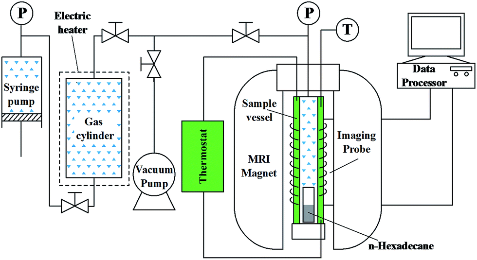 | ||
| Fig. 1 Schematic diagram of the experimental apparatus. | ||
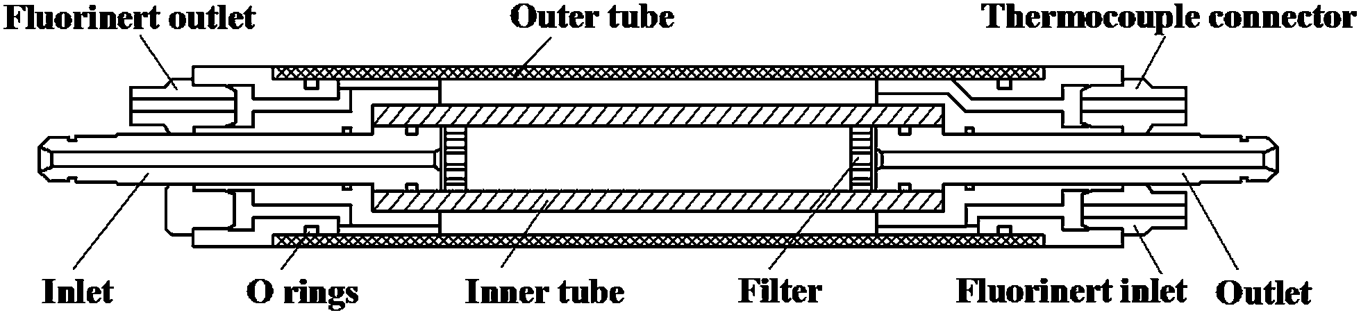 | ||
| Fig. 2 Vertical-section diagram of the sample vessel. | ||
Images were measured using a 400 MHz NMR spectrometer (Varian Inc., Palo Alto, CA, USA) with microimaging kits including a 55 mm i.d. gradient coil, which provided a maximum gradient magnetic field of 50 Gauss per cm, and a 40 mm i.d. 400 MHz 1H Millipede vertical imaging probe. All data and images were processed with a data processor.
n-Hexadecane of 98% purity was contained in a glass vial (i.d. = 8.8 mm) placed at the bottom of the sample vessel. Gaseous CO2 of 99.99% purity was injected via a syringe pump into the gas cylinder to attain the desired pressure.
2.2 Procedure
Prior to performing experiments, the diffusion cell and all connections were pressurized with nitrogen and tested for leakage at pressures up to 8 MPa for at least 24 h.All experiments were performed at 21 °C. The glass vial was filled to a height of about 20 mm with n-hexadecane. The system was placed under vacuum to extract air. After careful tuning, shimming and setting of the pulse parameters, the oil sample was scanned to obtain an initial MR image. CO2 was then injected into the gas cylinder with the syringe pump until the desired pressure was reached. The pressure in the gas cylinder was set between 3000 and 5000 kPa. The gas valves were then turned on to allow CO2 to enter the sample vessel and diffuse into the oil. The pressure was automatically recorded at the same time. Proton density images of the longitudinal planes along the diffusion direction were obtained while the pressure was being plotted. Each experiment was stopped after the pressure maintained a constant value (equilibrium pressure) for 1 h. When the experiment had finished, the system was slowly depressurized to atmospheric pressure, dismantled and cleaned before starting a new experiment.
The steps of applying MRI into diffusion experiment imaging are as follows:
(1) Put the n-hexadecane-containing glass vials upright in the sample vessel, and keep the height consistent with the imaging probe.
(2) Debug the MRI analyzer; open Vnmrj in the image acquisition computer, and enter “tune” into the dialog box; tune by regulating the knobs “tune” and “match” below the probe; then start to shim: click “manual shim” on the “start” interface, entering the shimming interface; regulate by using the right and left mouse buttons, until the images are appropriate.
(3) Setting of parameters. All images were acquired with a standard spin-echo multi-slice (SEMS) pulse sequence, because this sequence is free from the impacts of main magnetic field inhomogeneity, and produces high-quality images. The specific imaging parameters are as follows: echo time (TE) = 4.9 ms, repetition time (TR) = 5 s, image data matrix = 96 pixels × 96 pixels, field of view (FOV) = 40 × 40 mm2, slice thickness = 2 mm.
(4) Image acquisition. After gas injection was started, the MRIs were continuously acquired, until the end of the experiment.
2.3 Mathematical analysis
The diffusion process may be analyzed as shown in Fig. 3. This analysis can be derived based on the following assumptions:9,16,17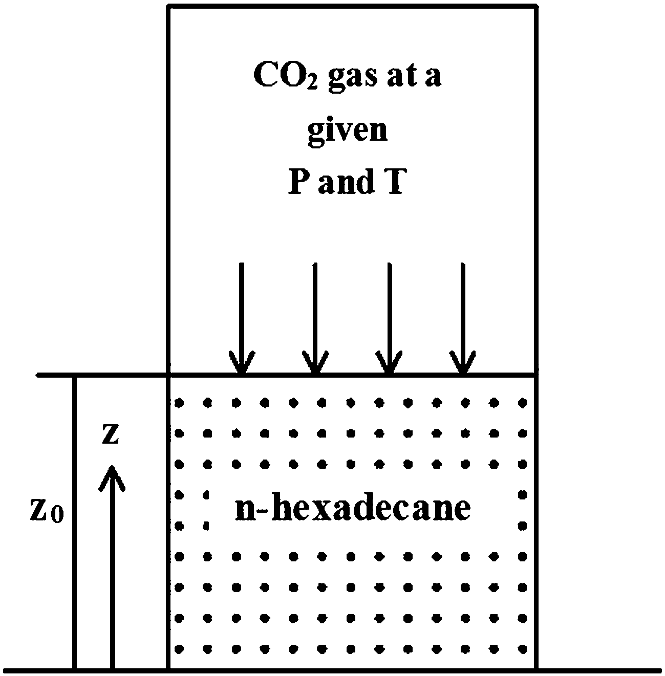 | ||
| Fig. 3 Schematic diagram of the diffusion model. | ||
(1) The swelling height of the oil is negligible, that is, z0 remains constant during the experiments.
(2) The temperature remains constant.
(3) The volatilization of n-hexadecane is negligible.
(4) The diffusion coefficient, D, does not change markedly with concentration during the experiments.
(5) The gas phase is a pure (single-component) gas.
The diffusion process can be expressed by Fick's law:9
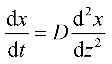 | (1) |
Based on this hypothesis and combined with Fick's law, the relationship between pressure P and time t can be acquired using semi-infinite boundary conditions and the matter balance principle. When the higher-order terms were ignored, a 2-order analytical expression was obtained as follows:9
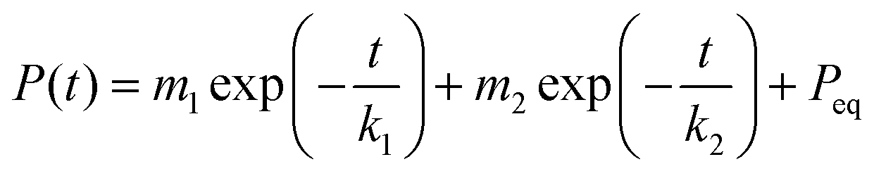 | (2) |
The first exponential term in eqn (2) fits the main part of the pressure–time curve (large k) and the second term fits the initial part of the pressure–time curve during the incubation period (small k). Numerical fitting is required to obtain the values of m1, m2, k1, k2 and Peq. Because the incubation period is relatively short, the first exponential term is more suitable to calculate the integral diffusion coefficient. The diffusion coefficient D is then given by:9
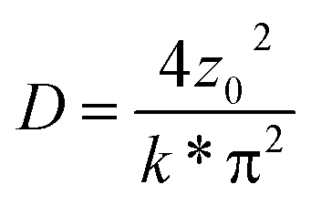 | (3) |
2.4 MR image analysis
An MR image of the proton density of n-hexadecane was obtained after one MRI scan. A region of interest (ROI) in the image was selected to analyze the diffusion process. Each ROI was 20 mm high, 7 mm wide and divided into ten segments of equal height. The image intensity of each segment at different times (defined as Iz,t) was then calculated. The initial I0 (at t = 0) and final Ieq (at t = teq) values are constant at all positions.CO2 has no signal in MRI. A previous study18 confirmed that the MR signal intensity at any location is proportional to the content of oil. When CO2 diffuses into the oil, the content of oil decreases. Thus, the signal intensity of the MR images will weaken as the concentration of CO2 increases. Therefore, the distribution of dimensionless CO2 concentration can be calculated based on the measured signal intensity distribution of the MR images at each time period. According to Fick's law, the diffusion coefficients related to time and displacement can be computed based on such dimensionless concentrations. The concentration of CO2 is related to the MR signal intensity. The normalized concentration of CO2 in oil at different positions, z, and times, t, can be written as:
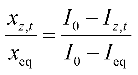 | (4) |
| xz,t = k(I0 − Iz,t) | (5a) |
| x0 = 0 | (5b) |
| xeq = k(I0 − Ieq) = const | (5c) |
Here, a non-iterative, finite-volume analytical technique was used to calculate the diffusion coefficient of CO2 in oil (n-hexadecane). It was assumed that there was a small composition gradient in the oil along the diffusion direction, and that the diffusion front did not move appreciably. Thus, Fick's Law (eqn (1)) can be used to relate the concentration and diffusion coefficient.
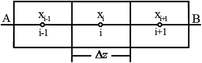
As shown in Fig. 4, the medium domain was discretized with mesh size Δz, time step Δt and grid points zi = i × Δz (i = 1, 2, …10) and tn = n × Δt (n = 0, 1, 2, …).19,20
 | ||
| Fig. 4 Model of the finite-volume analytical method used in this work. | ||
Assuming that the concentration in boundary A is constant and there is no flux in boundary B, the discretized equations are as follows:
Internal points
 | (6a) |
Boundary A
 | (6b) |
Boundary B
 | (6c) |
By arranging the discretized equations within the medium domain and at the boundary surfaces, eqn (6) can be written in matrix form as Ax = b. Vector b is composed of the concentration measurement at specified grid locations along the sample in the experiment. The components of vector x are the unknown diffusion coefficients.
3 Results and discussion
The pressure attenuation curves obtained with an initial pressure of 4377 kPa are shown in Fig. 5. A biexponential decrease in pressure is observed. Initially, the decrease was more pronounced, and became stable after an extended period. The decrease of pressure resulted from the dissolution of CO2, which demonstrates that the mass transfer rate during the initial stage of the experiment was relatively high. This suggests that high concentration and density gradients existed along the internal diffusion direction for n-hexadecane, which led to considerable convection and mass transfer. The initial stage of this high-mass-transfer diffusion is called the “incubation period”.21 With lengthening diffusion time, the internal concentration gradient and density gradient of hexadecane were gradually reduced. At the same time, the rates of mass transfer and pressure decay decreased. This result also indicates that after a long time, normal diffusion behavior occurs, and that mass transfer after the incubation period is dominated by molecular diffusion. Furthermore, it was found that a higher initial pressure resulted in a higher pressure decay rate and equilibrium being reached in less time than a lower initial pressure. It is evident that the incubation period has a considerable influence on the diffusion of CO2, and a high initial pressure increases this influence.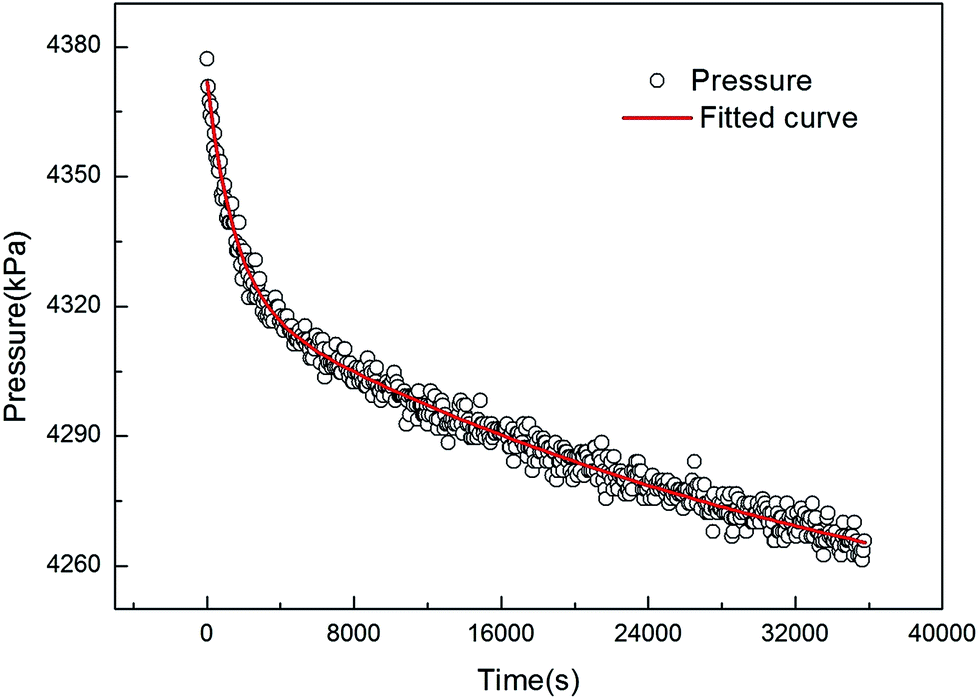 | ||
| Fig. 5 Pressure decay during diffusion of CO2 in n-hexadecane starting with an initial pressure of 4377 kPa. | ||
The collected pressure–time data were numerically fitted in origin with an accuracy of 0.974–0.993. The overall diffusion coefficient of CO2 in n-hexadecane can be obtained by the fitted k* using eqn (3). The overall diffusion data obtained under different pressures are listed in Table 1. We calculated that the overall diffusion coefficient of CO2 in n-hexadecane to be about 3.21–4.30 × 10⁻⁹ m2 s−1. Even though the exact experimental conditions used in the present work have not been used before, Wang et al.22 and Grogan23 obtained diffusion coefficients of 3.32–5.71 × 10⁻⁹ m2 s−1 and 1.8–3.21 × 10⁻⁹ m2 s−1, respectively, under similar conditions. Our diffusion coefficients are of the same order of magnitude as these.
| NO. | Oil height (mm) | Pressure of gas cylinder (kPa) | Initial pressure (kPa) | k* (s) | D (10−9/m−2 s−1) |
|---|---|---|---|---|---|
| 1 | 20 | 3000 | 2568 | 50![[thin space (1/6-em)]](https://www.rsc.org/images/entities/char_2009.gif) 537.3 537.3 |
3.21 |
| 2 | 20 | 3500 | 3093 | 49201.1 |
3.71 |
| 3 | 20 | 4000 | 3420 | 41170.2 |
3.94 |
| 4 | 20 | 4600 | 3980 | 39020.2 |
4.15 |
| 5 | 20 | 5000 | 4377 | 37680.5 |
4.30 |
The ROIs of MR images were cropped with image processing software. As an example, Fig. 6 shows a time series of ROIs of the diffusion process of CO2 in n-hexadecane starting with an initial pressure of 4377 kPa. As CO2 diffuses into n-hexadecane, a decrease in image intensity from top to bottom can be clearly observed because CO2 is invisible in MR images. The CO2 concentration distribution can be calculated from the image intensity data.
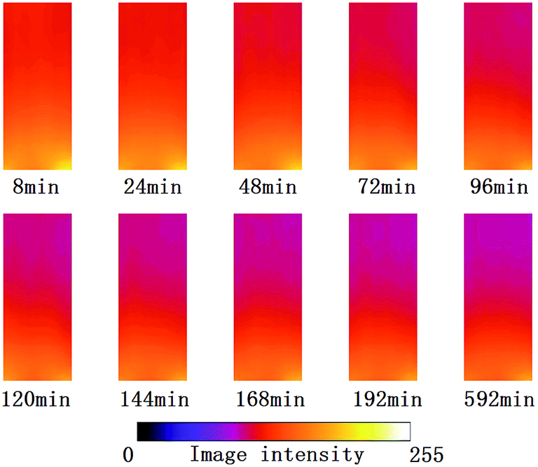 | ||
| Fig. 6 Series of ROI images at different times during the CO2 diffusion process for an initial pressure of 4377 kPa. | ||
The normalized concentration distribution of CO2 along the diffusion direction at different times calculated by eqn (4) for an initial pressure of 4377 kPa is shown in Fig. 7. Along the diffusion direction, the concentration of CO2 decreases gradually. For a given diffusion distance, the concentration of CO2 increases with time, which is consistent with the experimental results of Islas et al.24
 | ||
| Fig. 7 Normalized CO2 concentration distributions calculated from MR images with an initial pressure of 4377 kPa. | ||
The diffusion coefficients calculated from eqn (6) for initial pressures of 2568, 3093, 3420, 3980 and 4377 kPa are depicted in Fig. 8. All of the diffusion coefficients decreased as the diffusion distance increased. Moreover, the diffusion coefficients decreased dramatically near the gas/oil interface and gradually decreased to a stable value as the diffusion distance increased. The maximum diffusion coefficient was observed near the interface, indicating that concentration and density gradients exist in n-hexadecane during dissolution and diffusion of CO2. In addition, because the dissolution of CO2 is much higher at the gas/oil interface, the concentration gradient at this interface is relatively high, which causes convection and results in increased diffusion. Furthermore, Fig. 8 also reveals that the diffusion coefficient in a given region decreases over time, which suggests that the influence of the density gradient on convection and the incubation period decreases. In addition, diffusion coefficients increase with increasing initial pressure, which is consistent with the findings of the pressure decay method.
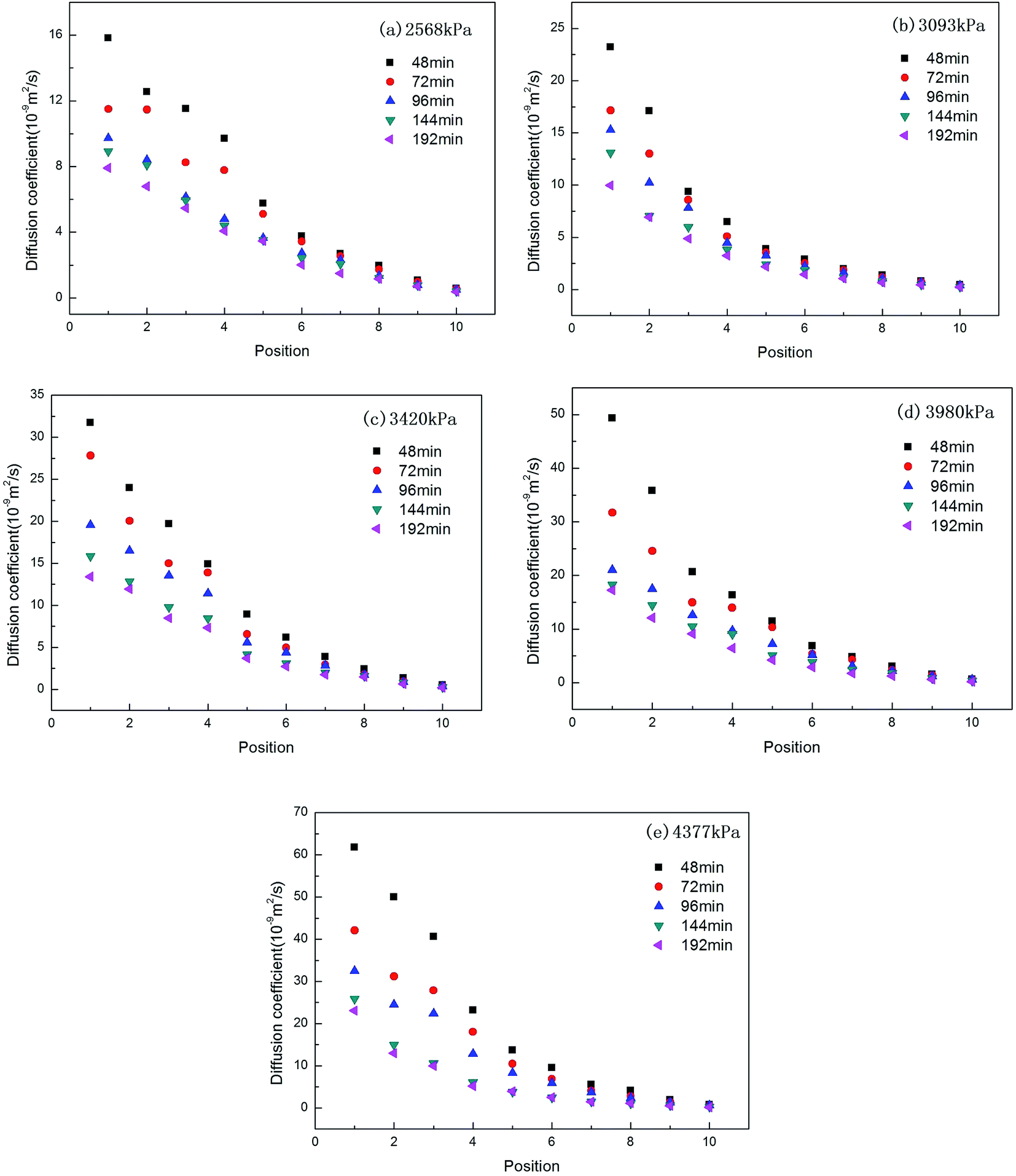 | ||
| Fig. 8 Diffusion coefficients measured at different diffusion distances and times for initial pressures of (a) 2568 kPa, (b) 3093 kPa, (c) 3420 kPa, (d) 3980 kPa and (e) 4377 kPa. | ||
MRI can be used to measure the overall diffusion coefficient of CO2 in n-hexadecane. The overall diffusion coefficient is the average of diffusion coefficients measured during the time it takes for diffusion to reach equilibrium. First, the computational time step was determined, and then the distribution of dimensionless CO2 concentration was calculated using eqn (4). The position dependence of diffusion coefficients was computed using eqn (6). According to the relationship between diffusion coefficient and position, the trapezoidal rule was used to obtain the averages that represent time-dependent diffusion coefficients. Likewise, considering the relationship between diffusion coefficient and time, the trapezoidal rule was then used to average values to calculate the overall diffusion coefficient for each initial pressure. Fig. 9 shows the overall diffusion coefficients for different initial pressures calculated from the experimental MRI data and by the PVT method. According to eqn (3), the error between the MRI and PVT is about 1.6–4.3%. The two results showed good agreement. MRI can measure both unsteady-state local diffusion coefficients and overall diffusion coefficients. In addition, according to the experimental results, the overall diffusion coefficients increase along with the increase of the initial pressure.
 | ||
| Fig. 9 Overall diffusion coefficients obtained by MRI and the PVT method. | ||
4 Conclusions
This study demonstrates that visualization and qualitative analysis of CO2 diffusion in n-hexadecane can be achieved by combining MRI with the dual-chamber pressure decay method. The non-iterative finite-volume analytical technique can be used to calculate CO2 diffusion coefficients from concentration profiles obtained from MR images. The results agree very well with the conventional pressure decay method in binary mixtures. MRI can determine both unsteady-state local diffusion coefficients and overall diffusion coefficients. Based on our experimental results, the following conclusions can be drawn:(1) The concentration of CO2 decreases along the diffusion direction and is higher during the later stages of the reaction than during the initial stages.
(2) The diffusion coefficient of CO2 is a function of both diffusion time and diffusion distance. The diffusion coefficient decreases as diffusion distance and time increase until an equilibrium state is reached.
(3) The initial pressure has an obvious effect on the diffusion coefficient of CO2, with pressure being directly proportional to diffusion coefficient.
(4) The influence of the incubation period decreases with increasing diffusion time. Mass transfer after the incubation period is mainly governed by molecular diffusion.
Nomenclature
| D | Diffusion coefficient (m2 s−1) |
| P(t), Peq | Measured and equilibrium pressure, respectively (kPa) |
| t | Time (s) |
| xeq(P) | Oil/gas interface molar concentration at a given time (mol cm−3) |
| z0 | Height of the oil in the vessel (cm) |
| xz,t | CO2 concentration at position z and time t from MRI images |
| xeq | CO2 concentration at equilibrium state |
| I0 | Initial image intensity at time t = 0 |
| Iz,t | Image intensity at position z and time t |
| Ieq | Image intensity at equilibrium state |
| x | Molar concentration of CO2 gas |
Acknowledgements
This study was supported by the National Natural Science Foundation of China (Grant no. 51206018, and 51106019), the Fundamental Research Funds for Central Universities, the Specialized Research Fund for the Doctoral Program of Higher Education (Grant no. 20120041120057), and the Ph. D. Startup Foundation of Liaoning Province (Grant no. 20121022).References
- C. F. Robert, N. Christopher and V. L. Kuuskraa, Storing CO2 with enhanced oil recovery, Energy Procedia, 2009, 1, 1989–1996 CrossRef PubMed.
- M. Nobakht, S. Moghadam and Y. Gu, Effects of Viscous and Capillary Forces on CO2 Enhanced Oil Recovery under Reservoir Conditions, Energy Fuels, 2007, 21, 3469–3476 CrossRef CAS.
- Y. P. Zhang, C. L. Hyndman and B. B. Maini, Measurement of gas diffusivity in heavy oils, J. Pet. Sci. Eng., 2000, 25, 37–47 CrossRef CAS.
- A. Boustani and B. B. Maini, The role of diffusion and convective dispersion in vapour extraction process, J. Can. Pet. Technol., 2001, 40(4), 68–77 CAS.
- J. C. Moulu, Solution-gas drive: experiments and simulation, J. Pet. Sci. Eng., 1989, 2(4), 379–386 CrossRef.
- H. Sheikha, D. M. Pooladi and A. K. Mehrotra, Development of graphical methods for estimating the diffusivity coefficient of gases in bitumen from pressure-decay data, Energy Fuels, 2005, 19(5), 2041–2049 CrossRef CAS.
- P. Guo, Z. H. Wang, P. P. Shen and J. F. Du, Molecular diffusion coefficients of the multicomponent gas–crude oil systems under high temperature and pressure, Ind. Eng. Chem. Res., 2009, 48(19), 9023–9027 CrossRef CAS.
- M. R. Riaziet, A new method for experimental measurement of diffusion coefficients in reservoir fluids, J. Pet. Sci. Eng., 1996, 14, 235–250 CrossRef.
- Y. P. Zhang, C. L. Hyndman and B. B. Maini, Measurement of gas diffusivity in heavy oils, J. Pet. Sci. Eng., 2000, 25, 37–47 CrossRef CAS.
- Y. Liu, Y. Zhao, J. Zhao and Y. Song, Magnetic resonance imaging on CO2 miscible and immiscible displacement in oil-saturated glass beads pack, Magn. Reson. Imaging, 2011,(29), 1110–1118 CrossRef CAS PubMed.
- Y. Song, N. Zhu, Y. Liu, J. Zhao, W. Liu, Y. Zhang, Y. Zhao and L. Jiang, Magnetic resonance imaging study on miscibility of a CO2/n-decane system, Chin. Phys. Lett., 2011, 28, 0964011–0964014 Search PubMed.
- H. Luo, D. Salama, S. Kryuchkov and A. Kantzas, The effect of volume changes due to mixing on diffusion coefficient determination in heavy oil and hydrocarbon solvent system, The 2007 SPE Annual Technical Conference and Exhibition held in Anaheim, California, USA, 11 November 2007 Search PubMed.
- L. Song, A. Kantzas and J. Bryan, Experimental measurement of diffusion coefficient of CO2 in heavy oil using X-ray computed-assisted tomography under reservoir conditions, The Canadian Unconventional Resources & International Petroleum Conference held in Calgary, Alberta, Canada, 19 October 2010 Search PubMed.
- D. Salama and A. Kantzas, Monitoring of diffusion of heavy oils with hydrocarbon solvents in the presence of sand, The 2005 SPE International Thermal Operations and Heavy Oil Symposium held in Calgary, Alberta, Canada, 1 November 2005 Search PubMed.
- D. B. Fisher, A. K. Singhal, S. K. Das, J. Goldman and C. Jackson. Use of magnetic resonance imaging and advanced image analysis as a tool to extract mass transfer information from a 2D physical model of the Vapex process, The 2000 SPE/DOE improved oil recovery symposium held in Tulsa, Oklahoma, 3 April 2000 Search PubMed.
- A. K. Tharanivasan, C. Yang and Y. Gu, Measurements of Molecular Diffusion Coefficients of Carbon Dioxide, Methane, and Propane in Heavy Oil under Reservoir Conditions, Energy Fuels, 2006, 20, 2509–2517 CrossRef CAS.
- Z. Li and M. Dong, Experimental Study of Carbon Dioxide Diffusion in Oil-saturated Porous Media, Ind. Eng. Chem. Res., 2009, 48, 9307–9317 CrossRef CAS.
- Y. C. Zhao, Y. C. Song, Y. Liu, L. L. Jiang and N. J. Zhu, Visualization of CO2 and oil immiscible and miscible flow processes in porous media using NMR micro-imaging, Pet. Sci., 2011, 8, 183–193 CrossRef CAS PubMed.
- U. Guerrero-Aconcha and A. Kantzas, Diffusion of hydrocarbon gases in heavy oil and bitumen, The 2009 SPE Latin American and Caribbean Petroleum Engineering Conference held in Cartagena, Colombia, 31 May–3 June 2009 Search PubMed.
- C. Chang and M. Chang, Non-iteration estimation of thermal conductivity using finite volume method, Int. Commun. Heat Mass Transfer, 2006, 33, 1013–1020 CrossRef PubMed.
- M. Jamialahmadi and M. Emadi, Diffusion coefficients of methane in liquid hydrocarbons at high pressure and temperature, J. Pet. Sci. Eng., 2006, 53, 47–60 CrossRef CAS PubMed.
- L. S. Wang, Z. X. Lang and T. M. Guo, Measurement and correlation of the diffusion coefficients of carbon dioxide in liquid hydrocarbons under elevated pressures, Fluid Phase Equilib., 1996, 117, 364–372 CrossRef CAS.
- A. T. Grogan, V. W. Pinczewski, G. Ruskauff and F. M. Orr Jr, Diffusion of CO2 at reservoir conditions: models and measurements, SPE Reservoir Eng., 1988, 2, 93–102 CrossRef PubMed.
- R. Islas-Juárez and F.-V. Samanego, Experimental Study of Effective Diffusion in Porous Media, The 2004 SPE International Petroleum Conference in Mexico held in Puebla, Mexico, 8–9 November 2004 Search PubMed.
| This journal is © The Royal Society of Chemistry 2014 |