DOI:
10.1039/C4RA07731H
(Paper)
RSC Adv., 2014,
4, 48516-48521
A novel probe for selective colorimetric sensing of Fe(II) and Fe(III) and specific fluorometric sensing of Fe(III): DFT calculation and logic gate application†
Received
28th July 2014
, Accepted 18th September 2014
First published on 24th September 2014
Abstract
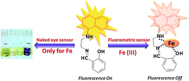
A novel fluorescent probe 1 (2-((2-(naphthalen-1-ylamino)ethylimino)methyl)phenol) has been synthesized and characterized by various spectroscopic methods. Probe 1 was found to be highly selective for iron over tested metal ions. The naked eye detection of iron is useful for the discrimination of the +2 and +3 oxidation state while fluorescence studies conclude selective and specific sensitivity towards Fe(III).
1. Introduction
The design and synthesis of probes or chemosensors for the detection of metal ions are important areas of chemical research.1 Iron is one of the most essential elements present in the biosystem and exhibits several crucial roles in different enzymatic and other biological activities;2 however, this metal ion could exhibit detrimental effects alone or in a combined state causing diseases like hemochromatosis, cancer etc.3 Hence detection of the presence of iron and its concentration as well as localizations are extremely important for the treatment of such diseases. The common oxidation states of iron are Fe(II) and Fe(III) which are important for ordinary and related chemistry.4 Naked eye detection for metal ions is important for qualitative identification as well as quantitative detection. Hence simple and easy to use colorimetric probes are highly demanding for metal ion sensing.5 In this regard, fluorescent chemosensors gained considerable attention in the recent years due to simplicity, sensitivity and easy handling.6 Different colorimetric as well as fluorimetric probes have been reported for iron sensing.7 We have recently reported a simple probe for fluorimetric detection of Fe(III).8 However, to the best of our knowledge, there is no report of a naphthyl-based probe which could detect iron by colorimetric as well as fluorometric way.9 Tedious methods, complicated structures as well as interference from several metals such as Cu(II) and Cr(III) were also observed in fluorescence probes for Fe(III).10 In this report we have synthesized a simple but novel probe 2-((2-(naphthalen-1-ylamino)ethylimino)methyl)phenol (1) for the colorimetric detection of Fe(II) and Fe(III). We have investigated the same probe for the fluorometric detection of Fe(III). The results will be discussed in the light of theoretical calculation via DFT calculations. Possible application to logic gates will also be scrutinized.
2. Experimental details
2.1 Materials
N-(1-Naphthyl)-ethylenediaminedihydrochloride purchased from Thomas Baker, India. Salicylaldehyde was purchased from SRL, Mumbai, India. All metal salts used in the synthesis were purchased from commercial source and used directly (without any purification). 1H-NMR and 13C-NMR spectra of 1 in CDCl3 were recorded using Bruker AVANCE, 500.13 MHz spectrometer; GC-MS spectrometry was recorded with Perkin Elmer. The UV-vis spectra were obtained by using Evolution 600, Thermo Scientific UV-visible spectrophotometer. Emission spectra were obtained from RF-5301PC with a 3 cm standard quartz cell.
2.2 UV-vis and fluorescence measurements and computational details
Stock solution of 1 (1 mM) and chloride salt (10 mM) of metals Zn(II), Sn(II), Ni(II), Mn(II), Mg(II), Hg(II), Fe(II), Fe(III), Cu(II), Co(II), Cd(II), Ca(II) and Ba(II) were prepared in methanol for metal sensing. All fluorescence spectra of 1 were recorded with the slit width 3/3. Fluorescence quantum yield (QY) was calculated by comparative method using 2-aminophenol solution in 0.1 N H2SO4 as the standard.11 DFT calculations for 1 and 1–Fe(III) was performed at the B3LYP level using LANL2DZ basis set for Fe center and 6-31G(d) basis set for non metal atoms.
2.3 Synthesis and characterization of 2-((2-(naphthalen-1-ylamino)ethylimino)methyl)phenol 1
N-(1-Naphthyl)-ethylenediaminedihydrochloride salt was dissolved in water and then neutralized using a saturated solution of KOH. Free amine was extracted with the help of dichloromethane and dried over Na2SO4. A solution of salicylaldehyde (122 mg, 1.00 mmol) in 5 mL of methanol was added to (186 mg, 1.00 mmol) of N-(1-naphthyl)-ethylenediamine in 10 mL methanol with continuous stirring. After refluxing of few hours, reaction was cooled to room temperature. An oily compound obtained after evaporation of solvent. GC-MS (MeOH, m/z): 290 M+. Selected IR data (KBr, νmax/cm−1): 3428,νN–H, 1631,νC
Nimine. UV-visible [CH3OH, λmax/nm (ε/M−1 cm−1)]: 213 (65
555), 247 (24
695), 325 (9730), 400 (1185). 1H NMR (CDCl3, δ/ppm, 500 MHz): 3.251 (t, 2H), 3.318 (dd, 2H), 4.586 (s, 1H), 6.702 (d, J = 7.5, 1H), 7.198 (d, J = 7.5, 1H), 7.267 (d, J = 7.5 Hz, 1H), 7.270–7.451 (m, 5H), 7.742 (d, J = 8.5, 1H), 7.799 (d, J = 8, 1H), 8.005 (d, J = 8, 1H), 8.345 (s, 1H), 13.260 (s, 1H). 13C NMR (CDCl3, δ/ppm, 500 MHz): 44.41, 58.12, 104.7, 117, 117.8, 118.73, 118.78, 119.82, 123.58, 124.97, 125.89, 126.54, 128.73, 131.53, 132.53, 134.42, 142.69
161.09, 166.7. Anal. calcd for C18H17N3: C, 78.52; H, 6.22; N, 15.26, found: C, 76.88; H, 6.46; N, 14.45.
3. Results and discussions
3.1 Characterization of 1
Fluorescent probe 1 has been synthesized by condensation of N-(1-naphthyl)-ethylenediaminedihydrochloride and salicylaldehyde and was characterized using spectral studies. Structure of probe 1 was authenticated by various spectroscopic techniques like IR, UV-vis, 1H-NMR, 13C-NMR spectral studies and GC-MS (data have been deposited in the ESI Fig. S1–S5†).
3.2 Naked eye detection
Unique color changes for iron metal ions with probe 1 were observed during investigation with other metal ions. Among the representative metal ions only Fe(II) (yellow-brown) and Fe(III) (purple) showed color changes that can easily be observed via “naked eye”(Fig. 1). Several fluorescent probes have been reported for the visible detection of iron but there is no report on naked eye detection for discrimination of Fe(II) and Fe(III) using naphthyl-based probe.
 |
| Fig. 1 Images showing naked eye visible changes of probe 1 in the presence of representative metal ions (10 equiv.). | |
3.3 UV-vis absorption studies
Absorption studies of probe 1 have been investigated using methanol as a solvent. UV vis spectrum in methanol exhibited typical naphthalene absorption band at 320 nm due to charge transfer transition between amine and naphthyl group.12 Probe 1 exhibited a band around 402 nm in visible region and slightly yellow in color. Addition of Fe(II) into the methanolic solution gave rise to yellow-brown color and showed an intense absorption band in the visible region around 536 nm. Addition of Fe(III) into the methanolic solution gave rise to purple color and showed an intense absorption band in the visible region around 590 nm (Fig. 2). The coloration was due to a CT band in the visible region.13 Insolubility of 1 in water prompted us to study this experiment in water–methanol mixed solvent media. In mixed aqueous media the band responsible for color generation completely disappeared. Changes in pH in mixed aqueous media were examined (Fig. S6 in ESI†) and in this case also we did not observe the band responsible for the color (Fig. S7 in ESI†). Hence the naked eye detection was not possible on moving from methanol to methanol–water solvent media as well as for pH variation. The titration experiments with Fe(II) and Fe(III) ions were performed and results are deposited in the ESI (Fig. S8–S9†).
 |
| Fig. 2 UV-vis spectral change of 1 (20 μM) in methanol on addition of 10 equivalent of Zn(II), Sn(II), Ni(II), Mn(II), Mg(II), Hg(II), Fe(II), Fe(III), Cu(II), Co(II), Cd(II), Ca(II) and Ba(II). | |
3.4 Fluorescence studies
Further, we have examined photophysical properties of probe 1 in methanol. Probe 1 was found to be fluorescent at λexc 320 nm. The wavelength of emission was around 420 nm. Probe 1 showed maximum emission intensity within 5 min and remains constant for more than half an hour (Fig. S10†). Clearly, the maximum excitation and emission are at 320 nm and 420 nm, respectively, showing a large Stokes shift (Fig. S11†). Addition of metal ions Zn(II), Sn(II), Ni(II), Mn(II), Mg(II), Hg(II), Fe(II), Cu(II), Co(II), Cd(II), Ca(II) and Ba(II) did not cause any appreciable change in emission intensity of probe 1. On the other hand, introduction of Fe(III) gave rise to quenching in emission intensity with the red shift of 13 nm which further kept on decreasing on further addition of Fe(III) (Fig. 3(A) and (B)).
 |
| Fig. 3 (A) Fluorescent emission spectra of 1 (20 μM) in methanol with 10 equivalent of Zn(II), Sn(II), Ni(II), Mn(II), Mg(II), Hg(II), Fe(II), Fe(III), Cu(II), Co(II), Cd(II), Ca(II) and Ba(II). (B) Fluorescent emission titration spectra of 1 (20 μM) in the presence of varying concentration of Fe(III) in MeOH at λex 320 nm (inset – change in emission intensity with number of equivalents of Fe(III). | |
Due to large value of stokes shift, fluorescence resonance energy transfer (FRET) is not a favorable pathway for the quenching mechanism (Fig. S11†). Hence the electron transfer could be a probable mechanism for the quenching as Fe(III) is a strong Lewis acid which can accept electron easily.14
Similar to absorption spectral studies the fluorescence properties of probe 1 were also investigated in mixed aqueous media as well as in different pH of the solutions (Fig. S12–S13†). The data obtained in mixed aqueous media were different to that of our results in methanol. Comparison of Fig. 3(A) and S12† clearly indicated the retention of quenching of Fe(III), however selectivity of iron sensing diminished (Fig. S14†). We would like to mention here that in Fig. 3(A) we found enhancement of emission intensity as compare to probe for all metal ions excluding Fe(III). On the other hand, we found enhancement as well as quenching of emission intensity for different metal ions (Fig. S14†). The Stern–Volmer plot, measurement of life time τ = 7.94 ns (probe only), 8.09 ns (probe + Fe(III)) and changes in absorption spectra clearly indicate the static nature of quenching.15 (Fig. 4(A) and (B)).
 |
| Fig. 4 (A) Fluorescence decay profile of 1 in the presence and absence of Fe(III) in methanol. (B) Stern–Volmer plots for titrations of 1 with different concentrations of Fe(III) in methanol. | |
Quantum yield values were calculated using 2 aminopyridine as standard. The values for relative quantum yield for probe as well as for probe + Fe(III) were found to be 0.03 and 0.005.11
3.5 Selectivity studies
Competitive binding studies were also performed to study the interference of the other metal ions Zn(II), Sn(II), Ni(II), Mn(II), Mg(II), Hg(II), Fe(II), Cu(II), Co(II), Cd(II), Ca(II) and Ba(II) in the detection of Fe(III) using spectrophotometry as well as spectrofluorometry. Selectivity of probe is very crucial as the interference of metal can affect its sensitivity. For the competitive binding studies equal concentration of Fe(III) and other metals were taken in methanolic solution. Anti-interference experiment showed that among the served metal ions no metal ion interfere with the Fe(III). Fig. 5 and 6 clearly indicates that Fe(III) acts as an excellent sensor even in presence of other metal ions. To investigate the effect of metal ions on the selectivity of Fe(III) ion mixed metal ion studies were performed using UV-vis as well as fluorescence spectral studies.
 |
| Fig. 5 Selectivity of metal ions at wavelength 565 nm in a solution having probe 1 + metal ions (black bar) and 1 + metal ions + Fe(III) (red bar) observed using UV-vis absorption studies. | |
 |
| Fig. 6 Selectivity of metal ions at wavelength 421 nm in a solution having probe 1 + metal ions (black bar) and 1 + metal ions + Fe(III) (red bar) observed using fluorescence spectral studies. | |
During absorption studies wavelength 565 nm was chosen for the mixed metal ion sensing. Bar diagram clearly represents the effect of Fe(III) on fluorescence of probe 1 (Fig. 5). Rest of the metal ions did not show any band around 565 nm but on addition of Fe(III) band arises at 565 nm only. During UV-vis studies only Cu(II) interfere with the band formation.
Fluorescence studies performed for the selectivity Fe(III) showed quenching without interference of any other metal ion (Fig. 6). Hence probe 1 can be used highly selective as well as specific fluorimetric sensor for Fe(III).
3.6 Binding stoichiometry of probe 1 and Fe(III)
The binding stoichiometry of 1 was obtained from the Job's plot measurement on the basis of fluorescence.16 Emission intensity was plotted against mole fraction of Fe(III) at 420 nm. The maxima clearly expressed 1
:
1 binding stoichiometry of 1 with Fe(III) (Fig. 7). Binding ratio of probe and Fe(III) ion was also calculated using Benesi–Hildebrand plot which supports the 1
:
1 stoichiometry obtained from Job's plot. Association constant was found to be 2.884 × 10³ M−1 (Fig. 8).17 We have also examined electrospray ionization-mass spectral (ESI-MS) studies on the probe in presence of Fe(III) ion. Results obtained from this experiments supported our observations described above and were deposited in the ESI (Fig. S15(A) and (B)†).
 |
| Fig. 7 Job's plot of 1 and Fe(III) in methanol, total concentration of 1 and Fe(III) were maintained 100 μM and observed at 420 nm. | |
 |
| Fig. 8 B–H plot for binding of Fe(III) with the probe 1. Association constant was found to be 2.884 × 10³. A good linear fit of the B–H plot supported the 1 : 1 binding stoichiometry. | |
Limit of detection for Fe(III) was also calculated for a linear range of 10–60 μM and found to be 20.85 μM (Fig. S16†).
3.7 Reversibility of probe 1
For better insight into the mechanism of Fe(III) sensing fluorescence titration were conducted in presence of EDTA. EDTA has a strong tendency to chelate Fe(III) and formation of Fe–EDTA complex stops the electronic interaction of probe and Fe(III) ion. Hence instead of the fluorescence quenching we observed enhancement of fluorescence in presence of EDTA. Further addition of Fe(III) ion to the solution afforded fluorescence quenching. Titration of probe 1 with EDTA suggested that binding of Fe(III) with probe to some extent found to be reversible as well as supports the electron transfer pathway for quenching of the probe (Fig. S17†). Hence probe 1 can be considered as a reversible sensor for Fe(III).7c
3.8 DFT calculation
In order to understand the mechanism of such processes, we performed theoretical calculations. DFT calculations for 1 and 1–Fe(III) complex was performed at the B3LYP level using LANL2DZ basis set for Fe center and 6-31G(d) basis set for non metal atoms.18 The highest occupied molecular orbital (HOMO) and the lowest unoccupied molecular orbital (LUMO) of 1 and 1–Fe(III) complexes has been depicted in Fig. 9. The energy gaps between HOMO and LUMO in the probe 1 and 1–Fe(III) complex were 0.10468 eV and 0.0102 eV respectively. Energy gap between HOMO and LUMO in complex decreases in 1–Fe(III) shows a favorable complexation according to proposed coordination. The optimized configuration showed suitable binding of Fe(III) with tridentate probe having NNO donor.19
 |
| Fig. 9 Energy diagrams of HOMO and LUMO orbitals of probe 1 and 1–Fe(III) complex calculated at the DFT level using a B3LYP/6-31+G(d,p) basis set. | |
3.9 Logic gate application
Logic gates have been attracted the attention of researchers due to their increasing application in molecular switches and molecular keypad devices.20
Here we have chosen Fe(III) and EDTA used for reversibility experiment as input.21 Truth table and logic gate have been shown in Fig. 10. Emission intensity at 100 has been taken as a threshold value at wavelength 420 nm. Emission intensity above threshold gives a state OUT = 0 and below OUT = 1.
 |
| Fig. 10 Change in emission spectra 1 upon chemical inputs of Fe(III) IN1, EDTA IN2. Truth table indicates and logic functions. | |
4. Conclusions
In this report a simple and novel probe 1 has been synthesized via a simple one step synthetic procedure and resultant compound was characterized by various spectroscopic techniques. Photo-physical properties of 1 have been investigated to study the sensing of metal ions in methanolic solution. Probe 1 selectively detected iron in both +2 and +3 oxidation states giving rise to yellow-brown and purple color respectively. Although there is interference of copper during absorption spectral studies the probe was highly sensitive and selective towards Fe(III) during fluorimetric detection because of fluoroscence quenching of the probe. Life time measurement, Stern–Volmer plot and UV-vis spectral studies indicated static nature of quenching. Binding stoichiometry was found to be 1
:
1 for Fe(III) and probe 1. DFT calculation provided that the decrease in the energy gap between HOMO and LUMO is probably responsible for the quenching of fluorescence. We have performed UV-vis spectra and fluorescence spectral studies in mixed (water and methanol) solvents. UV-vis experiments clearly showed the disappearance of bands near 500 nm which were responsible for the color. Modification of the probe for experiments in aqueous media and their biological applications are under progress.
Acknowledgements
KG is thankful to CSIR, India for financial assistance no. 01(2720)/13/EMR-II dated 17-APRIL-2013. KG is also thankful to DST-SERB, India for financial assistance no. SR/S1/IC-47/2012 dated 21-OCT-2013. SR is thankful to UGC for fellowship. We are thankful to DST-FIST program for providing us ESI-MS facility in our department. We are thankful to Vishal for his help.
Notes and references
-
(a) X. Chen, T. Pradhan, F. Wang, J. S. Kim and J. Yoon, Chem. Rev., 2012, 112, 1910 CrossRef CAS PubMed;
(b) H. N. Kim, W. X. Ren, J. S. Kim and J. Yoon, Chem. Soc. Rev., 2012, 41, 3210 RSC.
-
(a) J. A. Cowan, Inorganic Biochemistry: an Introduction, Wiley-VCH, New York, 1997, pp. 167–255 Search PubMed;
(b) I. Bertini, H. B. Gray, S. J. Lippard and J. S. Valentine, Bioinorganic Chemistry, University Science Books: South Asian Edn, 1998 Search PubMed;
(c) J. J. R. F. Da Silvaand R. J. P. Williams, The Biological Chemistry of the Elements: The Inorganic Chemistry of Life, Clarendon Press, Oxford, 1993 Search PubMed.
- N. K. Desai, G. B. Kolekar and S. R. Patil, New J. Chem., 2014, 38, 4394 RSC.
- F. A. Cotton, G. Wilkinson, C. A. Murillo and M. Bochmann, Advanced Inorganic Chemistry, Wiley, New York, 6th edn, 2003 Search PubMed.
- Y.-K. Yang, K.-J. Yook and J. Tae, J. Am. Chem. Soc., 2005, 127, 16760 CrossRef CAS PubMed.
- P. Das, N. B. Chandar, S. Chourey, H. Agarwalla, B. Ganguly and A. Das, Inorg. Chem., 2013, 52, 11034 CrossRef CAS PubMed.
-
(a) S. Sen, S. Sarkar, B. Chattopadhyay, A. Moirangthem, A. Basu, K. Dhara and P. Chattopadhyay, Analyst, 2012, 137, 3335 RSC;
(b) Y. Xiang and A. Tong, Org. Lett., 2006, 8, 1549 CrossRef CAS PubMed;
(c) V. Bhalla, N. Sharma, N. Kumar and M. Kumar, Sens. Actuators, B, 2013, 178, 228 CrossRef CAS PubMed;
(d) S. Bae and J. Tae, Tetrahedron Lett., 2007, 48, 5389 CrossRef CAS PubMed;
(e) N. R. Chereddy, K. Suman, P. S. Korrapati, S. Thennarasu and A. B. Mandal, Dyes Pigm., 2012, 95, 606 CrossRef CAS PubMed;
(f) S.-R. Liu and S.-P. Wu, Sens. Actuators, B, 2012, 171–172, 1110 CrossRef CAS PubMed;
(g) Z.-Q. Liang, C.-X. Wang, J.-X. Yang, H.-W. Gao, Y.-P. Tian, X.-T. Tao and M.-H. Jiang, New J. Chem., 2007, 31, 906 RSC;
(h) A. M. G. Silva, A. Leite, M. Andrade, P. Gameiro, P. Brandao, V. Felix, B. de Castro and M. Rangel, Tetrahedron, 2010, 66, 8544 CrossRef CAS PubMed.
- K. Ghosh, S. Rathi and R. Kushwaha, Tetrahedron Lett., 2013, 54, 6460 CrossRef CAS PubMed.
-
(a) S. Ghosh, C. K. Dey and R. Manna, Tetrahedron Lett., 2010, 51, 3177 CrossRef CAS PubMed;
(b) C. R. Lohani and K.-H. Lee, Sens. Actuators, B, 2010, 143, 649 CrossRef CAS PubMed.
-
(a) J. Mao, L. Wang, W. Dou, X. Tang, Y. Yan and W. Liu, Org. Lett., 2007, 9, 4567 CrossRef CAS PubMed;
(b) M. Xu, S. Wu, F. Zeng and C. Yu, Langmuir, 2010, 26, 4529 CrossRef CAS PubMed;
(c) J. P. Sumner and R. Kopelman, Analyst, 2005, 130, 528 RSC;
(d) X. Zhang, Y. Shiraishi and T. Hirai, Tetrahedron Lett., 2007, 48, 5455 CrossRef CAS PubMed;
(e) N. C. Lim, S. V. Pavlova and C. Bruckner, Inorg. Chem., 2009, 48, 1173 CrossRef CAS PubMed;
(f) C. Wolf, X. Mei and H. K. Rokadia, Tetrahedron Lett., 2004, 45, 7867 CrossRef CAS PubMed;
(g) J. L. Bricks, A. Kovalchuk, C. Trieflinger, M. Nofz, M. Buschel, A. I. Tolmachev, J. Daub and K. Rurack, J. Am. Chem. Soc., 2005, 127, 13522 CrossRef CAS PubMed;
(h) Y. Ma, W. Luo, P. J. Quinn, Z. Liu and R. C. Hider, J. Med. Chem., 2004, 47, 6349 CrossRef CAS PubMed;
(i) X. Wu, B. Xu, H. Tong and L. Wang, Macromolecules, 2010, 43, 8917 CrossRef CAS.
- R. Kagit, M. Yildirim, O. Ozay, S. Yesilot and H. Ozay, Inorg. Chem., 2014, 53, 2144 CrossRef CAS PubMed.
- M. Suresh, A. Ghosh and A. Das, Chem. Commun., 2008, 3906 RSC.
- S. Konetschny-Rapp, G. Jung, K. N. Raymond, J. Meiwes and H. Zahner, J. Am. Chem. Soc., 1992, 114, 2224 CrossRef CAS.
- M. Yanga, M. Sun, Z. Zhang and S. Wang, Talanta, 2013, 105, 34 CrossRef PubMed.
- J. R. Lakowicz, Principles of Fluorescence Spectroscopy, Springer, New York, USA, 3rd edn, 2006 Search PubMed.
- A. Senthilvelan, I. Ho, K. Chang, G. Lee, Y. Liu and W. Chung, Chem.–Eur. J., 2009, 15, 6152 CrossRef CAS PubMed.
-
(a) H. A. Benesi and J. H. Hildebrand, J. Am. Chem. Soc., 1949, 71, 2703 CrossRef CAS;
(b) S. Wu, T. Wang and S. Liu, Tetrahedron, 2010, 66, 9655 CrossRef CAS PubMed.
- M. J. Frisch, G. W. Trucks and H. B. Schlegel, et al.,Gaussian 03, Revision B.03, Gaussian, Inc, Wallingford, CT, 2004 Search PubMed.
- H. Xu, Z. Liu, L. Sheng, M. Chen, D. Huang, H. Zhang, C. Song and S. Chen, New J. Chem., 2013, 37, 274 RSC.
-
(a) M. Devi, A. Dhir and C. P. Pradeep, Dalton Trans., 2013, 42, 7514 RSC;
(b) M. Kumar, A. Dhir and V. Bhalla, Org. Lett., 2009, 11, 2567 CrossRef CAS PubMed;
(c) D. Margulies, C. E. Felder, G. Melman and A. Shanzer, J. Am. Chem. Soc., 2007, 129, 347 CrossRef CAS PubMed;
(d) A. Misra, P. Srivastava and M. Shahid, Analyst, 2012, 137, 3470 RSC;
(e) V. Bhalla, Roopa and M. Kumar, Org. Lett., 2012, 14, 2802 CrossRef CAS PubMed;
(f) B. Rout, P. Milko, M. A. Iron, L. Motiei and D. Margulies, J. Am. Chem. Soc., 2013, 135, 15330 CrossRef CAS PubMed.
- S. Wang, G. Men, L. Zhao, Q. Hou and S. Jiang, Sens. Actuators, B, 2010, 145, 826 CrossRef CAS PubMed.
Footnote |
† Electronic supplementary information (ESI) available: Characterization of probe 1, supplementary spectra and graphs. See DOI: 10.1039/c4ra07731h |
|
This journal is © The Royal Society of Chemistry 2014 |
Click here to see how this site uses Cookies. View our privacy policy here.