Dongli Fan,
ShuangShuang Wu,
Shu Tian*,
Jingyang Zhou,
Yun Ju,
Chunjuan Ma and
Jian Shi*
School of Chemistry and Chemical Engineering, Nantong University, Nantong 226007, P. R. China. E-mail: tian0429@ntu.edu.cn; shijianchuyu@163.com; Fax: +86-513-85012851; Tel: +86-513-85012856
First published on 16th September 2014
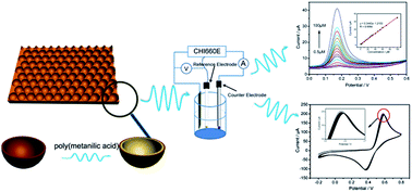
A shape-controllable, highly ordered, two-dimensional gold cavity array (GCA) electrode was prepared by electrodeposition using a closely packed monolayer of 1.2 μm-diameter polystyrene spheres as a template and was characterized by FESEM and XRD. A significant enhancement for the electrooxidation of dopamine at this nanostructured electrode was found due to the increased amount of the active surface area. By electropolymerizing a poly(metanilic acid) thin film on its surface, the enhanced electrochemical properties of the GCA electrode were maintained, and the antifouling property was improved. The SWV technique was used for the trace determination of DA, and the dependence of current vs. concentration was linear from 0.2 to 100 μM with a regression coefficient of 0.9988, and the detection limit of DA was ∼0.08 μM. Furthermore, the signals of DA and UA can be well distinguished at this poly(metanilic acid) modified GCA electrode. The proposed method was applied to the selective and precise analysis of DA in commercial injections.
Being an electroactive substance, DA can be easily detected by electrochemical methods through its oxidation at the surface of various electrodes. One of the primary problems is that, in real biological samples, the physiological concentration of DA is ultra-low (from 0.01 to 1 μM)8 and even in the nanomolar range for patients with Parkinson's disease, whereas the concentrations of the main detection interferences, e.g., ascorbic acid (AA) and uric acid (UA), are several orders of magnitude higher.17 A further complication is that both AA and UA are also electroactive and undergo oxidation within the same potential window as DA. To perform the DA detection with high sensitivity and selectivity, various chemically modified electrodes have been fabricated using metal or metal oxide nanoparticles,18 metal complexes,19 carbon materials,15 conductive polymers,17 and self-assembled monolayers.9 These modified electrodes show excellent properties in DA detection by decreasing the overpotential, which accelerates the electron and mass transfer rate and enlarges the electrode surface.
In recent years, porous metal electrodes with highly ordered nanostructures have attracted increasing research attention. The large active surface area and the ordered arrangement of pores of such two-dimensional or three-dimensional metal electrodes have found their important roles in many applications, such as electrocatalysis, spectroelectrochemistry, chemical and biological sensors, and microelectronic devices.20–22 For example, Kuhn's group reported the fabrication of a fluorenone-based mediator modified macroporous gold electrode to detect glucose in solution, and the currents increased by more than one order of magnitude relative to those achieved on flat electrode surfaces.23,24 Due to their uniform structures, macroporous electrodes can be also used to host enzymes and/or redox proteins at the monolayer level.25–27 Xia and co-workers25 prepared a three-dimensional macroporous gold electrode to enlarge the surface area and accelerate the direct electron transfer between the electrode and the adsorbed hemoglobin.
Although photolithography, electron beam lithography, X-ray lithography, scanning tunneling microscopy, and atomic force microscopy lithography are the most widely used methods to create defect-free nanostructured arrays, their high costs are beyond the purchasing power of most laboratories.28 Template-based methods have proven to be flexible and low-cost for preparing porous films. In this method, using highly ordered monodispersed nanoparticles (usually silica and polystyrene (PS) particles) as a template, functional materials can be filled into the interspaces of the template through vapor deposition, chemical deposition, and electrodeposition.29–31 A highly ordered macroporous film with the same shape and size as the template is obtained by removing the template through dissolution or thermal decomposition. The first example of electrochemical deposition through a colloidal template was reported by Braun and Wiltzius.32 In their excellent paper, the authors reported the electrodeposition of CdSe and CdS using 466 nm-diameter PS spheres as the template. Following this approach, the deposition materials have been extended to semiconductors, metals, metal oxides, and conducting polymers. Combined with electrodeposition, it is possible to control the morphology of the obtained macroporous film by employing different sizes of PS spheres, and by altering the electrochemical parameters during the deposition process. Furthermore, no shrinkage of the material occurs because electrodeposition ensures a high density of the deposited material within the interspaces of the template.33
Conductive polymers, such as polypyrrole, polythiophene, and polyaniline, have also been widely used to alleviate the above-mentioned problems with DA detection.34–36 Their widespread popularity arises not only because of their easier fabrication processes and good electrical conductivity, but also because it is possible to increase the functional density and facilitate electron exchange through chemical substitution on the polymer chain (doped conductive polymer). The multifunctionality of metal/conductive polymer composites is particularly useful, as they are endowed with enhanced electrochemical catalytic ability and better physical stability.37
In this work, we combine the benefits of a two-dimensional gold cavity array (GCA) electrode and poly(metanilic acid) to develop a sensitive and selective electrochemical DA sensor. The concave bowl-shaped morphology greatly increased the surface area available for the sensitive detection of the target analyte. A thin film of poly(metanilic acid) was then electropolymerized on the surface of the GCA electrode, and the electrocatalytic ability of the gold nanoparticles (AuNPs) and their strong interaction with the polyaniline backbone significantly improved the stability of the poly(metanilic acid). The conductivity and electrochemical activity in neutral solutions were greatly enhanced due to the –SO3H doped in the polymer chain. In this study, we demonstrate that by depositing this thin layer of poly(metanilic) onto the GCA electrode, DA concentrations as low as 0.08 μM with a linear range from 0.2 to 100 μM were detected with the square wave voltammetry (SWV) technique. Since the anti-fouling effect of the poly(metanilic) thin film, the problem of the adsorption of the DA oxidation product was avoided, ensuring the significant reproducibility and stability during DA detection. Moreover, DA and UA can be simultaneously detected when in binary mixtures.
The SEM images were obtained using a Hitachi S4700 (Japan) field emission scanning electron microscope. X-ray diffraction (XRD) patterns were recorded at a Bruker AXSD8 ADVANCE X-ray diffractometer (Cu Kα radiation, λ = 0.15406 nm) at room temperature. The electropolymerization of poly(metanilic acid) and all electrochemical measurements were recorded at a CHI 660E electrochemical station (Shanghai ChenHua Instruments CO. LTD., China). A bare Au or gold cavity array electrode was used as the working electrode. In order to maintain a constant surface area of the working electrode, the surface of the electrode was sheltered by a sticker and only a circular portion of 0.5 cm diameter was exposed to the solution. Platinum and Ag|AgCl (3 M KCl) electrodes were used as the auxiliary and reference electrodes, respectively. All electrochemical measurements were conducted in an argon-purged (at least 15 min and maintained under an argon atmosphere during whole course) solution and at room temperature.
:
1 water–ethanol) was then spread slowly and evenly across the silicon wafer treated as described above, which was then placed into the water at a 45° angle. The nanosphere suspension ran down the silicon wafer onto the water surface, where it formed a stable film. The monolayer was transferred to a gold coated ITO by placing it under the monolayer and withdrawing it slowly from the solution. Areas up to 1 cm2 were easily modified using this method with very high quality. The gold coated slides used in this work were prepared by electromagnetic sputtering of a 10 nm thick chromium layer, followed by a 200 nm thick gold layer onto ITO glass slides.
The PS template-coated substrate was then used as the working electrode for further electrodeposition of gold in a plating bath solution containing 0.029 M HAuCl4, 0.017 M EDTA, 1.267 M Na2SO3 and 0.173 M K2HPO4 according to a method previously reported in the literature.39 The gold arrays were produced using multi-current pulse plating with the first pulse to a current density of 20 mA cm−2 for 100 ms, followed by a train of pulses of 5 mA cm−2 for 60 ms separated by a rest time of 1 s (zero current). Then, the electrode was immersed into toluene to dissolve the PS spheres, leaving an ordered array of interconnected sphere segment voids.
Multi-current pulse plating is a preferable method for preparing uniformly sized metal nanoparticles that firmly adhere to the conducting substrate. Electrochemical deposition occurs from the electrode surface out through the overlying template, AuNPs gradually fill the interstices among the PS spheres, and the growth of AuNPs on the electrode surface is directed by the ordered structure of the assembled PS particles. As a result, the cavity arrays are obtained after removal of the template. Fig. 1b shows a tilted SEM image of a hemispheric cavity array grown through the template made up of a monolayer of 1.2 μm-diameter PS spheres (as shown in Fig. 1a). It can be clearly seen that the resulting gold cavities match the size of the PS particles and the array retains the two-dimensional close-packed ordering. The first pulse produced “gold seeds.” Fig. S1a† shows the SEM image of the “gold seeds” produced under a 20 mA cm−2 current density. As observed, these nanoparticle-like dots were evenly distributed on the electrode surface, which was the key factor to ensure good adhesion and uniform morphology. An appropriate increase in the current density of the first pulse resulted in the increased density of the gold nanoparticles (AuNPs) on the electrode and made the growth of the gold seeds more controllable. However, as shown in Fig. S1b,† the pulse current density was increased to 50 mA cm−2, the size distribution of the deposited gold seeds became uneven, thus affecting the morphology of the cavity array and further applications. The film thickness can be easily controlled by regulating the total amount of charges passed during the electrodeposition. As shown in Fig. 1c–f, the depth of the gold cavities increased with an increase in the repeat times of the second pulse. Moreover, it was found that when the interstices of the assembled PS particles were fully filled with AuNPs, the growth of AuNPs was no longer directed by the shape of the PS particles, showing up as irregular shapes of the pore mouths. Further increase in the number of the second pulses led to a continuous increase in the thickness until the PS template was completely covered.
The further magnified image (the inset of Fig. 1c) shows that the as-prepared gold cavities mainly consist of AuNPs in the size range of 10 to 20 nm and these AuNPs are in contact with each other, resulting in the final structure. The smooth inner surface of the cavities seems to indicate that the bottom site of each cavity is also uniformly covered with a thin layer of AuNPs. The UV-vis reflection spectra of the films further characterized the special morphology of the gold cavity array. As shown in Fig. S2,† the reflection spectrum of the flat Au substrate, the only dip that can be observed is at ca. 480 nm, which originates from the well-known surface plasmon resonance of AuNPs. The different dips in the reflection spectrum of the gold cavity array suggest the variation of the localized surface plasmon resonance (LSPR), as the plasmonic properties of metal nanostructures are critically dependent on their shape and size (void diameter, film thickness).41
![]() | ||
| Fig. 2 CVs obtained at the bare Au and GCA electrode in 0.1 M PBS (pH 7.0) containing 0.5 mM DA. Scan rate: 50 mV s−1. | ||
Fig. 3 shows the X-ray diffraction (XRD) pattern of the obtained gold cavities, which exhibit characteristic diffraction peaks of metallic Au with the face-centered cubic structure (Joint Committee on Powder Diffraction Standards (JCPDS) file: 04-0784, the peaks marked with asterisks result from the bulk ITO electrode).43,44 Moreover, the intensity ratio between the [200] and [111] diffraction peaks for the GCA electrode was 0.04, which was much lower than that in the standard file (0.37). This indicates that the electrochemically deposited gold nanoparticles grew in a favorable manner on the [111] crystal facet. It was suggested that metallic Au with an enriched [111] have a specific catalytic activity to some electrochemical reactions. For example, Pineda and coworkers reported the systematical research on Au(111) electrodes, and demonstrated that metallic Au with an enriched [111] facet have some specific electrochemical properties.45,46 More recently, the same group experimentally verified that AuNPs|ODT-SAM|Au(111) ensembles could efficiently promote electron transfer (ET) across an ODT insulating monolayer, which opened up a new way of building fast and sensitive electrochemical sensors.46 Moreover, the adsorption of OH− at the (111) facet of the gold particles surface may play a critical role in mediating electrocatalytic oxidation.47 We think that, in the present work, the formation of negatively charged incipient oxide species would be more facile at the surface of the gold cavity structures, resulting in the enhanced current levels for electrooxidation of DA. However, further research must be carried out to confirm this.
It has to be noted that even at the GCA electrode, the electrochemical process for the oxidation of 0.5 mM DA is still irreversible (considering that n × ΔEp = 2 × 140 mV > 200 mV; n = 2, the number of electrons transferred).48 This could be caused by the adsorption of the large quantities of producing dopaminequinone on the electrode surface, resulting in a sluggish electron transfer.49,50 When the concentration of DA decreased to 0.05 mM, the CV response showed a couple of quasi-reversible redox peaks with a peak separation of 65 mV, and the oxidation and reduction peak potentials were located at 0.227 V and 0.162 V, respectively (see Fig. S3, ESI†). The result might indicate that, at lower concentration ranges, the electron transfer between DA and Au surface was more facile at the GCA electrode.
To alleviate the fouling of the electrode surface by the oxidation products, a poly(metanilic acid) thin film was electropolymerized on the surface of the GCA electrode. Fig. 4a shows the typical SEM image of the poly(metanilic acid) modified GCA surface (for clarity, we chose a GCA electrode with a relative small depth for visual observation of the polymer thin film). Comparing Fig. 4a with the SEM images in Fig. 1b–f, the surface of the former was covered by a dense and compact film, which is particularly clear in the inset of Fig. 4a. The obvious differences in the surface morphologies confirmed that the GCA electrode was coated by poly(metanilic acid). Because the –SO3H group is immobilized irreversibly in the chain of the conducting polymer, the electroactivity of the film was very stable over a wide pH-range, and had a reproducibility to DA detection. The reproducibility and stability of this poly(metanilic acid) modified GCA electrode were also tested. The oxidation peak currents in a 2 mM DA solution were measured for twenty cycles of consecutive detection using CV technique. As shown in Fig. 4b, the oxidation current is almost unchanged during the scanning process, and only a 4.5% decrease appears after the completion of the twentieth cycle. The good reproducibility of this electrode could be associated with closely arranged poly(metanilic acid) thin film occupying most of the Au surface, making it difficult for the adsorption of unwanted oxidized product of DA. For the five different modified electrodes, the RSD was 8.6%. The modified electrode was stored at 4 °C over 30 days, and 92% of its original peak current was maintained. These results showed excellent stability of this electrode towards the DA oxidation and its excellent antifouling property against the oxidation products.
The diffusion coefficient of DA was calculated according to the Randles–Sevcik equation,51
Taken together, the better electrochemical response exhibited at the poly(metanilic acid) modified GCA electrode is ascribed to the combination of the larger surface area of the GCA, excellent anti-fouling property of the poly(metanilic acid) thin film against the oxidation products, easy diffusion of dopamine through the thin film to the surface of GCA, and the possibility of the electrostatic interaction between the negatively charged sulfonic group and the positively charged DA (DA is positively charged at pH 7.4 or in a neutral environment).9 Thus, the composite film with unique structural and electrochemical properties could be a promising candidate for the construction of sensitive and selective sensors.
It is well known that ascorbic acid (AA) and/or uric acid (UA) usually have a higher concentration than DA in biological samples and are oxidized at a similar potential, resulting in an overlapped voltammetric response.17 The oxidation of co-existing DA and UA at the poly(metanilic acid) modified GCA electrode was carried out by CV and SWV. Fig. S5 (see ESI†) shows the CVs for the oxidation of 0.5 mM DA, 1 mM UA at the GCA electrode, and a mixture of 50 μM DA + 1 mM UA at the poly(metanilic acid) modified GCA electrode. As observed, at the GCA electrode, the oxidation peak potentials for DA and UA were located at 0.32 and 0.45 V, respectively, with a certain degree of overlap between these two peaks. Compared to the individual detection of DA and UA, the peak potential of DA in the mixture showed a 72 mV negative shift, whereas for UA, the peak potential positively shifted ∼50 mV, the peak potential difference increased to 270 mV. Therefore, at the poly(metanilic acid) modified electrode, the electrochemical signals of DA and UA can be well distinguished. This electrochemical behavior is similar with an earlier report based on single-walled carbon nanotube modified electrodes,46 and graphene/size-selected Pt nanocomposite modified GC electrodes.55 Fig. 7a shows part of the SWV curves at the modified electrode obtained for the oxidation of various concentrations of DA in the presence of 0.2 mM UA in PBS solution. As observed, the oxidation peak currents of DA at + 0.17 V increased with the increasing DA concentration from 2 to 25 μM in the presence of UA, while the oxidation peak currents of UA at + 0.46 V remained unchanged. The signal of DA can be clearly detected, even when the concentration of UA is 500 times higher than that of DA (figure not shown). Fig. 7b shows the SWV obtained at the modified electrode for the different concentrations of UA in the presence of 20 μM DA. The UA peak current increased with increasing concentration of UA from 2 to 100 μM and the DA peaks were essentially unchanged. These results verified that DA and UA could be individually and simultaneously detected at the modified electrode with high sensitivity and selectivity. The interference of AA in the electrochemical detection of DA was also investigated. The SWVs of standard additions of AA into 0.1 M pH 7.0 PBS solution containing 25 μM DA are shown in Fig. S6.† As observed, the electrochemical response of AA was less sensitive than those of DA and UA, and showed a nonlinear increase with the increasing AA concentration from 0.05 to 0.5 mM, while the peak currents of DA almost remained unchanged. This result indicates that AA did not present an obvious interference in the determination of DA.
| No. | Original (μM) | Added (μM) | Found (μM) | RSD (%) | Recovery (%) |
|---|---|---|---|---|---|
| 1 | 10.55 | 0 | 10.61 | 2.87 | 100.57 |
| 2 | 10.55 | 4.45 | 15.37 | 3.81 | 102.47 |
| 3 | 10.55 | 9.90 | 20.19 | 2.65 | 98.73 |
| 4 | 21.10 | 14.78 | 37.26 | 4.16 | 103.85 |
| 5 | 21.10 | 19.61 | 39.74 | 3.35 | 97.62 |
| 6 | 21.10 | 24.39 | 43.46 | 5.62 | 95.53 |
Footnote |
| † Electronic supplementary information (ESI) available: Fig. S1–S6. See DOI: 10.1039/c4ra07649d |
| This journal is © The Royal Society of Chemistry 2014 |