
Complexation of Bi(III) with 3-mercaptopropionic acid in aqueous solutions: a combined experimental and theoretical study†
A. I. Petrov*a,
I. D. Dergacheva,
S. V. Trubinab,
S. B. Erenburgb,
M. A. Lutoshkina,
N. N. Golovneva,
A. A. Kondrasenkoc and
V. D. Dergachevd
aInstitute of Non-Ferrous Metals and Materials Science, Siberian Federal University, 79 Svobodny Prospekt, 660041 Krasnoyarsk, Russian Federation. E-mail: sfupetrov@gmail.com
bNikolaev Institute of Inorganic Chemistry, Siberian Branch, Russian Academy of Sciences, 3 Lavrentyev Prospekt, 630090, Novosibirsk, Russian Federation
cInstitute of Chemistry and Chemical Technology, Siberian Branch, Russian Academy of Sciences, Krasnoyarsk, Russian Federation
dSiberian State Aerospace University named after academician M. F. Reshetnev, 31 Krasnoyarskiy Rabochiy Prospekt, Krasnoyarsk, 660014, Russian Federation
First published on 13th October 2014
Abstract
This paper deals with complex formation of Bi(III) with 3-mercaptopropionic acid (H2MPA, H2L) in aqueous perchloric solutions. An extensive investigation on stoichiometry, stability and geometry of complex species in aqueous solutions and of solid H[Bi(MPA)2]·H2O complexes has been carried out by UV-Vis, 1H-NMR, Raman, ICP-AES and EXAFS spectroscopic methods combined with ab initio research. All complex species have cyclic S,O-chelate structures. An ab initio simulation on the thermodynamics of complexation of Bi(III) by H2MPA has been carried out aiming to choose an appropriate quantum chemical methodology. The most accurate results have been achieved with M06 density functional, a SMD solvation model and the Def2-TZVPP basis set with discrepancies of stability constants within ± 1.0 logarithmic units.
Introduction
As well as other mercapto acids, 3-mercaptopropionic acid, HSCH2CH2COOH (H2MPA) is a biologically active reagent.1–4 It serves as a traceless linker for chemical and enzymatic synthesis of oligosaccharides5 and as an inhibitor of neurulation movements in amphibian larvae.6 Also 3-mercaptopropionic acid is a potent inhibitor of fatty acids oxidation in rat heart mitochondria.7There are multiple reports focused on complexation of H2MPA with Zn(II), Ni(II), In(III), Cd(II), Hg(II), Co(III) Lanthanides(III), Pd(II) and Pt(II).8–21 The reason for the choice of bismuth(III) ion is essentially due to the fact that bismuth compounds have attracted attention recently because of their multiple applications in diverse areas including medicine materials and material sciences. The medicinal application of bismuth compounds is focused in two fields: antimicrobial and anticancer.22,23
Previous works have shown that interaction of bismuth(III) with H2MPA in neutral pH media leads to formation of cluster compounds.24,25 Current research is dedicated to study of complexation of H2MPA with Bi(III) in strong perchloric solutions. This condition is able to provide simpler model with contribution from the poly-nuclear species being rather negligible and consequently more detailed description.
The complexation of H2MPA with Bi(III) ions in aqueous perchloric solutions has been investigated by using a combination of experimental (UV-Vis, 1H-NMR, Raman, ICP-AES and EXAFS spectroscopic methods) and computational (density functional theory) tools in order to attain structural and electronic properties of the resulting complex species.
Systematic research on thermodynamics of complex formation of Bi(III) with S-donor organic polyfunctional ligands in aqueous solutions and on structure of such complex species is important not just in terms of coordination chemistry. A detailed information on bismuth(III) speciation in aqueous solutions is necessary in many application fields. For instance, these thermodynamic and structural data might seem useful for optimization of nanoparticles availability from various sulfur containing bismuth(III) precursor complex species in aqueous solutions.26–29
One of the most powerful ways to characterize interactions between metal ions and bio-relevant ligands is the integration of experimental methods with quantum chemical methodologies, which provides accurate calculations of the spectroscopic and physicochemical properties of bioinorganic systems by wave-function (ab initio) and DFT methods.30
The aim of the current research was to find an appropriate quantum chemical methodology, which could provide high agreement between results of experimental and theoretical investigations of complexation of Bi(III) with 3-mercaptopropionic acid in perchloric aqueous solutions.
Experimental
The chemicals
All chemicals were of analytical grade: Bi2O3, HClO4, Na2CO3 and disodium EDTA. H2MPA was available from Sigma-Aldrich.The stock solution of bismuth(III) triperchlorate was prepared by dissolution of accurate weight of Bi2O3 in concentrated HClO4 had been standardized before with 0.1 N Na2CO3 solution. The acid was taken in excess to provide CH+ = 1.5 M in the Bi(III) stock solution. The accurate concentration of Bi(III) was determined by complexometric titration with 0.05 M disodium EDTA solution (pyridylazoresorcin served as indicator). The excess acidity of stock bismuth triperchlorate solution was determined by Gran titration.31
The NaClO4 solution was obtained by neutralization of Na2CO3 with perchloric acid. The concentration of sodium perchlorate was determined gravimetrically as Na2SO4. 1 ml of NaClO4 solution was dissolved in 1 ml of concentrated sulphuric acid and evaporated then. The dry salt was subsequently calcined at 650 °C to constant mass.
The stock solution of 3-mercaptopropionic acid was obtained by dilution of accurate aliquot of pure reagent in adjusted volume. The H2MPA stock solutions also contained 0.5 M HClO4.
Equipment
The UV-Vis electronic absorption spectra (EAS) were measured with an Evolution 300 scanning spectrophotometer (ThermoScientific, England) using 1 cm quartz cells. Cell thermostating (±0.1 K) was performed with a Haake K15 thermostat connected to Haake DC10 controller. The absorbance was measured within 220–450 nm.The ICP-AES has been performed with an iCAP 6500 spectrometer (ThermoScientific, UK). TGA had been carried out on the simultaneous SDT-Q600 TA Instruments thermal analyzer in the argon atmosphere within 20–200 °C at the scan rate of 10 °C min−1.
The Raman spectra were recorded with a Nicolet Almega XR Raman spectrometer at 785 nm excitation wavelength and 4 cm−1 resolution.
The Hi-Res 1H-NMR spectra of aqueous solutions of Bi(III)–H2MPA compounds were measured with a Bruker Avance III spectrometer (600 MHz 1H-resonance gradient coil). The D2O/H2O mixture served as external standard with deuterium signal being the reference one. The spectra were obtained with 17 millisecond pulses, water suppression was performed by selective saturation with gradients based on a conventional Bruker pulse sequence (zgesgp). The Fourier spectrum was obtained by transformation of 32K points and consequent convolution with exponential function with 0.8 Hz broadening.
The EXAFS spectra in transmission mode both of a solid compound and of aqueous Bi(III)–H2MPA solution were collected at the synchrotron radiation channel of the VEPP-3 storage ring (The Budker Institute of Nuclear Physics of SB RAS). The electrons energy and current of the storage ring were set to 2 GeV and 70–140 mA respectively. The measurements were conducted beyond Bi(III) LIII-edge at 800 eV using the Si(111) double-crystal monochromator. The ionization chambers were filled with Ar/He gas mixture and with xenon gas as monitoring and final detectors respectively. The solid complex was grinded with cellulose powder filler and pelletized. The aqueous solution was placed into a 10 mm Teflon cell with Mylar windows. Four scans were taken for each sample at 298 K and averaged to improve the statistics.
Sample preparation
The stoichiometry of Bi–H2MPA species was determined by isomolar ratio method (Table 1). Perchloric acid was taken in excess to prevent bismuth(III) hydrolysis.| Method | Conditions (298 K) |
|---|---|
| Raman | CM = 0.1 M, CH2L = 0.3 M, CHClO4 = 0.8 M |
| 1H-NMR | CM = 0.1 M, CH2L = 0.3 M, CHClO4 = 0.8 M |
| EXAFS | CM = 0.1 M, CH2L = 0.6 M, CHClO4 = 1.0 M |
| UV-Vis: stoichiometry and stability constants for M(HL)2–3 species | CM = 2.5 × 10⁻⁴ M, CH2L = 1.5 × 10⁻⁴ M–4.5 × 10⁻³ M, CHClO4 = 0.5–2.0 M |
| Stability constants for M(HL) species | CM = 1.5 × 10⁻³ M–2.0 × 10⁻² M, CH2L = 1.5 × 10⁻⁴, CHClO4 = 0.5–2.0 M, I = 0.5–2.0 (NaClO4) |
Stability constant for M(HL) species has been determined under the condition of metal excess. Each set of process solutions included a solution of ligand (CH2L), a series of solutions containing bismuth(III) (CM) and a series of solutions containing both bismuth(III) and ligand (CM + CH2L).
Samples for Raman, 1H-NMR and EXAFS spectroscopy have been prepared according to Table 1.
The solid complex was obtained from mixing 4.5 ml of 0.25 M Bi(ClO4)3 stock solution containing excess acidity of 1.1 M (HClO4) and 0.1 ml of pure H2MPA. The yellow precipitate appeared instantly after mixing the reagents. The precipitate was collected; a dry weight of 20 mg was dissolved in 100 ml of 10% nitric acid solution. The ICP-AES analysis found 49.5% of Bi and 15.0% of S (calculated for H[Bi(MPA)2]·H2O![[thin space (1/6-em)]](https://www.rsc.org/images/entities/char_2009.gif) :Bi = 47.9%, S = 14.7%). TGA-measurements of H[Bi(MPA)2]·H2O compound showed up to 200 °C the weight loss of 4.8% (calc. −4.3%). Thus, analysis of the complex salt has indicated that the compound contains one molecule of crystallization water.
:Bi = 47.9%, S = 14.7%). TGA-measurements of H[Bi(MPA)2]·H2O compound showed up to 200 °C the weight loss of 4.8% (calc. −4.3%). Thus, analysis of the complex salt has indicated that the compound contains one molecule of crystallization water.
Uv-Vis study
Process of complex formation causes change of electronic absorption spectra: ΔA = Acomplex − AM − AH2L (A − absorbance, L2− = MPA2−). Mathematical processing of UV-Vis EAS has been carried out with Scilab 5.5 software.32 The number of absorbing species N contributing to the absorbance matrix has been estimated with the factor indication function (IND).33 Calculation of stability constants has been performed by the non-linear LSR analysis. The A values served as raw data for calculation of cumulative conditional stability constants β′ (temperature, ionic strength and medium were fixed)34 and have been enclosed in the ESI (Tables S1 and S2†). The values εH2L267 = 4 and εBi267 = 2 have been used during computation of Acalc. The optimal values for β′n and εnλ were found from the condition of minimum net discrepancy:34,35| Σ(Aexp − Acalc)2 → min. | (1) |
The common A – λ and ΔA – λ plots for Bi(III)–H2MPA perchloric aqueous solutions are shown in Fig. 1A and C and S1†). Examples calculations on eqn (1) are shown in Fig. 1B and D.
 | ||
| Fig. 1 The A versus λ (A) and (Aexp − Acalc) versus CBI (B) plots for Bi(III)-H2MPA system. CBi × 10−3 M: 1.5 (1); 2.0 (2); 3.0 (3); 4.0 (4); 5.0 (5); 10.0 (6); 20.0 (7). CH2MPA = 1.5 × 10⁻⁴ M. 0.5 M HClO4. I = 0.5 (HClO4). The A versus λ (C) and (Aexp − Acalc) versus CH2MPA/CBi plots at different acidities (D): 1–0.5 M HClO4, 2–1.0 M HClO4, 3–2.0 M HClO4. CBi = 2.5 × 10⁻⁴ M. CH2L × 10−4 M: 1.5 (1); 2.5 (2); 5.0 (3); 10.0 (4); 15.0 (5); 20.0 (6); 25.0 (7); 30.0 (8); 40.0 (9); 45.0 (10) 298 K. l = 1 cm. | ||
Ab initio study
Calculations were carried out using the GAMESS US 2013 R1 program package36 with the Super-computer of Institute of Space and Information Technologies (SFU).37 Geometry optimization was performed by density functional theory (DFT) with the hybrid functional PBE038 under Grimme's empirical correction39,40 and M06,41 M06-L,42 M11,43 M11-L44 density functionals. The Def2-SVP or Def2-TZVPP45 basis set including ECP pseudopotential for Bi was applied to every atom in the complex during every computational procedure. The solvent effects were evaluated using the SMD solvation model46 (water built-in parameters were used). The UV-Vis EAS of complex species were reproduced from the vertical excitation energies for the first 11 singlet excited states (TD-DFT/PBE0/SMD). The optimized geometries and molecular orbitals were visualized with the ChemCraft software.47 The MO's percentage composition was found with the QmForge software.48EXAFS spectra analysis
The XAS Bi-LIII spectra were processed in several stages including extrapolation of pre-edge absorption region into the EXAFS region, separation of smooth and oscillating components, construction of the normalized oscillating component of the absorption coefficient on the scale of photoelectron wavevectors and consequent Fourier transformation to construct the atomic radial distribution function (Fig. 2). | ||
| Fig. 2 Bi LIII EXAFS-spectra k2χ(k) (A) and their Fourier transformation modules (B) for the solid complex and its aqueous solution. | ||
All of these spectrum processing procedures were performed using VIPER software.49 The local environment of the Bi(III) atom was modeled with EXCURV 98 software50 for Fourier-filtered (ΔR – 1.2–2.5 Å) k-weighed data within 3–10 Å−1 range of wavevectors for solid compound and k2-weighed data within 3–11 Å−1 range of wavevectors for liquid solution.
During the data processing the phase and amplitude properties were calculated using von-Bart and Hedin approximation. The amplitude damping factor S02, caused by multi-electron effects, was taken to be 0.8 and fixed during the spectrum modelling of solutions allowing R (bond distance), σ2 (Debye–Waller parameter) and sometimes N (coordination number) to float. Coordination numbers have been fixed during final modelling.
Results and discussion
A spectroscopic and an ab initio investigation of complex formation in aqueous solutions
The number of absorbing species of 2 under the condition of CM > CH2L within 245–320 nm is equivalent of metal ion and mono complex species being the only absorbing species and consistent with Fig. S1† since the ligands contribution in the absorbance matrix is of negligible quantity.
Though, according to the method of isomolar series (Fig. 1D) at H2L:M mole ratio ≥ 3.0, the dominating species under such conditions has M:H2L stoichiometry = 1:3. There are five absorbing species contributing to the UV-Vis EAS of solution under the condition of CH2L > CM within 230–330 nm and such species are metal ion, free ligand, mono, bis and tris complex species. The EAS of bis and tris species have the same absorption maximum at 267 nm – likewise the EAS of mono complex species.
In general, process of interaction of Bi(III) with H2MPA in strongly acidic (CH+ = 0.5 M) solutions is described with eqn (2):
| Bi3+ + nH2L ⇌ Bi(H2−iL)n(3−ni)+ + niH+. | (2) |
The β′–[H+] relationship (n is equal to 1 in the case of monocomplex species) has shown displacement of one proton being substituted by bismuth(III) upon complexation (i = 1). The stepwise stability constants (logKn) for complex species of H2MPA with Ni2+,8 Zn2+,8,10 Cd2+,14 In3+,11,12 Ln3+ (ref. 13) and protonation constants (KH)51 of 3-mercaptoproionic acid in water solutions are given in Table 2.
Kn and KH values at 298 Ka
| Species | Ni2+ | Zn2+ | Cd2+ | In3+ | Ln3+ | H+ (ref. 1) |
|---|---|---|---|---|---|---|
| a Confidence limits for logKn and KH do not exceed ± 0.1 and ± 0.005, respectively. |
||||||
| ML | 5.2 | 6.8 | 8.8 | 11.87 | 1.40–1.74 | 10.03, I1 9.96, I2 |
| ML2 | 4.4 | 6.5 | 4.8 | 7.66 | — | 4.26, I1 4.09, I2 |
| ML3 | — | — | — | 6.25 | — | — |
| Coordination | S, O | S, O | S, O | S | O | L = S−; L2 = COO− |
| Background, I | 0.2 | 0.2 | 0.1 | 0.1 | 2.0 | I1 = 2.0; I2 = 0.5 |
Calculation of β values (“true” stability constant) was carried out using pKH2MPA values from Table 1 by the following expression:
| β = β′(1 + KH[H+]). | (3) |
The averaged logβ′ and logβ values are given in Table 3. The “±” values represent confidence limits (P = 0.95) throughout the article.
| Ionic strength, I | Background, M | N | logβ′n |
ε267 | logβn |
|---|---|---|---|---|---|
| 0.5 | 0.5 HClO4 | 1 | 3.04 ± 0.02 | 4388 ± 150 | 13.1 ± 0.1 |
| 2 | 5.21 ± 0.14 | 3600 ± 250 | 15.2 ± 0.1 | ||
| 3 | 8.28 ± 0.08 | 5500 ± 200 | 17.8 ± 0.1 | ||
| 1.0 | 0.5 HClO4 + 0.5 NaClO4 | 1 | 2.99 ± 0.02 | 4491 ± 150 | 13.1 ± 0.1 |
| 1.0 HClO4 | 1 | 2.66 ± 0.05 | 4327 ± 150 | 13.0 ± 0.1 | |
| 2 | 4.81 ± 0.1 | 4200 ± 250 | 15.2 ± 0.1 | ||
| 3 | 7.42 ± 0.08 | 5550 ± 200 | 17.8 ± 0.1 | ||
| 2.0 HClO4 | 1 | 2.51 ± 0.02 | 4128 ± 150 | 13.2 ± 0.1 | |
| 2 | 4.34 ± 0.1 | 4000 ± 250 | 15.2 ± 0.1 | ||
| 3 | 6.51 ± 0.08 | 5450 ± 200 | 17.8 ± 0.1 | ||
| 2.0 | 1.0 HClO4 + 1.0 NaClO4 | 1 | 2.77 ± 0.02 | 4560 ± 150 | 13.2 ± 0.1 |
| 0.5 HClO4 + 1.5 NaClO4 | 1 | 3.08 ± 0.06 | 4864 ± 150 | 13.2 ± 0.1 |
The logβn and ε267Bi(HMPA)n(3−n)+ have been considered as constants within the range of I = 0.5–2.0. The very similar logβ1 value is observed for triple-charged In(III) while double-charged Zn(II), Ni(II) and Cd(II) metal ions form weaker species with H2MPA. Despite the lack amount of data, one may conclude that strength of complex species of transition metals with H2MPA is governed by ionic potential of metal ion. The experimental “true” and conditional stability constants have been used for the construction of distribution diagrams (Fig. 3 and S2†). It can be seen that mono- and tris-ligand complexes are dominating species.
 | ||
| Fig. 3 Distribution diagram obtained using experimental “true” stability constants; 1 – [M(HL)], 2 – [M(HL)2], 3 – [M(HL)3]. | ||
Raman spectroscopy
Raman spectroscopy has indicated apparent formation of Bi–S bond, hence 3-mercaptopropionic acid coordinates via sulfur atom of thiol group (Fig S3†). The band at 2585 cm−1 corresponding to ν(S–H) vibrations was found in the Raman spectrum of H2MPA solution. No such band was present in the spectrum of H2MPA-Bi(III) solution while the new band at 304 cm−1 corresponding to ν(Bi–S) vibrations clearly indicated binding of bismuth(III) to sulfur.1H-NMR spectroscopy
The decrease in intensity of the peak located at 2.24 ppm in the 1H-NMR spectrum of H2MPA-Bi(III) aqueous solution (Fig. S4 and S5†) assigned to the S–H group supports the coordination of the ligand via sulfur of thiol group.Extended X-ray absorption fine structure and DFT calculations
The distribution diagram (Fig. 3) evidently shows tris-ligand complex being the most dominating species under the condition of ligand excess. Thus, EXAFS investigation has been performed to establish the structure of the complex species.According to EXAFS data, bismuth(III) under condition of ligand excess (molar ratio H2L:M ≥ 3) is coordinated by three oxygen and three sulfur atoms. The experimental Fourier-filtered and simulated EXAFS spectra in aqueous perchloric solution are compared in Fig. 4A.
 | ||
| Fig. 4 The experimental Fourier-filtered (1) and simulated Bi LIII EXAFS spectra (2) (A) of Bi(HMPA)3 complex species (B) in aqueous perchloric solution. | ||
Density functional theory (DFT) calculations were carried out in the present work to interpret the thermodynamic data. Besides the DFT study has highlighted the relationship between the intrinsic affinity of a given metal ion for the target ligand and the solvating effects.
Previous research52 dedicated to study of S,O-chelate complex formation of Bi(III) with 3-mercaptoethansulfonic (H2MES) and 3-mercaptopropanesulfoic acids (H2MPS) has found Bi(H2O)3(HL)2+·H2O species to be more stable rather than Bi(H2O)4(HL)2+ one. The solvation model SMD was found the best to fit the thermodynamics calculations for complexation of main groups elements.
In strong acidic medium the ligand coordinates via sulfur atom of thiol group, while carboxyl group remains protonated being involved in formation of Bi–O ionic bond via carbonyl oxygen atom. Thus a bidentate O,S-chelation of 3-mercaptopropionic ligands has been suggested during the current research. Optimized geometries of complexes are shown in Fig. 5 and Fig. 4B.
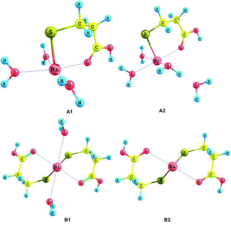 | ||
| Fig. 5 Optimized geometries of Bi(H2O)3(HMPA)2+ (A1), Bi(H2O)3(HMPA)2+·H2O (A2), Bi(HMPA)2(H2O)2+ (B1) and Bi(HMPA)2+ (B2) complex species. | ||
Thermodynamic properties of the complex species were theoretically investigated aiming to check consistence with experimental stability constants. The calculation was based on the thermodynamic cycle (Scheme 1) suggested by Bryantsev et al.53 The best calculation of stability constant for Bi(HL)2+ complex species has been achieved using H7O3+ leaving group contrary to mono and tris complex species. It is worth separating the reaction free energy in aqueous solution as the sum of three parts: electronic plus nuclear repulsion energy (ΔEele), thermal contribution (ΔGT), and solvation free energy (ΔΔGsolv), as given in eqn (4). The thermal contribution is estimated using the ideal gas model, the calculated harmonic vibrational frequencies to estimate the zero point energy correction (ZPE), and the correction due to the thermal population of vibrational levels (Table S3†).
| ΔGtotaq = ΔEele + ΔGT + ΔΔGSolv. | (4) |
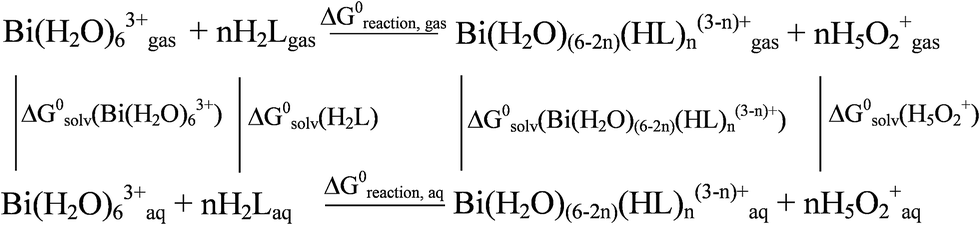 | ||
| Scheme 1 Thermodynamic cycle for interaction of Bi(III) with H2MPA in aqueous solution. | ||
According to the previous research,52 the PBE0 hybrid functional has been chosen as the reference one. Also Zhao et al. recommend Minnesota group density functionals, especially M06, for calculation of molecular structures and thermochemistry of main group elements.54 Results have been obtained using various density functionals on DFT/Def2-TZVPP level are compared in Table 4.
| Species | DFT | ΔEele | ΔGT | ΔΔGSolv | ΔGtot | logβcalc |
logβexp |
|---|---|---|---|---|---|---|---|
| a a,b,c,d correspond to A1, A2, B1 and B2 ofFig. 5. | |||||||
| M(HL)a | PBE0 | −822.5 | 8.8 | 721.8 | −91.9 | 16.1 | 13.1 |
| PBE0 | −816.8 | 7.5 | 721.6 | −87.6 | 15.3 | ||
| M06 | −792.6 | 5.2 | 711.3 | −76.1 | 13.3 | ||
| M(HL)b | M11 | −803.3 | −4.4 | 730.0 | −77.7 | 13.6 | |
| M06L | −783.6 | 5.6 | 715.6 | −62.4 | 10.9 | ||
| M11L | −802.0 | 3.8 | 739.2 | −59.0 | 10.3 | ||
| M(HL)2c | M06 | −1009 | 13.6 | 925.6 | −69.8 | 12.2 | 15.2 |
| M(HL)2 d | −1095 | 12.1 | 1001 | −81.6 | 14.3 | ||
| M(HL)3 | −912.4 | 15.6 | 792.6 | −104.2 | 18.3 | 17.8 | |
Obviously, M06 density functional and def2-TZVPP basis set provide the most accurate calculation of complexation thermodynamics. Literally – the least discrepancy between logβcalc and logβexp values. Thus DFT/M06/def2-TZVPP level has been chosen for further calculations.
The final sequence of cumulative stability constants β1 < β2 < β3 agrees both with theoretical and empirical data. It should be noted that logβ1 value for Bi(H2O)3(HMES)2+·H2O52 with the similar structure (sulfonic group instead of carboxylic) (11.7) is close to one for Bi(H2O)3(HMPA)2+·H2O (13.1).
The DFT calculations provide increasing Bi–O bond distance from 2.40 Å (M(HL)) to 2.49 Å (M(HL)2) and up to 2.94 (M(HL)3). This fact is consistent with decreasing charge of complex species: MHL2+ < M(HL)2+ < M(HL)30.
The combination of EXAFS and DFT data (Table 5) on Bi(HMPA)2+, Bi(HMPA)3 structure provided quite consistent Bi–S bond distances matching with previously reported Bi–S distances for bismuth-thiolate complexes in aqueous solutions – 2.5–2.6 Å.55,56 On the contrary, the Bi–O bond distances rather mismatched. The similar deference in experimental and calculated Bi–O bond distances was observed in.56
| Bond | N | 2σ2 (Å2) | Interatomic distance (Å) | |
|---|---|---|---|---|
| EXAFS | DFT | |||
| a 2σ2: Debye–Waller factor. | ||||
| Bi–S | 3 | 0.012 ± 0.008 | 2.51 ± 0.01 | 2.53 |
| Bi–O | 3 | 2.70 ± 0.02 | 2.94 | |
Discrepancy between Bi–O bond distances from EXAFS (2.70) and DFT calculations (2.94) for tris complex species can be explained in assumption of contribution of bi-ligand complex species (2.43 from DFT). Theoretical methods have been widely applied to interpret experimental spectroscopic data. Since authors of57,58 consider PBE0 density functional appropriate for calculations of vertical excitation spectrum, it has been chosen for TD-DFT simulations. The optimized geometry for TD-DFT study was taken from DFT/M06 calculation. A TD-DFT study has been carried out on electronic structure of the every complex species to investigate charge transfer process (Table 6).
| Species | λexp, nm | λcalc, nm | f | MO | Transition |
|---|---|---|---|---|---|
| M(HL) | 267 (max) broad | 270 | 0.009 | H → L (83.2%) | LMCT |
| 267 | 0.01 | H − 1 → L + 1 (36.8%) + H → L + 2 (47.1%) | |||
| M(HL)2 | 273 | 0.008 | H → L + 1 (70.5%) | ||
| 289 | 0.014 | H → L (30.9%) + H → L + 1 (45.6%) | |||
| M(HL)3 | 271 | 0.011 | H − 1 → L (27.4%) + H → L + 2 (35.0%) | ||
| 265 | 0.013 | H − 1 → L + 1 (60.4%) |
The band at 267 nm corresponds to the main electronic transition occurring from the HOMO to the LUMO for M(HL). The spreading of absorbance band for M(HL)3 complex species has been confirmed by TD-DFT calculations. Fig. 6 exhibits MO's (SMD/DFT/PBE0) contributing to charge transfer for M(HL) (see Fig. S6 and S7† for Bi(HL)2+ and M(HL)3, respectively). Atomic orbital population analysis is given in the Table S4† providing evidence of electronic transition from sulfur to bismuth. These data give solid evidence of π → π* charge transfer from thiol sulfur atom to bismuth(III) in both complex species. Recently we reported of the same π → π* S → Bi charge transfer at 268 nm in the Bi(H2O)3(HMPS)2+·H2O and Bi(HMPS)3 species in aqueous perchloric solutions [52, 58].
 | ||
| Fig. 6 Rendered HOMO and LUMO of Bi(H2O)3(HMPA)2+·H2O. | ||
Hence, SMD/TD-DFT/PBE0 model and Def2-TZVPP basis set provide good accordance of theoretical and experimental EAS. Actually more accurate structural, spectroscopic and thermodynamic data might be obtained by direct calculation of relativistic and specific solvation effects or by application of different diffusion basis sets. Nevertheless, such calculations were considered as a very resource-intensive operation and thus had not been performed during our current research.
Solid H[Bi(MPA)2]·H2O complex
The final product of complexation of Bi(III) by 3-mercaptopropanesulfonic acid in perchloric acid aqueous solutions are governed by initial mole ratio of reagents. The current study revealed yellow colored precipitate in solutions at H2L:M ratio = 2.0. This precipitate is found to dissolve at higher H2L:M ratios.
A structure of H[Bi(MPA)2]·H2O was consistent with and resulted from elemental analysis (ICP-AES), Raman and EXAFS spectroscopy. This complex was found to be X-ray amorphous according to XRD analysis.
The Raman spectra (Fig. S8†) have given evidence of ligand coordination via sulfur atom of thiol functional group. The band at 304 cm−1 referring to ν(Bi–S) vibrations was present in the Raman spectrum in the case of solid H[Bi(MPA)2]·H2O and was absent in the case of free ligand. On the contrary, the band at 2585 cm−1 corresponding to ν(S–H) vibrations was found in the Raman spectra of the ligand and was absent in the Raman spectra of the complex.
According to EXAFS data, the central Bi(III) atom is surrounded by four neighbor oxygen atoms and by two sulfur atoms. The experimental Fourier-filtered and simulated EXAFS spectra for the H[Bi(MPA)2]·H2O solid complex are compared in Fig. 7A.
 | ||
| Fig. 7 The experimental Fourier-filtered (1) and simulated Bi LIII EXAFS spectra (2) of the solid [Bi(MPA)2]− (A) and optimized fragment of polymer −[Bi(MPA)2]− (B). | ||
3-mercaptopropionic acid commonly forms complex species of cyclic S,O-chelate structures.8,18 However, it also forms polymeric S-bridged structures with Pd(II) and Pt(II)16,17 and O-bridged polymers with Gd(III), Eu(III) and Tb(III).15
No bidentate coordination of carboxylic functional group in [Bi(MPA)2]− is observed (DFT) so this complex is likely to have polymeric structure. However, this assumption can not be confidently confirmed by EXAFS data, because the objective conditions do not allow with certainty to detect the appropriate feature on the curve of Fourier transform module of corresponding spectrum. The suggested geometry has been optimized by DFT/M06/Def2-SVP computations (Fig. 7B). Fig. 7B shows that MPA2− ions are bound to bismith(III) as bidentate (S,O) chelating ligands forming a polymeric structure. Coordination number of six is achieved by binding of Bi(III) with other two complex fragment by bridging COO– group. Such ligand coordination is consistent with previous reports.8,15–18 The optimized geometry corresponds to the C2 point symmetry group. The results of EXAFS spectroscopy investigation and of DFT simulation are given in Table 7.
| Bond | 2σ2 (Å2) | N | Interatomic distance (Å) | |
|---|---|---|---|---|
| EXAFS | DFT | |||
| Bi–S | 0.023 ± 0.002 | 2 | 2.50 ± 0.01 | 2.65 |
| Bi–O | 2 | 2.36 ± 0.02 | 2.35 | |
| 2 | 2.75 ± 0.02 | 2.80 | ||
There are two types of Bi–O bonds in the [Bi(MPA)2]− complex according to EXAFS data analysis: the covalent (2.36 ± 0.02 Å) and the ionic (2.75 ± 0.02 Å) ones. The DFT simulation has provided Bi–O bond distances of 2.35 Å and 2.80 Å, respectively. Hence, EXAFS and DFT study provided Bi–O and Bi–S bond distances in the solid [Bi(MPA)2]− being consistent with previous report for Bi(III)-thiolate complexes: 2.4–2.7 Å and 2.5–2.6 Å long respectively.58,59
Some curious behaviour can be observed making comparison between M(HL)3 and H3O+[ML2]− complex species: tris complex has not participated from solution during any time, despite the fact that this complex has no charge. It may be associated with specific contributions of solvation effects in total Gibbs energies. The similar pattern has been reported previously56 for Bi(III)-2-mercaptopropanesulfonic acid (H2MPS). Further investigations on biological activity seem to be curious and valuable.22,23
Conclusions
The aim of the current research has been achieved successfully and the following conclusions have been drawn:1. Complex formation of Bi(III) with 3-mercaptopropionic acid has been studied in the aqueous perchloric solutions at CH+ = 0.5–2.0 M. Equilibrium study had been performed by spectrophotometry at 298 K within I = 0.5–2.0. Formation of Bi(HMPA)3 species under ligand dominance (CH2L ≫ CM) condition has been vindicated by Raman, 1H-NMR, UV-Vis and EXAFS spectroscopy. Each 3-mercaptopropionato ligand (HMPA−) is coordinated to bismuth(III) via sulfur atom of thiol group and via carbonyl oxygen atom of carboxylic group.
The novel solid complex (H[Bi(MPA)2]·H2O) has been obtained and characterized by Raman and EXAFS spectroscopy and DFT simulation. The polymeric structure formation with Bi–O bridging via carboxyl group of MPA2− species has been suggested.
2. Among the PBE0, M06, M06-L, M11 and M11-L density functionals which have been tested, the M06 has been chosen as the most appropriate one for calculations of H2MPA complexation thermodynamics and molecular geometries. The SMD/M06/def2-TZVPP model provided the most accurate prediction of stability constants of bismuth(III)-thiolate species with discrepancies within ± 1.0 logarithmic unit. The TD-DFT (PBE0) study proved that 267 nm bands in the UV-Vis EAS of [Bi(H2O)3(HMPA)2+]·H2O, Bi(HMPA)2+ and Bi(HMPA)3 species correspond to π → π* charge transfer from thiol sulfur atom to bismuth(III).
Acknowledgements
The research has been funded and carried out in terms of state contract of Ministry of Education and Science of Russian Federation. Authors also thank SFU CEJU for technical support.Notes and references
- M. Netopilová, J. Dršata, H. Kubová and P. Mareš, Original Research Article Epilepsy Research, 1995, 20, 179 CrossRef.
- S. G. Fan, J. P. Zhou, H. Xu and J. S. Han, Brain Res., 1985, 37, 184 CrossRef.
- R. R. Traut, A. Bollen, T. T. Sun, J. W. B. Hershey, J. Sundberg and L. R. Pierce, Biochemistry, 1973, 12, 3266 CrossRef CAS.
- D. Cuebas, J. D. Beckmann, F. E. Frerman and H. Schulz, J. Biol. Chem., 1985, 260, 7330 CAS.
- N. Merbouh, F. K. Wallner, O. M. Cociorva and P. H. Seeberger, Org. Lett., 2007, 9, 651 CrossRef CAS PubMed.
- S. E. Blackshaw and A. E. Warner, J. Physiol., 1976, 255, 231 CAS.
- E. Sabbagh, D. Cuebas and H. Schulz, J. Biol. Chem., 1985, 260, 7337 CAS.
- Q. Fernando and H. Freiser, J. Am. Chem. Soc., 1958, 80, 4928 CrossRef CAS.
- H. F. Brabander, H. S. Creyf, A. M. Goeminne and L. C. Van Poucke, Talanta, 1976, 23, 405 CrossRef.
- R. S. Saxena and K. C. Gupta, J. Inorg. Nucl. Chem., 1968, 30, 3373 CrossRef.
- I. Toth, L. Zekany and E. Brucher, Polyhedron, 1984, 3, 871 CrossRef CAS.
- R. Sarin and K. N. Munshi, Aust. J. Chem., 1972, 25, 929 CrossRef CAS.
- G. Choppin and L. Martixez-Perez, Inorg. Chem., 1968, 7, 2657 CrossRef CAS.
- M. A. Vairavamurthy, W. S. Goldenberg, S. Ouyang and S. Khalid, Mar. Chem., 2000, 70, 181 CrossRef CAS.
- H. A. Philips and N. Burford, Inorg. Chem., 2008, 47, 2428 CrossRef PubMed.
- M. B. Mishra, H. L. Nigam and A. Mehra, J. Inorg. Nucl. Chem., 1968, 30, 881 CrossRef CAS.
- S. Bagchi, D. Mandal, D. Ghosh and A. K. Das, J. Phys. Chem. A, 2013, 117, 1601 CrossRef CAS PubMed.
- M. Belcastro, T. Marino, N. Russo and E. Sicilia, J. Phys. Chem. A, 2004, 108, 8407 CrossRef CAS.
- E. R. Souza, I. O. Mazali and F. A. Sigoli, J. Fluoresc., 2014, 24, 203 CrossRef CAS PubMed.
- A. I. Frenkel, M. A. Vairavamurthy and M. Newville, J. Synchrotron Radiat., 2001, 8, 669 CrossRef CAS.
- M. Chandrasekharan, M. R. Udupa and G. Aravamudan, J. Inorg. Nucl. Chem., 1974, 36, 1153 CrossRef CAS.
- K. D. Mjos and C. Orvig, Chem. Rev., 2014, 114, 4540 CrossRef CAS PubMed.
- D. Gaynor and D. M. Griffith, Dalton Trans., 2012, 41, 13239 RSC.
- H. A. Phillips, M. D. Eelman and N. Burford, J. Inorg. Biochem., 2007, 101, 736 CrossRef CAS PubMed.
- B. J. McCormick and G. Gorin, Inorg. Chem., 1963, 2, 928 CrossRef CAS.
- D. S. Yoo, S. Y. Ha, I. G. Kim, M. S. Choo, G. W. Kim, E. S. Lee, S. J. Cho and B. C. Lee, Nucl. Instrum. Methods Phys. Res., Sect. B, 2011, 269, 1350 CrossRef CAS PubMed.
- J. Wen-Ping, L. Sheng, L. Jun and Y. Wen-sheng, Chem. Res. Chin. Univ., 2008, 24, 353 CrossRef.
- C. N. R. Rao, H. S. S. Ramakrishna Matte, R. Voggu and A. Govindaraj, Dalton Trans., 2012, 41, 5089 RSC.
- Y.-J. Zhu and F. Chen, Chem. Rev., 2014, 114, 6462 CrossRef CAS PubMed.
- T. A. Rokob, M. Srneca and L. Rulíšek, Dalton Trans., 2012, 41, 5754 RSC.
- D. Midgley and K. Torrance, Potentiometric Water Analysis, John Wiley & Sons, New York, 1978 Search PubMed.
- http://www.scilab.org/.
- M. Meloun, J. Čapek, P. Mikšík and R. G. Brereton, Anal. Chim. Acta, 2000, 423, 51 CrossRef CAS.
- D. J. Leggett, Computational methods for the determination of formation constants, Plenum Press, New York, 1985 Search PubMed.
- S. A. Grebenyuk, I. F. Perepichka and A. F. Popov, Spectrochim. Acta, Part A, 2002, 58, 2913 CrossRef.
- M. W. Schmidt, K. K. Baldridge, J. A. Boatz, S. T. Elbert, M. S. Gordon, J. H. Jensen, S. Koseki, N. Matsunaga, K. A. Nguyen, S. Su, T. L. Windus, M. Dupuis and J. A. Montgomery, J. Comput. Chem., 1993, 14, 1347 CrossRef CAS.
- http://www.cluster.sfu-kras.ru, accessed 1 July 2014.
- C. Adamo and V. Barone, J. Chem. Phys., 1999, 110, 6158 CrossRef CAS PubMed.
- S. Grimme, J. Antony, S. Ehrlich and H. Krieg, J. Chem. Phys., 2010, 132, 154104 CrossRef PubMed.
- R. Peverati and K. K. Baldridge, J. Chem. Theory Comput., 2008, 4, 2030 CrossRef CAS.
- Y. Zhao and D. G. Truhlar, Theor. Chem. Acc., 2008, 120, 215 CrossRef CAS.
- R. Peverati and D. G. Truhlar, J. Phys. Chem. Lett., 2011, 2, 2810 CrossRef CAS.
- Y. Zhao and D. G. Truhlar, J. Chem. Phys., 2006, 125, 194101 CrossRef PubMed.
- R. Peverati and D. G. Truhlar, J. Phys. Chem. Lett., 2012, 3, 117 CrossRef CAS.
- F. Weigend and R. Ahlrichs, Phys. Chem. Chem. Phys., 2005, 7, 3297 RSC.
- A. V. Marenich, C. J. Cramer and D. G. Truhlar, J. Phys. Chem. B, 2009, 113, 6378 CrossRef CAS PubMed.
- G. A. Zhurko, ChemCraft, version 1.6, http://www.chemcraftprog.com, accessed 1 July 2014 Search PubMed.
- A. L. Tenderholt, QMForge: A Program to Analyze Quantum Chemistry Calculations 2.1, 2007, http://qmforge.sourceforge.net Search PubMed.
- K. V. Klementev, J. Phys. D: Appl. Phys., 2001, 34, 209–217 CrossRef CAS , http://www.cells.es/Beamlines/CLAESS/software/viper.html.
- S. Tomic, B. G. Searle, A. Wander, N. M. Harrison, A. J. Dent and J. F. W. Mosselmans, EXCURV, http://www.cse.clrc.ac.uk/cmg/EXCURV/ Search PubMed.
- C. Bretti, C. De Stefano, C. Foti, O. Giuffrè and S. Sammartano, J. Solution Chem., 2009, 38, 1225 CrossRef CAS.
- A. I. Petrov, N. N. Golovnev, I. D. Dergachev and A. A. Leshok, Polyhedron, 2013, 50, 59 CrossRef CAS PubMed.
- V. S. Bryantsev, M. S. Diallo and W. A. Goddard, J. Phys. Chem. B, 2008, 112, 9709 CrossRef CAS PubMed.
- Y. Zhao and D. G. Truhlar, Theor. Chem. Acc., 2008, 120, 215–241 CrossRef CAS.
- J. Naslund, I. Persson and M. Sandstrom, Inorg. Chem., 2000, 39, 4012 CrossRef CAS.
- A. I. Petrov, N. N. Golovnev, S. V. Trubina, S. B. Erenburg and I. D. Dergachev, J. Coord. Chem., 2013, 66, 4188 CrossRef CAS.
- D. Jacquemin, V. Wathelet, E. A. Perpete and C. Adamo, J. Chem. Theory Comput., 2009, 5, 2420 CrossRef CAS.
- L. M. Reith, M. Stiftinger, U. Monkowius, G. Knör and W. Schoefberger, Inorg. Chem., 2011, 50, 6788 CrossRef CAS PubMed.
- G. G. Briand, N. Burford, M. D. Eelman, N. Aumeerally, L. Chen, T. S. Cameron and K. N. Robertson, Inorg. Chem., 2004, 43, 6495 CrossRef CAS PubMed.
Footnote |
| † Electronic supplementary information (ESI) available: Listing of 1H-NMR, Raman and UV-Vis spectral data, distribution diagram, Cartesian coordinates for DFT/M06-optimized geometries and energies, atomic percentage and rendered molecular orbitals. See DOI: 10.1039/c4ra07572b |
| This journal is © The Royal Society of Chemistry 2014 |