Photo-responsive reversible micelles based on azobenzene-modified poly(carbonate)s via azide–alkyne click chemistry

Ding Hu, Yefei Li, Yile Niu, Ling Li, Jingwen He, Xiangyu Liu, Xinnian Xia, Yanbing Lu*, Yuanqin Xiong and Weijian Xu
Institute of Polymer Science and Engineering, College of Chemistry & Chemical Engineering, Hunan University, Changsha 410082, China. E-mail: yanbinglu@hnu.edu.cn

Received 20th July 2014 , Accepted 5th September 2014

First published on 5th September 2014


Abstract

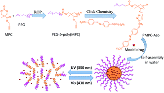
Photo-induced reversible amphipathic copolymer PMPC-azo was click conjugated by connecting amphiphilic poly(ethylene glycol)-modified poly(carbonate)s (PEG-b-poly(MPC)) and azide-functional trifluoromethoxy-azobenzene (azo-N3). The resulting copolymer self-assembled into spherical micelles with a hydrophobic azo core stabilized by a hydrophilic PEG corona in aqueous solution. As characterized by time-resolved UV-vis spectroscopy, dynamic light scattering (DLS) and transmission electron microscopy (TEM), these micelles showed reversible self-assembly and disassembly in aqueous solution under alternating UV and visible light irradiation. The model drug Nile Red (NR) was then successfully encapsulated into the micelles. Light-controlled release and re-encapsulation behaviors were demonstrated by fluorescence spectroscopy. The cell cytotoxicity of PMPC-azo micelles was also evidenced by MTT assay. This study provides a convenient method to construct smart nanocarriers for controlled release and re-encapsulation of hydrophobic drugs.

Introduction

During the last decades, stimuli-responsive block copolymers (BCP) have been widely investigated for “on–off” drug delivery and “on-demand” nanomedicines because of their switch properties.1–5 The most frequently used stimuli are pH, temperature, light, redox potential, ultrasound, charge, gases, biomolecules and enzymes.6–14 Compared with other stimuli-responsive systems, light-responsive BCP do not require any changes in the surroundings. In addition, the wavelength and intensity of illumination can be accurately adjusted and the time, direction and area of illumination are easily controlled.15–18 Light-responsiveness is usually provided by photochromic molecules attached to the polymers. Azobenzene (azo) is a well-known and well-used compound because it can undergo transcis photo isomerization in response to UV and visible light. Azo isomerization is a facile method for changing the hydrophobicity of BCP micelles since the cis form of azobenzene is more polar than the trans form.15 There are several reports on amphipathic BCP decorated with azobenzenes.19–23 Chen et al. reported the construction of photo-responsive micelles from azobenzene-modified hyperbranched polyphosphates, which showed reversible self-assembly and disassembly behaviors via irradiating with UV and visible light.24 Recently, Blasco et al. reported a series of light-responsive vesicles based on linear-dendritic amphiphiles, which tailored the photo-responsive properties of the vesicles and consequently the release rate via adjusting the percentages of azo and hydrocarbon chains.25

Amphiphilic copolymers have been widely studied for their potential applications in biomedical and pharmaceutical fields. Among them, block copolymers of aliphatic poly(carbonate)s combined with poly(ethylene glycol) (PEG) have been widely investigated for drug delivery because of their low toxicity, biocompatibility, and biodegradability.26–30 PEG is a hydrophilic and nonionic polymer that has been widely used as a biocompatible polymer in both academia and industry for chemical and biological applications.31–33 Polymeric micelles containing PEG as the hydrophilic shell are able to form a palisade preventing the adsorption of proteins and enzymes and subsequent non-specific uptake by the reticuloendothelial system (RES) after intravenous injection.34,35 Several groups have grafted PEG onto polyesters using different synthetic pathways. Yang et al. reported the ring opening copolymerization of acryloyl carbonate (AC) and ε-caprolactone (CL) using methoxy PEG as an initiator.36 Hydrophilic and amphiphilic PEG-poly(trimethylene carbonate) hydrogels were prepared using PEG-bisazides via “click” chemistry by Truong et al.37

However, there have been limited reports for the synthesis of stimuli-responsive poly(carbonate)s. Zhong's group prepared pH-responsive biodegradable polymers, which were comprised of a novel acid-labile polycarbonate hydrophobe and PEG.38 They also developed reduction-responsive poly-(carbonate)s via ring-opening copolymerization of ε-CL and pyridyl disulfide-functionalized cyclic carbonate monomers.39,40 However, there is no report on light-responsive biodegradable biomaterials based on poly(carbonate)s.

In this study, we developed a facile method to prepare amphiphilic block copolymers with poly(carbonate)s as a hydrophobic chain using a ring opening polymerization of cyclic carbonates. Then, copper-catalyzed Huisgen 1,3-dipolar cycloaddition of azides with alkynes was employed as the coupling reaction to form reversible light-responsive biodegradable micelles based on hydrophobic poly(carbonate)s. This amphiphilic copolymer can self-assemble into micelles consisting of a hydrophobic core surrounded by a hydrophilic shell. Such core–shell polymeric micelles allow the properties of reversible transition via irradiation with UV and visible light. The micelle formation, reversible light-responsive self-assembly and disassembly of the micelles, and cytotoxicity of the copolymer micelles, as well as light-responsive model drug release were investigated.

Experimental section

Materials

Monomethoxy poly(ethylene glycol) (PEG5k, Mn = 5000), 4-(trifluoromethoxy)-aniline, ethyl chloroformate, 1,4-dibromobutane and 2,2-bis(hydroxyl methyl) propionic acid were obtained from Sigma-Aldrich and used as received. Dichloromethane (DCM), dimethylformamide (DMF), 1,8-diazabicyclo[5.4.0]undec-7-ene (DBU), and triethylamine were dried over calcium hydride for 24 h at room temperature and distilled under reduced pressure. The thiourea catalyst (TU) was synthesized as previously reported and recrystallized from dry DCM.26,41 Tetrahydrofuran (THF) was dried by refluxing over a benzophenone–sodium mixture until a deep blue color appeared and distilled. All the other reagents and solvents were purchased from Sinopharm Chemical Reagent Co. Ltd, China and used as received.

Characterization

1H and 13C NMR spectra were recorded on an INOVA-400 NMR spectrometer in chloroform-d (CDCl3). Mass spectroscopy analyses were conducted using an LCQ-Advantage mass spectrometer (Thermo Finnigan, United States). The number-average molecular weight (Mn) and molecular distribution (polydispersity index, PDI = Mw/Mn) of the polymers were determined at room temperature using a Waters GPC (Waters, USA) equipped with four TSK HXL series polystyrene divinylbenzene gel columns (300 × 7.8 mm). Calibration was established with polystyrene standards from Polymer Laboratories. THF was used as solvent with a flow rate of 1 mL min−1. Transmission electron microscopy (TEM) images were obtained on an H-7000 NAR transmission electron microscope (Hitachi) with a working voltage of 100 kV. A drop of the micelle aqueous solution (0.5 mg mL−1) was deposited onto a 230 mesh copper grid coated with carbon and allowed to dry at room temperature before measurement. The mean size of the micelles was determined by dynamic light scattering (DLS) using a Malvern Nano S instrument (Malvern, UK). Measurements of the solution of micelles (0.5 mg mL−1) were performed at a scattering angle of 90° and at room temperature (25 °C). UV-vis spectra were carried out using a Hitachi UV-4100 UV-vis spectrophotometer and the spectra were collected within the range of 200–800 nm.

Synthesis of 4-hydroxy-4′-trifluoromethoxyazobenzene (1)

4-(Trifluoromethoxy)aniline (4.5 g, 25.4 mmol) was dissolved in 1.5 M aqueous H2SO4 (70 mL) and placed in an ice bath at 0 °C. Sodium nitrite (2.35 g, 34.0 mmol) in water (15 mL) was added dropwise to the former solution and stirred for 3 h. Then, the mixture was added dropwise to a solution of sodium carbonate (15.7 g, 147.9 mmol), sodium hydroxide (1.0 g, 25.0 mmol) and phenol (2.5 g, 26.6 mmol) in water (100 mL) at 0 °C and stirred for 3 h. The resultant mixture was poured into deionized water, and then neutralized with 2 M aqueous HCl. The crude product was filtered and washed with a large volume of deionized water, and then dried in a vacuum oven at 60 °C. Yield: 5.7 g (79.6%).

1H NMR (CDCl3): δ = 6.95–7.94 (m, 8H, ArH), 5.30 (s, 1H, ArOH);

13C NMR (CDCl3): δ = 158.50, 150.93, 150.45, 146.99, 125.15, 123.99, 121.39, 115.87.

Synthesis of 4-(4-bromobutyloxy)-4′-trifluoromethoxy-azobenzene (2)

To a 100 mL three-neck round bottomed flask was added 1,4-dibromobutane (12.9 g, 60 mmol), K2CO3 (2.76 g, 20 mmol), KI (0.12 g, 0.7 mmol) and acetone (30 mL). To a 50 mL pressure equalizing dropping funnel, compound 1 (5.6 g, 20 mmol) in 40 mL acetone was added. The solution was added dropwise to the flask. The reaction mixture was magnetically stirred at 60 °C for 12 h. The salts formed were filtered and washed with acetone. The solvent of the filtrate was evaporated using a rotary evaporator. The crude product was washed with a very small amount of ethanol and chloroform, and then recrystallized from methanol and dried in a vacuum oven at 50 °C. Yield: 5.8 g (69.5%).

1H NMR (CDCl3): δ = 6.99–7.94 (m, 8H, ArH), 4.08–4.11 (t, 2H, CH2CH2CH2CH2Br), 3.50–3.53 (t, 2H, CH2CH2CH2CH2–Br), 1.96–2.14 (m, 4H, CH2CH2CH2CH2Br);

13C NMR (CDCl3): δ = 161.74, 151.11, 150.38, 146.99, 125.07, 124.08, 121.27, 114.72, 67.29, 33.41, 29.62, 27.81.

Synthesis of 4-(4-azidebutyloxy)-4′-trifluoromethoxy-azobenzene (azo-N3)

Compound 2 (5 g, 12.0 mmol) and sodium azide (1.56 g, 24.0 mmol) were dissolved in DMF (15 mL), and the mixture was stirred at room temperature for 24 h. The solution was added to DCM (100 mL) and reverse-extracted three times with water (100 mL × 3). The organic phase was dried with anhydrous magnesium sulfate, filtered and evaporated to dryness. The product was collected as a yellow powder. Yield: 4.1 g (90.1%).

1H NMR (400 MHz, CDCl3): δ = 7.01–7.96 (m, 8H, ArH), 4.09–4.12 (t, 2H, CH2CH2CH2CH2N3), 3.40–3.43 (t, 2H, CH2CH2CH2CH2N3), 1.81–1.98 (m, 4H, CH2CH2CH2CH2N3);

13C NMR (400 MHz, CDCl3): δ = 161.66, 151.10, 150.39, 147.00, 125.36, 124.06, 121.27, 114.72, 67.63, 51.01, 26.44, 25.57.

Synthesis of 2,2-bis(hydroxyl methyl)propionate (3)

In a 250 mL round-bottom flask, 2,2-bis(hydroxyl methyl)-propionic acid (19.21 g, 143.3 mmol), KOH (8.6 g, 153.6 mmol) and DMF (100 mL) were added. The mixture was stirred at 100 °C for 2 h, and then propargyl bromide (18.28 g, 153.6 mmol) was added dropwise over 30 min. After 72 h of reaction, the reaction mixture was filtered, the solvent was evaporated under reduced pressure, and the residues were dissolved in 200 mL of DCM and extracted three times with saturated salt water (100 mL × 3). The organic phase was concentrated to yield the crude product, which was purified by column chromatography (eluent: ethyl acetate–petroleum ether = 1/5, v/v). Yield: 10.68 g (43.4%).

1H NMR (400 MHz, CDCl3): δ = 4.76–4.77 (d, 2H, CHCCH2CO), 3.91–3.94 (d, 2H, CH2OH), 3.72–3.75 (d, 2H, CH2OH), 2.51–2.52 (t, 1H, CHCCH2CO), 1.11 (s, 3H, CH3CC).

13C NMR (400 MHz, CDCl3): δ = 175.01, 75.20, 67.82, 52.43, 49.29, 16.95.

Synthesis of 5-methyl-5-propargylxycarbonyl-1,3-dioxane-2-one (MPC)

Compound 3 (8.60 g, 50 mmol) was mixed with ethyl chloroformate (10.86 g, 100 mmol) and THF (100 mL) in a sealed vessel that was purged with nitrogen and cooled in an ice bath. After stirring for an hour, triethylamine (10.14 g, 100 mmol) was added dropwise over 30 min under a nitrogen atmosphere. The reaction was allowed to stir for 3 h, after which it was allowed to warm to 25 °C and stirred overnight. The solution was then filtered and evaporated to dryness, and the product was precipitated in a mixture of ethyl acetate and diethyl ether (1[thin space (1/6-em)]:[thin space (1/6-em)]1) as white crystals. Yield: 9.24 g (93.3%).

1H NMR (400 MHz, CDCl3): δ = 4.79–4.80 (d, 2H, CHCCH2CO), 4.71–4.74 (d, 2H, CH2OCO), 4.22–4.25 (d, 2H, CH2OCO), 2.54–2.55 (t, 1H, CHCCH2CO), 1.37 (s, 3H, CH3CC).

13C NMR (400 MHz, CDCl3): δ = 170.33, 147.22, 76.36, 75.95, 72.71, 53.47, 40.18, 17.39.

Typical procedure for the synthesis of PEG-b-poly(MPC)

The ring-opening polymerization of MPC was carried out under an inert atmosphere of nitrogen using standard Schlenk-line techniques. In a typical experiment, MPC (0.594 g, 3 mmol), PEG5k (0.601 g, 0.12 mmol), TU (0.055 g, 0.15 mmol), DBU (0.005 g, 0.03 mmol) and dried DCM (10 mL) were placed in a dried Schlenk tube, fitted with a rubber septum. The solution was further degassed by three freeze–pump–thaw cycles. The resulting mixture was stirred at room temperature for 7 h, followed by precipitation in ice-cold diethyl ether and centrifugation. The resulting product was collected by filtration and dried under vacuum to yield a white powder. Yield: 1.087 g (91.0%).

1H NMR (400 MHz, CDCl3): δ = 4.71 (m, OCH2CCH), 4.25–4.33 (m, OC(O)OCH2), 3.62 (m, OCH2CH2O), 3.36 (s, CH3O), 2.53 (s, CH2CCH), 2.30 (br s, OH), 1.27 (s, CH3).

GPC (THF, RI): Mn (PDI) = 10[thin space (1/6-em)]544 g mol−1 (1.11).

Synthesis of PMPC-azo via “click” chemistry

In a Schlenk tube, PEG-b-poly(MPC) (302.9 mg, propargyl group, 0.74 mmol), azo-N3 (289.2 mg, 0.74 mmol), sodium ascorbate (17.1 mg, 0.074 mmol), and DMF (4 mL) were introduced. The tube was fitted with a rubber septum. The solution was further degassed using three freeze–pump–thaw cycles. A DMF solution of copper sulfate (10.55 mg, 0.037 mmol) was then added to the Schlenk tube. The solution was stirred at room temperature for 24 h. The crude material was purified by dialysis (dialysis tubing 3500 MWCO) against deionised water, which was regularly renewed. After 3 days, the final product, PMPC-azo, was obtained by lyophilization. Yield: 450.0 mg (76.0%).

1H NMR (400 MHz, CDCl3): δ = 7.84–7.87 (m, ArH), 7.69 (br s, N3CHC), 7.28–7.30 (d, ArH), 6.92–6.94 (d, ArH), 5.24 (s, C(O)OCH2), 4.43 (m, NCH2), 4.24 (m, OC(O)OCH2), 4.02 (m, CH2CH2OAr), 3.64 (m, OCH2CH2O), 3.38 (s, CH3O), 2.11 (m, NCH2CH2CH2CH2), 1.82 (m, NCH2CH2CH2CH2), 1.20 (s, CH3).

GPC (THF, RI): Mn (PDI) = 16[thin space (1/6-em)]285 g mol−1 (1.12).

Preparation of micelles

Micelles of PEG-b-poly(MPC) and PMPC-azo were prepared by a dialysis method. 10.0 mg copolymer was dissolved in DMF (2 mL), and then DI water (20 mL) was slowly added with vigorous stirring. After vigorous stirring for another 2 h at room temperature, the micelles were obtained and further dialyzed against DI water for 24 h to remove DMF (MWCO 1000 Da). The final polymer concentration was adjusted by adding DI water to 0.5 mg mL−1.

Fluorescence measurement of the critical micellar concentration

The critical micellar concentrations (CMC) of PEG-b-poly(MPC) and PMPC-azo amphiphiles were determined by a dye solubilization method using Nile Red (NR) as a probe molecule. NR in THF (0.1 mg mL−1, 30 μL) was added to a glass vial using a microsyringe. After THF was evaporated, a micellar solution (2 mL) was added. The concentration of the micellar solution was varied from 0.1 to 5 × 10⁻⁴ mg mL−1. Then, the solution was stirred for 5 h. The fluorescence measurements were taken at an excitation wavelength of 550 nm and the emission was monitored from 570 nm to 750 nm.

Light-responsive release and re-encapsulation of NR

NR was selected as a hydrophobic model drug to be encapsulated into the PMPC-azo micelles and fluorescence spectroscopy was used to investigate the release and re-encapsulation behaviours of the micelles. NR in THF (0.1 mg mL−1, 30 μL) was added to a glass vial via a microsyringe. After THF was evaporated, a 0.5 mg mL−1 micellar solution (4 mL) was added. Then, the solution was stirred for 5 h. After the encapsulation process, the fluorescence spectrum of the micelles was immediately recorded. The micelles were then exposed to 365 nm UV light for 15 min and subsequently irradiated with 450 nm visible light for 80 min. Fluorescence measurements were performed at an excitation wavelength of 550 nm and the emission was monitored from 570 to 750 nm.

In vitro cytotoxicity assay

HeLa cells were used for studying the cytotoxicity of the micelles. They were seeded in a 96-well plate at a density of 9.6 × 10³ cells per well and incubated in DMEM at 37 °C in 5% CO2 for 24 h. Then, the medium was removed and replaced with 200 μL polymer micelles. The aggregate concentrations of each formulation were prepared by serial dilution with DMEM medium. After treatment for 24 h, 20 μL of fresh medium containing 10% of MTT of 5 mg mL−1 stock was replaced in each well. The plates were incubated for 4 h, and 230 μL of DMSO was added to each well to dissolve intracellular MTT formazan crystals, followed by absorbance measurement at 490 nm using a microplate reader (Varioskan Flash, Thermo Scientific). Experiments were performed in triplicate.

Results and discussion

Synthesis and characterization of amphiphile PMPC-azo

The monomers of azo-N3 and MPC were synthesized according to the design route shown in Scheme 1. The PMPC-azo amphiphile was prepared via a two-step method (Scheme 2). First, the PEG-b-poly(MPC) block copolymer was synthesized through the ROP of MPC with PEG5k as a macroinitiator. The 1H NMR spectrum of PEG-b-poly(MPC) in CDCl3 is shown in Fig. 1C. The degree of polymerization (DP) of the polycarbonate backbone was determined to be 24 by comparing the integrals of peaks at δ = 3.36 (CH3O–, methyl protons of poly(ethylene glycol) end group) with δ = 4.28 (–C(O)OCH2CCH2O–, methylene protons of the carbonate units), which were close to the theoretical value (Table 1). GPC analysis showed that the obtained copolymer had a narrow polydispersity index (PDI) of 1.11 and a Mn of 10[thin space (1/6-em)]544 g mol−1, which was in agreement with those determined by 1H NMR end group analysis.
image file: c4ra07345b-s1.tif
Scheme 1 Synthesis of azo-N3 and MPC.

image file: c4ra07345b-s2.tif
Scheme 2 Synthesis of amphiphilic block copolymer PMPC-azo and facile formation of reversible light-responsive micelles for drug packaging and release.

image file: c4ra07345b-f1.tif
Fig. 1 1H NMR (in CDCl3) spectra of N3-azo (A), PMPC-azo (B) and PEG-b-poly(MPC) (C).
Table 1 Molecular characteristics of amphiphiles PEG-b-poly(MPC) and PMPC-azo
Entry Mw/Mna Mn,GPCa Mn,NMRb CMCc (mg mL−1)
a Both molecular weight (Mn,GPC) and the polydispersity (Mw/Mn) of the amphiphiles were determined by GPC.b Mn,NMR was determined by 1H NMR.c CMC: the critical micellar concentration of the amphiphiles was determined by fluorescence spectroscopy (Fig. 3).
PEG-b-poly(MPC) 1.11 10[thin space (1/6-em)]544 9752 0.0145
PMPC-azo 1.12 16[thin space (1/6-em)]285 15[thin space (1/6-em)]005 0.0157


Second, the modification of PEG-b-poly(MPC) to prepare the PMPC-azo amphiphile was performed in DMF with CuSO4/sodium ascorbate as the catalytic system at room temperature for 24 h via click reaction (Scheme 2). The crude copolymer was purified by dialysis in order to remove the copper salt. Fig. 1 compares the 1H NMR spectra of azo-N3, PMPC-azo and PEG-b-poly(MPC). The signal at δ = 2.54 in Fig. 1C was assigned to the protons of the alkynyl groups of PEG-b-poly(MPC), but this signal disappeared in Fig. 1B after click chemistry modification, indicating that the propargyl groups of PEG-b-poly(MPC) have been completely grafted with azo-N3. Compared to Fig. 1A and C, Fig. 1B revealed that signals assignable to azo-N3 were detected at δ 1.75–2.15, 6.90–7.34 and 7.87, while signals at δ 1.22, 3.37, 3.65 and 4.26 owing to PEG-b-poly(MPC) were also found in the PMPC-azo amphiphile. Moreover, the weak signal at δ 7.69, assigned to the proton of the triazole ring, evidenced the attachment of azo-N3 branches to the polyester backbone. GPC results revealed that the PMPC-azo copolymer had a narrow PDI of 1.12 and an Mn of 16[thin space (1/6-em)]285 g mol−1, close to that calculated by end group analysis from 1H NMR (Table 1). The GPC trace of the graft copolymer showed a slight shift to lower retention time, while maintaining narrow distributions with dispersities similar to that of the unmodified copolymer PEG-b-poly(MPC) (Fig. S6). It is evident that PMPC-azo graft copolymer can be readily prepared from propargyl-functionalized polycarbonate via the copper catalyzed azide–alkyne cycloaddition (CuAAC) click reaction.

Self-assembling behavior of amphiphilic copolymers

The amphiphilic copolymers PEG-b-poly(MPC) and PMPC-azo self-assembled into hydrophobic cored micelles stabilized with hydrophilic PEG coronae (Scheme 2), and their self-assembly behaviors were investigated in detail using fluorescence spectroscopy, DLS and TEM. Using hydrophobic Nile Red as a probe, fluorescence spectroscopy can conveniently monitor the micellar self-assembly and determine the critical micellar concentration (CMC) of the amphiphiles.24,41 As shown in Fig. 2, the emission fluorescence intensity gradually increased with increasing amphiphile concentration, suggesting the spontaneous self-assembly of micelles. PMPC-azo showed a similar CMC value to PEG-b-poly(MPC) (0.0157 mg mL−1 vs. 0.0145 mg mL−1), suggesting that the self-assembled micelles are thermodynamically stable in aqueous solution. These values are consistent with literature data reported for graft copolymers.24,35,41 Then, the size and morphology of the self-assembled micelles were measured by DLS (Fig. 3A and B) and TEM (Fig. 4A and B), respectively. From the TEM images, spherical morphology was observed with an average size of 30 nm for PEG-b-poly(MPC) micelles and an average size of 80 nm for PMPC-azo micelles. Despite having a common spherical morphology, PMPC-azo micelles have a larger DLS-determined diameter than PEG-b-poly(MPC) micelles, which can be attributed to the expanded hydrophobic nucleus. These results demonstrate that the PMPC-azo amphiphiles self-assembled in aqueous solution into spherical micelles approximately 150 nm in size, which contained an azo core stabilized by a PEG corona.
image file: c4ra07345b-f2.tif
Fig. 2 Fluorescence emission spectra of NR in PEG-b-poly(MPC) micelles (A) and PMPC-azo micelles (C) of varying concentrations and the relevant emission intensity at 630 nm versus the log of concentration for PEG-b-poly(MPC) micelles (B) and PMPC-azo micelles (D).

image file: c4ra07345b-f3.tif
Fig. 3 Mean size distributions of the micelles determined by DLS: PEG-b-poly(MPC) (A), PMPC-azo (B), PMPC-azo after 15 min of 365 nm irradiation (C) and PMPC-azo after subsequent 80 min of 450 nm irradiation (D).

image file: c4ra07345b-f4.tif
Fig. 4 TEM photographs of the micelles: PEG-b-poly(MPC) (A), PMPC-azo (B), PMPC-azo after 15 min of 365 nm irradiation (C), subsequent 80 min of 450 nm irradiation (D).

Photo-induced reversible self-assembly and disassembly

In order to investigate the photo-responsive self-assembly and disassembly behaviors of the micelles, the PMPC-azo micelles were examined by time-resolved UV-vis spectroscopy (Fig. 5). DLS and TEM measurements of the samples were then conducted and the images are shown in Fig. 3 and 4. Without irradiation, the amphiphile PMPC-azo formed spherical micelles with a diameter of approximately 150 nm in solution (Fig. 3B) and approximately 80 nm in the dry state (Fig. 4B). The diameters determined by TEM were smaller than those determined by DLS analysis, which was probably because of the shrinkage of the PEG shell upon drying.38,42 Upon irradiation with 365 nm UV light for 20 min, the diameter of the micelles became smaller and the polydispersity was larger (Fig. 3C). At the same time, the spherical micelles were disassembled as shown in Fig. 4C. This phenomenon was because of the changes in the hydrophilic-hydrophobic balance of PMPC-azo because the isomerization of trans-azobenzene into cis-azobenzene under 365 nm UV irradiation.15,20,23 Moreover, UV-vis spectroscopy was also employed to monitor this irradiation process (Fig. 5A). The intensity of the characteristic absorption peak of the π–π* transition of trans-azobenzene at around 340 nm gradually decreased with increased irradiation time, and then did not change. In addition, the absorption band at 440 nm, which is ascribed to the n–π* transition of cis-azobenzene became gradually stronger. These results suggested the completion of isomerization of trans-azobenzene into cis-azobenzene within 15 min. As mentioned in the introduction, the cis form of azobenzene is more polar than the trans form, resulting in the destruction of the hydrophilic–hydrophobic balance of PMPC-azo. As a result, the micelles disassembled.
image file: c4ra07345b-f5.tif
Fig. 5 UV-vis spectra of the PMPC-azo micelles: (A) 365 nm UV light irradiation induced trans-to-cis transition, and (B) 450 nm visible light irradiation induced cis-to-trans transition.

After further irradiating the sample with visible light with a wavelength of 450 nm for 80 min, the copolymer formed micelles with an diameter of approximately 190 nm and a polydispersity of 0.035 (Fig. 3D and 4D). This phenomenon occurred because of the isomerization of cis-azobenzene into trans-azobenzene. Fig. 5B showed the changes of the corresponding UV-vis spectrum for the sample in the visible light irradiation process. It can be seen that the absorption band at around 340 nm, which corresponds to trans-azobenzene showed a regular decrease in the n–π* absorption of cis-azobenzene suggesting the isomerization of cis-azobenzene into trans-azobenzene.17,19,21 Because of the recovery of the hydrophilic–hydrophobic balance of PMPC-azo, the micelles reassembled after 80 min of 450 nm irradiation. When comparing Fig. 5A and B, after irradiating with UV light and visible light, the intensities of absorption at 340 nm and 450 nm were almost identical to the original sample. These results indicated that the PMPC micelles could undergo photo-induced reversible self-assembly and disassembly. In order to further confirm that the process is completely reversible, the PMPC-azo micelles were irradiated with alternating UV and visible light (Fig. S7). It can be seen that the photo-isomerization of azobenzene groups in PMPC-azo can occur reversibly many times. After three cycles, the characteristic absorption peak of the π–π* transition of trans-azobenzene at 340 nm was still the same as that of the original micelles.

Model drug release and reload of NR-loaded micelles

Nile Red (NR) was used as a hydrophobic model drug to demonstrate the concept of photo-triggered drug release because the fluorescence intensity of NR is known to substantially increase in hydrophobic environments such as the interior of micelles.42 After loading the NR, the fluorescence spectrometry of the samples was immediately carried out. As shown in Fig. 6, the NR-loaded PMPC-azo micelles exhibited a photo-triggered drug release profile. During the 15 min of 365 nm irradiation, the fluorescence emission intensity of NR gradually decreased with the increase in irradiation time, indicating that the model drug NR was released into water from the disintegration of micelles (Fig. 6A).
image file: c4ra07345b-f6.tif
Fig. 6 Fluorescence emission spectra of PMPC-azo micelles with encapsulated NR after irradiation with: (A) 365 nm UV light for 15 min and (B) subsequent 450 nm visible light for 80 min.

By comparing fluorescence emission intensities at 613 nm, we found that 20% and 82% of drug molecules were released into water after irradiation with UV light for 1 min and 15 min, respectively. This phenomenon is attributed to the isomerization of azobenzene under UV irradiation, which resulted in the collapse of the micelles. Contrastingly, the characteristic peak of NR became considerably stronger in Fig. 6B when the solution was irradiated by 450 nm visible light. Obviously, some fluorescent dye molecules were re-encapsulated into the hydrophobic cores of the micelles. It is noted that the emission peak did not increase further after 80 min of irradiation, suggesting that not all NR molecules were encapsulated in the process. The reason for the incomplete re-encapsulation might be that the rate of micelle formation is higher than that of drug loading.43 These results proved that the drug release and re-encapsulation behaviors of the PMPC-azo micelles can be controlled by irradiating with UV or visible light.

Cell cytotoxicity of PMPC-azo micelles

In order to test the cell cytotoxicity of PMPC-azo micelles, the MTT assay was used to evaluate the cytotoxicity of the micelles against HeLa cells after 48 h of culture. As shown in Fig. 7, when the micelle concentration of PMPC-azo was 500 mg mL−1, the cell viability still remained at about 95%, demonstrating the low cytotoxicity. This result can be mainly attributed to the excellent biocompatibility of PEG and PC. It has been reported that biocompatible nanocarriers with diameters of less than 200 nm may avoid reticuloendothelial system (RES) recognition.1,2,4 Therefore, the micelles fabricated in this study have potential to be an ideal drug carrier system.
image file: c4ra07345b-f7.tif
Fig. 7 Cell viability of the HeLa cell line against different concentrations of micelles after being cultured for 48 h: (A) PEG-b-poly(MPC); (B) PMPC-azo.

Conclusions

In this paper, we present an azo-decorated block poly(carbonate) copolymer, PMPC-azo, via a convenient click conjugation of photo-responsive azobenzene molecules to propargyl-functionalized poly(carbonate)s. DLS and TEM measurements revealed that the polymer can self-assemble into spherical micelles with an average diameter of 150 nm in aqueous solution. The CMC of the micelles was determined as 0.0157 mg mL−1 by fluorescence spectroscopy using Nile Red as a fluorescence probe. Under alternative UV and visible light irradiation, the amphiphile PMPC-azo carried out a reversible micelle transition in water. Controlled release of model drug NR under 365 nm UV light and re-encapsulation under 450 nm visible light were confirmed by fluorescence spectroscopy. The reversibly light-responsive micelles with excellent biocompatibility, biodegradability and an appropriate size are highly promising as smart carriers for controlled delivery of hydrophobic molecules for biomedical applications.

Acknowledgements

Financial support from Planned Science and Technology Project of Human Province (2012WK4017) and the Fundamental Research Funds for Central Universities are gratefully acknowledged.

Notes and references

  1. N. Rapoport, Prog. Polym. Sci., 2007, 32, 962–990 CrossRef CAS PubMed.
  2. Y. Bae and K. Kataoka, Adv. Drug Delivery Rev., 2009, 61, 768–784 CrossRef CAS PubMed.
  3. J. F. Lovell, T. W. Liu, J. Chen and G. Zheng, Chem. Rev., 2010, 110, 2839–2857 CrossRef CAS PubMed.
  4. C. Oerlemans, W. Bult, M. Bos, G. Storm, J. F. W. Nijsen and W. E. Hennink, Pharm. Res., 2010, 27, 2569–2589 CrossRef CAS PubMed.
  5. N. Kamaly, Z. Xiao, P. M. Valencia, A. F. Radovic-Moreno and O. C. Farokhzad, Chem. Soc. Rev., 2012, 41, 2971–3010 RSC.
  6. J. Z. Du, X. J. Du, C. Q. Mao and J. Wang, J. Am. Chem. Soc., 2011, 133, 17560–17563 CrossRef CAS PubMed.
  7. L. Marta, M. G. G. Jose, Q. G. Isabel, G. Alberto and P. Rodrigo, Macromolecules, 2011, 44, 3739–3745 CrossRef.
  8. K. K. Venkata, S. F. Antoni, R. Janne and M. Raffaele, Macromolecules, 2011, 44, 4569–4573 CrossRef.
  9. Y. L. Cheng, C. L. He, C. S. Xiao, J. X. Ding, K. X. Ren, S. J. Yu, X. L. Zhuang and X. S. Chen, Polym. Chem., 2013, 4, 3851–3858 RSC.
  10. N. Rapoport, Z. Gao and A. Kennedy, J. Natl. Cancer Inst., 2007, 99, 1095–1106 CrossRef CAS PubMed.
  11. Y. Wang, S. Gao, W. H. Ye, H. S. Yoon and Y. Y. Yang, Nat. Mater., 2006, 5, 791–796 CrossRef CAS PubMed.
  12. Q. Yan and Y. Zhao, J. Am. Chem. Soc., 2013, 135, 16300–16303 CrossRef CAS PubMed.
  13. G. D. G. Bruno, M. J. Alain, D. Joseph and C. D. S. Stefaan, Langmuir, 2006, 22, 5070–5074 CrossRef PubMed.
  14. C. Wang, Q. S. Chen, Z. Q. Wang and X. Zhang, Angew. Chem., Int. Ed., 2010, 49, 8612–8615 CrossRef CAS PubMed.
  15. J. M. Schumers, C. A. Fustin and J. F. Gohy, Macromol. Rapid Commun., 2010, 31, 1588–1607 CrossRef CAS PubMed.
  16. Y. Zhao, Macromolecules, 2012, 45, 3647–3657 CrossRef CAS.
  17. A. Goulet-Hanssens and C. J. Barrett, J. Polym. Sci., Part A: Polym. Chem., 2013, 51, 3058–3070 CrossRef CAS.
  18. S. Swaminathan, J. Garcia-Amoros, A. Fraix, N. Kandoth, S. Sortino and F. M. Raymo, Chem. Soc. Rev., 2014, 43, 4167–4178 RSC.
  19. G. Wang, X. Tong and Y. Zhao, Macromolecules, 2004, 37, 8911–8917 CrossRef CAS.
  20. S. Kadota, K. Aoki, S. Nagano and T. Seki, J. Am. Chem. Soc., 2005, 127, 8266–8267 CrossRef CAS PubMed.
  21. F. D. Jochuma and P. Theato, Chem. Commun., 2010, 46, 6717–6719 RSC.
  22. B. Yu, X. Jiang, R. Wang and J. Yin, Macromolecules, 2010, 43, 10457–10465 CrossRef CAS.
  23. O. Boissière, D. Han, L. Tremblay and Y. Zhao, Soft Matter, 2011, 7, 9410–9415 RSC.
  24. C. J. Chen, G. Y. Liu, X. S. Liu, D. D. Li and J. Ji, New J. Chem., 2012, 36, 694–701 RSC.
  25. E. Blasco, J. L. Serrano, M. Piñol and L. Oriol, Macromolecules, 2013, 46, 5951–5960 CrossRef CAS.
  26. W. Chen, H. Yang, R. Wang, R. Cheng, F. Meng, W. Wei and Z. Zhong, Macromolecules, 2010, 43, 201–207 CrossRef CAS.
  27. W. Su, X. Luo, H. Wang, L. Li, J. Feng, X. Zhang and R. Zhuo, Macromol. Rapid Commun., 2011, 32, 390–396 CrossRef CAS PubMed.
  28. J. A. Edward, M. K. Kiesewetter, H. Kim, J. C. A. Flanagan, J. L. Hedrick and R. M. Waymouth, Biomacromolecules, 2012, 13, 2483–2489 CrossRef CAS PubMed.
  29. J. Feng, R. X. Zhuo and X. Z. Zhang, Prog. Polym. Sci., 2012, 37, 211–236 CrossRef CAS PubMed.
  30. S. Tempelaar, L. Mespouille, O. Coulembier, P. Dubois and A. P. Dove, Chem. Soc. Rev., 2013, 42, 1312–1336 RSC.
  31. Y. Yamamoto, Y. Nagasaki, Y. Kato, Y. Sugiyama and K. Kataoka, J. Controlled Release, 2001, 77, 27–38 CrossRef CAS.
  32. R. Duncan, Nat. Rev. Drug Discovery, 2003, 2, 347–360 CrossRef CAS PubMed.
  33. R. B. Greenwald, Y. H. Choe, J. McGuire and C. D. Conover, Adv. Drug Delivery Rev., 2003, 55, 217–250 CrossRef CAS.
  34. P. F. Gou, W. P. Zhu and Z. Q. Shen, Biomacromolecules, 2010, 11, 934–943 CrossRef CAS PubMed.
  35. F. Coumes, V. Darcos, D. Domurado, S. Li and J. Coudane, Polym. Chem., 2013, 4, 3705–3713 RSC.
  36. R. Yang, F. Meng, S. Ma, F. Huang, H. Liu and Z. Zhong, Biomacromolecules, 2011, 12, 3047–3055 CrossRef CAS PubMed.
  37. V. Truong, I. Blakey and A. K. Whittaker, Biomacromolecules, 2012, 13, 4012–4021 CAS.
  38. W. Chen, F. Meng, F. Li, S. J. Ji and Z. Zhong, Biomacromolecules, 2009, 10, 1727–1735 CrossRef CAS PubMed.
  39. W. Chen, Y. Zou, J. Jia, F. Meng, R. Cheng, C. Deng, J. Feijen and Z. Zhong, Macromolecules, 2013, 46, 699–707 CrossRef CAS.
  40. W. Chen, Y. Zou, F. Meng, R. Cheng, C. Deng, J. Feijen and Z. Zhong, Biomacromolecules, 2014, 15, 900–907 CrossRef CAS PubMed.
  41. R. C. Pratt, B. G. G. Lohmeijer, D. A. Long, P. N. P. Lundberg, A. P. Dove, H. Li, C. G. Wade, R. M. Waymouth and J. L. Hedrick, Macromolecules, 2006, 39, 7863–7871 CrossRef CAS.
  42. L. Sun, B. Zhu, Y. Su and C. M. Dong, Polym. Chem., 2014, 5, 1605–1613 RSC.
  43. C. J. Chen, Q. Jin, G. Y. Liu, D. D. Li, J. L. Wang and J. Ji, Polymer, 2012, 53, 3695–3703 CrossRef CAS PubMed.

Footnote

Electronic supplementary information (ESI) available. See DOI: 10.1039/c4ra07345b

This journal is © The Royal Society of Chemistry 2014