Stability and interaction forces of oil-in-water emulsions as observed by optical tweezers – a proof-of-concept study

Julie Nilsen-Nygaard*a, Marit Sletmoenb and Kurt Ingar Drageta
aNorwegian Biopolymer Laboratory (NOBIPOL), Department of Biotechnology, Norwegian University of Science and Technology (NTNU), 7491 Trondheim, Norway. E-mail: julie.nilsen-nygaard@ntnu.no; Tel: +47 73591689
bDepartment of Physics, Norwegian University of Science and Technology (NTNU), 7491 Trondheim, Norway

Received 15th July 2014 , Accepted 9th October 2014

First published on 9th October 2014


Abstract

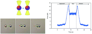
Increased insight into the interactions occurring between emulsion droplets is important to a range of applications from the food and pharmaceutical industries to oil recovery and mineral flotation. These interactions are often modified by the adsorption at the oil–water interface of surface-active species such as small molecule surfactants, proteins or polymers, in order to meet functional requirements of the emulsions. However, the experimental challenges faced when attempting to study these forces acting between emulsion droplets have hampered the progress in the understanding of the fundamental forces and to which extent these forces influence the destabilizing processes. In this paper we describe emulsion droplet studies by applying optical tweezers. By capturing two emulsion droplets in separate optical traps and bringing them into proximity, the forces acting between them can be measured as a function of separation distance. In this proof-of-concept study the force versus distance curves of emulsion droplets of different stabilization was obtained. Focus has been placed on the relative differences between micro- and macromolecular stabilization of emulsion droplets. Effects on depletion interaction, relaxation behaviour of the interfacial polymer layer during compression of the droplets and electrostatic screening have been observed. The present article documents the suitability of optical tweezers in studies aiming at revealing the forces acting between individual emulsion droplets as well as limiting factors of the technology.


Introduction

The destabilizing processes of emulsions have been extensively studied.1–8 However, much is still to be discovered related to the forces acting between single emulsion droplets and how these forces make destabilization progress. Optical tweezers (OT) represent a potential technique to identify and quantify the small forces exerted between single dispersed droplets. In optical tweezers studies, individual emulsion droplets can be confined in separate optical traps and the forces between them measured as they are forced closer together.

Emulsions are multiphase systems where one or more non-miscible phases are forced to blend by dispersing one or more phase(s) into a continuous phase. Well known emulsion systems are oil-in-water (O/W)-emulsions, water-in-oil (W/O)-emulsions, or water-in-oil-in-water (W/O/W)-emulsions. Emulsions are utilized in a vast number of products; pharmaceuticals, nutraceutics, cosmetics and food articles being the most important fields of application. The application of emulsions can be challenging considering that these systems are generally thermodynamic unstable. Within a given time, the emulsion will separate into its component phases. This occurs through different destabilizing mechanisms such as creaming/sedimentation, flocculation, coalescence and Ostwald ripening.9 The phase separation will lead to changes in physical and rheological properties of a material, and the material may totally fail to exhibit the desired properties or to meet the demands for shelf life of a product. The rate of phase separation in an emulsion is to a large degree determined by the stabilizing agents present. Stabilizers are defined as all substances with the ability to increase the stability of an emulsion and are further divided into emulsifiers and texture modifiers. Emulsifiers stabilize emulsion droplets by reducing the interfacial tension between the oil–water interface and form an adsorbed layer that prevents emulsion droplets from aggregating. Both low molecular weight surfactants and high molecular weight polymers and proteins can function as emulsifiers.10,11

When two emulsion droplets approach in an aqueous continuous phase a number of colloidal interactions come into play, the most important being the van der Waals, steric, electrostatic, hydrophobic and hydrodynamic interactions. The dominating colloidal interactions between emulsion droplets are to a large degree a function of the properties of the surface active molecules coating the oil–water interface. The main mechanisms for emulsion droplet stabilization are electrostatic and steric interactions. Steric interactions are a result of mixing and/or compression of the emulsifiers layers covering emulsion droplets upon approach.12 Electrostatic interactions on the other hand will be the predominant contribution to repulsion between charged emulsion droplets and are the result of overlap of the electrical double layers.13 In the case of emulsion droplets stabilized by charged emulsifiers with some extension out from the oil surface, one often talks of electrosteric stabilization where both electrostatic and steric interactions will contribute significantly to the overall emulsion droplet stabilization.11 For small non-ionic surfactants such as polysorbate 80, the steric contribution is expected to be the most pronounced, however, for small charged surfactants such as CITREM and SDS the electrical double layer (EDL) repulsion between droplets of equal charge is expected to be the determining factor at longer range separation, whilst the steric contribution come into play at short separations. The picture is more complex for large charged surface active polymeric emulsifiers, such as gelatin and sugar beet pectin. In these cases the electrostatic contribution is assumedly predominant at long separations; analogous to the Debye screening length. The steric contribution is expected to come into play at shorter surface separations corresponding to the extension of the polymer chains out from the droplet surface.

In previous publications the atomic force microscope (AFM) has been applied in combination with force spectroscopy to measure the forces acting between pairs of emulsion droplets,14–19 as well as between a colloidal probe and an emulsion droplet.20 Emulsion droplet studies on the AFM have included studies of depletion effects in the presence of adsorbing and non-adsorbing random coil polymers in the continuous phase17 and polymer bridging.17 However, there are some drawbacks with applying the AFM for emulsion droplet measurements. For instance the mechanical contact between droplets and cantilever/surface imply a deviation in the experimental setup from the situation in a bulk emulsion; where droplets randomly collide due to Brownian motions in a continuous phase. The size of the droplets studied using AFM, typically ranging from 30 to 100 μm, are also relatively large compared to the typical size of applied emulsion droplets18 and the measured forces are in the nanonewtons range.

On the other side of the force scale, the magnetic chaining technique introduced in 1994[thin space (1/6-em)]21 has been used to probe the force–distance interactions of nanometer sized magnetic particles such as ferrofluid oil droplets. Advantages with the technique is a relatively simple experimental set-up and also that the force profiles of a system are obtained from an average of a large number of particles/droplets. The technique is quantitative in both force and drop separation, and results of screened electrostatic forces,21 steric forces, as well as forces in polymer–surfactant stabilized systems22 have been reported. However, the fact that the technique requires the use of magnetic particles of relatively small size (100–500 nm) limits its use for studying more commonly applied emulsions, e.g. food emulsions.

For the study of micrometer sized emulsion droplets, the technique of colloidal particle scattering in a shear field (CPS) needs to be considered. In the technique the trajectories of particles in a shear field pre and post controlled collision with a stationary particle are imaged and used to describe the colloidal interactions.23 Through simulation studies24–27 and experimental measurements26,27 the method has been applied for the study of colloidal interactions, such as the van der Waals attractive forces between latex spheres.28 However, the interpretation of the collision data requires extensive computer analysis and the experimental design is quite specialized, making CPS less accessible as a routine technique for emulsion scientists.

Optical tweezers (OT) is based on the principle of transfer of photon momentum when light is refracted/reflected at surfaces, enabling the trapping of dielectric colloidal particles in the optical traps of a highly focused laser beam. A prerequisite for trapping is that the refractive index of the particles to be trapped is higher than that of the continuous phase.29 In a dual-beam optical tweezers set-up two particles can be trapped in separate optical traps. This makes it possible to bring the particles together and apart at defined velocities while measuring the forces acting between the particles.30 The optical tweezers have been applied for studying biological systems e.g. with regard to binding forces between macromolecules e.g. enzyme and substrate,31–33 elongation forces of macromolecules34,35 and manipulation of living cells.36 Optical tweezers have also previously been applied to measure the interaction between particles in the micro- and nano-scale such as polystyrene beads and colloidal particles with different surface coatings.37–41 In a study from 2005 mainly focusing on colloidal interactions between polystyrene beads, Mellor and coworkers performed some preliminary experiments on emulsion droplets using oscillating optical tweezers.38

Emulsion droplet studies applying optical tweezers is a novel approach to study droplet–droplet interactions and the forces involved. By capturing two emulsion droplets in separate optical traps and bringing them into proximity, the forces acting between them can be measured as a function of distance. The method is non-invasive as the droplets merely are held in place by the radiation pressure of light in the optical traps. It thus provides new opportunities for studying the interactions between emulsion droplets, and through this its use can potentially increase the understanding of the mechanisms that are important for the stability of bulk emulsions.

The scope of this study was firstly to determine whether it would be possible to measure the interactions between emulsion droplets with optical tweezers and secondly to apply this method to obtain and compare force–deformation curves for emulsion droplets stabilized by small-molecular surfactants and droplets stabilized by macro-molecular biopolymers.

Experimental

Materials

Corn oil (Sigma-Aldrich, St. Louis, USA) was used as the oil phase for all emulsions. The following surfactants/stabilizers were used:

• Charged surface active biopolymer emulsifier: highly methylated sugar beet pectin (SBP) with a degree of methylation of 62–68% (Danisco, Copenhagen, Denmark). 2% (w/w) in the aqueous phase of the original emulsion.

• Non-ionic surfactant: polysorbate 80 (Sigma-Aldrich, St. Louis, USA). 5% (w/w) in the aqueous phase of the original emulsion.

• Anionic surfactant for depletion experiments: sodium dodecyl sulphate (SDS) (Sigma-Aldrich, St. Louis, USA). 200 mM in the aqueous phase of the original emulsion.

• Anionic surfactant: citric acid ester of mono- and diglycerides (Grindsted CITREM N 12 Veg kosher, Danisco, Copenhagen, Denmark). 5% (w/w) in the aqueous phase of the original emulsion.

The O/W emulsions were prepared using an Ultra turrax homogenizer (IKA, Staufen, Germany). For all the prepared O/W emulsions the initial weight fraction of the corn oil was 20% (w/w) and these emulsions were diluted 1[thin space (1/6-em)]:[thin space (1/6-em)]5000–1[thin space (1/6-em)]:[thin space (1/6-em)]10[thin space (1/6-em)]000 in Milli Q water or buffer of choice to a droplet concentration appropriate for measurements on the optical tweezers.

Sulphated polystyrene beads with an average size between 2.7–2.9 μm were purchased from Spherotech (Illinois, USA) and applied for compression experiments on the optical tweezers for comparative purposes.

Methods

The optical tweezers instrument Nanotracker from JPK Instruments (Berlin, Germany) was applied for measurements on pairs of emulsion droplets. The instrument is mounted on an inverted light microscope (Zeiss Axio Observer A1, Carl Zeiss AG, Oberkuchen, Germany). The laser is a continuous wave (CW) infrared (IR) laser with a wavelength of 1064 nm (Gauss beam, TEM00, M-squared <1.1), and a maximum emitting power equal to 3 W. The beam waist of the traps has been estimated by JPK Instruments to 0.64 μm. The numeric aperture (NA) of the traps is 1.2.

The accessible force of the instrument is between 1–100 pN and the position sensitivity of the trapped object is in the nanometer range. The instrument is a dual beam OT with a built in beam splitter enabling the emulsion droplets to be trapped in separate optical traps and led into proximity at a controlled speed. The sample chambers utilized consisted of a circular borosilicate glass (35 mm diameter, VWR International, Pennsylvania, USA) as the bottom glass, two pieces of double sided tape as side walls and a regular square cover glass (22 × 22 mm, VWR International, Pennsylvania, USA) for attachment on top. In order to prevent evaporation of the continuous phase of the diluted emulsion, the sample chamber was sealed with nail polish. Immersion oil (Carl Zeiss AG, Oberkuchen, Germany) was applied to both sides of the sample chamber before approach of the objectives. Prior to sample chamber assembly, the top cover glass was treated with a solution of 1 mg mL−1 of bovine serum albumin (BSA, Sigma-Aldrich, St. Louis, USA) in order to delay adhesion of the droplets to the sample chamber surface. All measurements were performed at room temperature. The optimal droplet size for stable trapping in the optical traps on the Nanotracker has been suggested to be between 1 and 3 μm (oral communication with JPK Instruments). For each measurement series it was strived to find droplets of comparable size; ±0.25 μm, the droplets selected for the measurements were typically in the size range 2–3 μm. Calibration was performed based on recordings of the thermal fluctuations of the droplets when trapped, as specified in the instrument software (JPK Software, Berlin, Germany). The laser strength was held constant at 1.5 W on each trap for all measurements. The droplets were trapped in solution in the middle of the liquid chamber, always with a significant distance to the top plate. This implies that artifacts due to surface adhesion can be ruled out. The measurements were performed by moving emulsion droplet in trap 1 towards the emulsion droplet in trap 2 which was held steady, as displayed in Fig. 1. In some of the experiments a hold time of 5 seconds was practiced at maximum contact between the droplets to screen for time-dependent effects. If not otherwise specified, both the approach and retract speed was 0.5 μm s−1.


image file: c4ra07140a-f1.tif
Fig. 1 Emulsion droplets in the optical traps on the JPK Nanotracker. The droplet in trap 1 (left) is moved towards the droplet in trap 2 (right) at a specified distance and speed. The size of the droplets was approximately 2.3 μm.

Malvern Zetasizer Nano (Malvern Instruments Ltd., Worcestershire, England) was applied to measure the zeta potentials of the polystyrene beads as well as the droplets in the respective emulsions at a defined pH and ionic strength. The measurements were performed on diluted emulsions in MQ water or specified buffer.

The slopes of the force–distance curves were estimated for an assembly of independent measurements (different pairs of beads/droplets) for the polystyrene beads as well as for the emulsion systems of different stabilization. The linear area directly after onset of significant increase in repulsive force was consistently chosen for slope estimation for the force–distance curves.

For the force–distance curves with the signature of depletion interaction the comparisons of the works required to disconnect the droplets and the attractive forces of the interaction were based on an assembly of independent measurements, obtained for different pairs of emulsion droplets.

Results and discussion

Hard sphere vs. soft sphere measurements on OT

Fig. 2A presents two representative force–distance approach curves obtained using the optical tweezers, one obtained for two sulphated polystyrene beads and the other obtained for two emulsion droplets stabilized by polysorbate 80. There is a pronounced difference in the slope of the two curves as the beads/droplets come into contact. The reduced slope of the repulsive force at approach between the emulsion droplets relative to the slope observed for the polystyrene beads visualizes the higher deformability of the emulsion droplets compared to the polystyrene beads. A comparison of the slopes of a collection of representative curves obtained for both the sulphated polystyrene beads and the polysorbate 80, displayed in Fig. 2B, confirms the differences in slope of the force–distance curves for nondeforming and deforming droplets on the OT.
image file: c4ra07140a-f2.tif
Fig. 2 (A) Approach curves for compression of a pair of sulphated polystyrene beads (2.7 μm) and a pair of polysorbate 80 stabilized emulsion droplets (2.5 μm) in MQ water. Approach speed 0.5 μm s−1. (B) Box-plot comparing the slopes of a collection of independent force–distance curves for the two systems obtained at an approach speed of 0.5 μm s−1.

Zeta potential measurements showed that the zeta potential at neutral pH for the polysorbate 80 stabilized droplets in MQ water was −14.1 ± 1.1 mV (pH measured to 7.2), while for the sulphated polystyrene beads in MQ water it was measured to −49.7 ± 0.5 mV (pH measured to 7.1). The larger negative charge of the sulphated polystyrene beads implies that a larger electrical double layer force will be present between the polystyrene beads than for the emulsion droplets. This relative difference in surface charge of the emulsion droplets and the polystyrene beads must be taken into account when interpreting the curves, however the general characteristics of the deforming versus the non-deforming sphere repulsion are clearly depicted despite of this.

Though the exact distance between the optical traps is accurately known, the surface separation is a function of the droplet sizes and requires an accurate measure of this. Additionally, for deformable droplets, deformation of the droplets might start prior to physical contact due to long-range interactions, although this contribution is most likely insignificant compared to the magnitude of the full – contact force. Due to the limitations faced when working with the emulsion droplets, the force–deformation curves presented here should be viewed from a qualitative point of view with the differences in the curves shape being the focus of discussion. For the hard polystyrene spheres, the situation is less complex; the diameter of the beads is well defined and the beads can be considered as non-deformable under the forces applied in the OT experiments.

In the case of hard contact between non-deforming polystyrene beads, the force–distance curve would be vertical at contact, in this case it has a slope. The force–distance curve for the polystyrene beads have a slope that could imply that the beads surfaces in fact never are in contact. The measured force could potentially be merely an electrical double layer (EDL) force that arises due to the osmotic pressure caused by the higher charge density near the beads surfaces compared to in the continuous phase.42 Assumedly, the trap stiffness of the optical traps (and the measured force of 50 pN) on OT is not adequate to overcome the EDL forces of this system. However, a weakness with the OT method is that at a high degree of compression the beads are assumed to be displaced from their position in the optical traps center, resulting in the artificial softening of the force–distance curve. Preliminary tests performed on the PS curves with a computer analysis tool aiming to correct for the displacement of the colloids out of the traps on the basis of trap stiffness and the measured force, resulted in a steeper curve. Optimization of the method and the computer analysis will shed more light on this issue in future work.

The curves obtained for the polystyrene beads contain signatures of attractive forces prior to the onset of the repulsive forces. This has previously been reported as the van der Waals attractive forces. In particular, several AFM studies on the interactions between e.g. silica particles, polystyrene beads and latex particles with flat surfaces have reported measuring the van der Waals interaction.43–47 Also, total internal reflection microscopy (TIRM)44,48 and colloidal particle scattering (CPS)28 are methods that have been successfully applied for the measurement of these interactions. In a work from 2011 Lockie and co-workers detected the signatures of the van der Waals attractive interaction between a bromodecane droplet over a silica plate through the measurement of potential energy by TIRM.49 In the work with emulsion droplets on optical tweezers the van der Waals interaction has not been clearly observed, however by optimization of the method the interaction could potentially be possible to detect.

A comparison of the slopes of a collection of force–distance curves for emulsion droplets of comparable size stabilized by a small molecular surfactant of low surface charge (polysorbate 80), a charged surfactant (CITREM) and a high-molecular stabilizer (SBP) (Fig. 3) showed no significant differences. This observation is supported by studies using AFM that have shown that the slope of the force–deformation curve for deformable droplets is simply dependent on the diameter of the droplet (i.e. the Laplace pressure) and the interfacial tension and therefore not a measure of the interaction forces between the droplets.50,51 Considering that the droplet sizes applied in the OT measurements are ten- to hundred-fold smaller than the ones applied in comparable AFM studies, the Laplace pressures will be significantly higher, meaning that the droplet are less deformable. The results from AFM studies are therefore not directly transferable to the ones obtained in this study. Still, some degree of deformation is expected. Interfacial tension measurements are required to discuss the relation in more detail, however this is beyond the scope of this proof-of-concept study, and it will be elaborated on in future work.


image file: c4ra07140a-f3.tif
Fig. 3 Box plot comparing the distribution of slopes for the approach portion of the force–distance curves for assemblies of independent measurements on emulsion droplets stabilized by polysorbate 80, CITREM and SBP. All measurements were performed at an approach speed of 0.5 μm s−1.

Micro-molecular stabilization of emulsion droplets

Depletion effect. Fig. 4 shows the approach and retract curve for a measurement on two SDS stabilized emulsion droplets in a continuous phase with SDS micelles. The original emulsion was prepared with an excess of SDS, so the droplet surfaces were completely saturated by surfactant. Thereafter, the emulsion was diluted in a 90 mM solution of SDS. This is a concentration approximately tenfold the critical micelle concentration of SDS at 25 °C in water (0.0081 M).52 The approach portion of the force–distance curve shows a gradual increase in the repulsive force as the droplets approach, before entering the area of the curve characterized by a linear relationship between force and separation distance. The retract portion of the curve shows a clear hysteresis compared to the course of the approach portion. There is an attractive interaction between the droplets before they disconnect due to the depletion effect.
image file: c4ra07140a-f4.tif
Fig. 4 Approach and retract curve for SDS stabilized emulsion droplets with 90 mM SDS in MQ water present in the continuous phase. The approach and retract speed: 0.5 μm s−1.

The underlying mechanism of the depletion interaction is the exclusion/depletion of the micelles in the continuous phase from a volume surrounding the droplets, called the depletion layer. The thickness of this layer corresponds to the radius of the micelles. In the depletion layer the concentration of micelles is effectively zero. This creates a thermodynamically unfavorable concentration gradient between the depletion layers around the emulsion droplets and the surrounding continuous phase. The only way the system can reduce this osmotic pressure gradient is if two or more droplets combine by flocculation (or coalescence) and thereby reduce the volume of the depletion layers. The disconnection of the droplets is therefore thermodynamically unfavorable and requires a force exceeding the entropic attraction so that micelles can flow back into the volume between the droplets.9 In Fig. 4 the depletion effect manifests itself as an attractive force upon retraction (Δ17 pN) followed by a sudden disconnection of the droplets.

The depletion interaction for emulsion droplets has already been extensively studied by AFM.17,18,53 In their study from 2010, Gromer and co-workers reported on depletion interaction arising from both surface-active SBP and the non-surface active polymer random coil polymer PSS in the continuous phase. The study concluded that the observed depletion effect for the systems was the result of droplets being trapped in an attractive well (i.e. secondary energy minimum) where they are prevented from coalescing due to the steric barrier of the emulsifier layer. Increasing concentrations of polymer in the continuous phase was tested. At an intermediate SBP concentration hysteresis between the approach and retract curves was observed; a jump-to-contact upon approach and a larger attractive interaction followed by a jump-out upon retraction. The effect was attributed to liquid structural correlations in the thin liquid film separating the droplets due to the distribution of non-adsorbed polymer. The jump-in effect was assumed to be the manifestation of black spot formation when all solute diffuses away from a region of the interface.17 The depletion interaction between emulsion droplets in a concentrated SDS-micelle system has been investigated by Tabor and co-workers. The characteristic hysteresis behaviour in the force–distance curves was observed. The group reported on several jump-ins, i.e. oscillatory structural forces, upon approach of the droplets as well as the attractive well in the retract portion of the curve. The oscillatory structural forces were associated with the subsequent expulsion of layers of micelles from the droplet interface upon approach. Modelling revealed that the final layer of micelles were not squeezed out of the interface because of deformation/flattening of the droplets and that this remaining layer acted as a strong steric barrier preventing the droplets from coalescence when residing in the attractive well.18,53 Interestingly, the depletion interaction has also been observed with for non-deforming spheres in the presence of smaller PS-beads in the continuous phase with oscillatory optical tweezers in an early work by Crocker et al.54

The fact that the same attractive well aka secondary energy minimum of the depletion interaction as described by AFM studies also can be measured between emulsion droplets on optical tweezers is an interesting observation. The jump-in effect and the structural oscillatory forces reported on in the mentioned AFM studies have not been observed in this work on the OT. However, the concentrations of micelles and non-adsorbed polymer used in the different studies vary and an increase in the concentration of SDS-micelles in the continuous phase would increase the osmotic pressure and potentially manifest similar effects in this system as well.

A comparison of the work required to free the droplets from the attractive well for a collection of measurements for this emulsion produced at the same approach and retract speed and at the same concentration of micelles is presented in Fig. 5A. There is a positive correlation between the emulsion droplet size and the amount of work required to disconnect the droplets upon retraction. The magnitude of the depletion force is proportional to droplet size.55 Also, the attractive force for the corresponding curves correlate well and increase as the droplet size increase, as expected (Fig. 5B).


image file: c4ra07140a-f5.tif
Fig. 5 (A) Box plot displaying the distribution of the works required to disconnect the droplets residing in the secondary minimum (J) as a function of droplet size. (B) Box plot displaying the distribution of the force (pN) as a function of droplet size. Estimated from independent retract curves after collision of SDS coated emulsion droplets in a continuous phase of 90 mM SDS on OT. Approach and retract speed: 0.5 μm s−1.

The depletion effect was highly pronounced and reproducible in all measurements performed on this system. The effect was not observed for measurements on SDS stabilized droplets without the presence of micelles. Also, the depletion interaction was reduced at lower concentration of micelles in the continuous phase; i.e. at a lower osmotic pressure gradient (results not included).

Macro-molecular stabilization of emulsion droplets – surface active biopolymers

Fig. 6 shows the force versus time curve obtained from optical tweezers measurement on a pair of emulsion droplets stabilized by highly methoxylated SBP. The pectin is assumed to form a brush-like structure on the droplet surfaces with the protein and acetyl-rich areas adsorbing at the oil–water interface while the polar parts of the molecule extend into the continuous phase, presumably taking up a large volume. The high acetyl and protein content in SBP is assumed to explain its good emulsifying properties.56 The interfacial structure of highly methylated SBP has been studied using AFM. The imaging at an air–mica interface revealed a loosely packed film of protein and pectin chains interspersed with holes. Surface shear rheology performed showed an elastic structure of the SBP at an air–water interface.17
image file: c4ra07140a-f6.tif
Fig. 6 Approach and retract curve for SBP stabilized droplets in MQ water. The approach and retract portion of the curve are separated by a 5 seconds hold time at maximum contact between the droplets. Approach and retract speed: 0.5 μm s−1.

In the present optical tweezers measurements the SBP coated droplet pairs were led into contact at a speed of 0.5 μm s−1, held together at maximum contact for 5 seconds and then pulled apart at the same speed of retraction. The curve displayed in Fig. 6 shows the characteristic and reproducible response of the force signal for droplet 2 in the x-direction for this system. When the droplets approach there is a steady increase in the repulsive force, due at first to overlap of the electrical double layers, and subsequently to direct contact between the polymer layers. However, at a certain point in the approach segment of the curve, the force suddenly drops to a lower level and stabilizes there during the last part of the approach as well as during the hold time at this position (5 seconds). Upon retraction the force is re-established at the same maximum level before it drops as a result of the increased separation and then stabilizes at baseline level at large separation.

The fact that the force reduction is reversible upon retraction demonstrates that the changes occurring in the interface are restored when the strain on the system is reduced. One plausible explanation for the characteristic course of progress for these measurements is the rearrangement of the polymer layer at the interface as the droplets are compressed. When the strain on the system is increased further as the separation reaches its minimum, the polymer layer is assumed to partially collapse, but not followed by coalescence of the droplets. This could be the result of a partial reorganization of the SBP layer. The shift in the system to a lower energy level causes the force to drop as the static potential that has built up during compression is released when the entanglements are released. No further structural changes in the interface are observed until the retraction of the droplets starts. As illustrated in Fig. 7, the force is reestablished at a slightly lower level as prior to the entanglement relaxation indicating that some degree of entangled structure in the emulsifier layer is re-established as space is made available. The energy dissipation in the system upon retraction is surprisingly small. The time scale of reorganization in the approach and retract portion of the measurements was identical (data not included). This assumed relaxation behavior was reproducible and observed only for droplet pairs stabilized by macromolecular emulsifiers; SBP, highly acetylated chitosan (results not included) or alkaline pretreated gelatin (results not included), and not for systems stabilized by small-molecular surfactants.


image file: c4ra07140a-f7.tif
Fig. 7 Comparison of the maximum force in the approach portion of the force–time curve (A) and max force in the retract portion (B) for 8 individual observations of SBP stabilized emulsion droplets on OT.

However, it can be discussed that the clear steric repulsion between the droplets indicate a high coverage of SBP on the emulsion droplet surfaces and that this makes a potential reorganization of the polymer layer in the interface less likely. At low polymer coverage bridging behavior would be likely to occur and this has not been observed in the work with this system on the OT. Previous studies of polymer-coated droplets on the AFM, with larger droplet deformation, have not reported on similar effects.17,57 An alternative explanation for the observed tendencies of this system is droplet deformation in the interface. In their AFM study from 2012 on polymer coated decane drops Manor and co-workers noted that the polymer brush is relatively stiff compared to the deformable oil droplet interface. From this they conveyed that for droplets of low Laplace pressures and at low loading rates (negligible hydrodynamic effects) the drop deformation i.e. flattening of the interface was likely to be more important in the repulsive interaction than steric stabilization and reorganization of the polymer layer.57 Again, this will be a function of the droplet size and the droplets applied in this OT study have Laplace pressures significantly higher than the 30–40 μm droplets used in the discussed paper. Polymer layer rearrangements are potentially more prone to occur in systems of smaller emulsion droplets. Also, it is difficult to explain the characteristic course of the force–time curve of the SBP-system simply on the basis of surface deformation. The curves display an abrupt fall in force at maximum contact and this is not translatable to the gradual flattening of the emulsion droplet interface at increasing contact.

Another alternative explanation for the observed change in repulsive force during the holding phase is the potential manifestation of drop deformation in response to the drops moving to minimum energy positions in the traps. Preliminary tests have been performed on the system, taking into account the displacement of the droplets in the optical traps in all directions during the measurements. The data analysis reveals no significant effect on the force profile of the system when all directions are accounted for.

In 2005 Mellor and coworkers reported on an artifact observed in measurements on solid polystyrene beads on oscillating OT where the particles could roll around each other and out of the plane when compressed too hard. This shift out of the plane of motion was naturally accompanied by a drop in the repulsive force.38 The group also performed experiments with emulsion droplets, but these did not to our knowledge reveal any such slip-effects. Writing this observation off as a slip-effect, does not explain why the reduction in repulsive force at contact is only observed for macro-molecularly stabilized emulsion droplet pairs. A more in-depth-study of this effect is necessary in order to map out the underlying mechanisms.

In the work with emulsion systems stabilized by surface active biopolymers it was attempted to perform velocity series for the same droplet pair in order to investigate the effect of loading rate on the observed effect. No clear effect of the approach and retract speed could be mapped out. Additionally, experiments with increased hold time at maximum contact were performed for several droplet pairs, from 0 to 45 second. These revealed that no further change in the repulsive force between the droplets occurred after the initial reduction, the force stabilized at a constant level independent of the holding time at maximum contact. Coalescence was not observed during the work with this emulsion system.

Methodical limitations

In order to fully utilize the potential of the optical tweezers as a tool for studying emulsion droplet interactions, some methodical challenges need to be addressed. Measurements on soft deformable spheres such as emulsion droplets involves that the droplets are deformed when residing in the optical trap, as described in recent studies.58–60 The absolute surface distance between two emulsion droplets trapped in the optical tweezers is therefore not clearly defined. The absolute trap distance is known and the droplet size for the trapped droplets in each measurement series is measured manually. Making the assumption that the droplets are placed in the center of the optical traps, an adjustment is performed based on droplet radius when generating the force–distance curves. However, this approach is inaccurate; the discrepancies in the positioning of the curves on the x-axis relative to the zero point of contact and to each other observed in the present work reveal that in order to in a more robust way compare different force–deformation curves, an improved approach to this issue is needed. Application of a combination of position detection techniques involving direct imaging of the droplets when residing in the trap could be an alternative way of improving the position sensitivity in work with deformable droplets.

The need for surface treatment of the walls of the sample chamber early became evident when starting this project on optical tweezers. Emulsion droplets will cream in the continuous phase and tend to adhere to the top cover glass. Depending on the degree of surfactant/polymer coverage at the oil–water interface, the charge of the emulsifier layer, the sign of the charge and the ionic strength of the continuous phase, the droplets had a variable tendency of adhering to the glass surface. To delay this adhesion, coating of the cover glass was necessary. Several different coatings were screened for the emulsion droplets investigated and it turned out to be challenging to identify a coating that worked optimally for all of the emulsion systems. However, a coating with a 1 mg mL−1 solution of BSA functioned adequately for most systems of negative charge and was applied for the systems presented here. The BSA coating facilitated work with the same sample for approximately 60 minutes, however the identification of a more universal coating would be preferable.

For stable trapping of emulsion droplets in the optical traps, there are requirements to the droplet sizes. The optimal droplet size for OT studies are in a size regime 10–20 times smaller than what have been used on the AFM, optimally with a diameter between 1 and 3 μm for the instrument applied here. The relatively wide droplet size distribution obtained by setting up an emulsion with a homogenizer poses another challenge; even though the largest fraction of the droplet are in the correct size regime, there will unavoidably be a fraction of very small droplets in the range of a couple of hundred nm in diameter present. These have a tendency of being pulled into the optical traps during measurements. Better control of droplet size may be obtained by applying a membrane emulsification technique.

In future work the use of a flow cell enabling the exchange of continuous phase during the experiment will hopefully address some of the described challenges, making it possible to use the same pair of emulsion droplets whilst varying the ionic strength, pH or micelle concentration of the continuous phase.

Conclusion

In this study we have demonstrated the possibility for applying optical tweezers for emulsion droplet measurements by capturing two emulsion droplets in separate optical traps and leading them into proximity at a controlled loading rate. Also, force–deformation curves generated from measurements on emulsion droplets stabilized by small-molecular surfactants have been compared to macro-molecular stabilization using a biopolymer emulsifier, sugar beet pectin.

One of the major observations is the demonstration of the assumed reorganization of the polymer layer of sugar beet pectin covering the emulsion droplet surfaces during compression. This displays the dynamics of this macromolecular emulsifier layer to adapt to the lowest attainable energy conformation when subjected to strain, still stabilizing the droplets from coalescence and flocculation.

Considering the comparative measurements on non-deforming polystyrene beads, an interesting observation is that the attractive van der Waals forces actually can be measured with the OT.

With regard to micro-molecular stabilization, a distinct effect observed by OT measurement was the depletion interaction. The characteristic force–deformation curves of the depletion interaction was generated when SDS covered droplets were compressed in the presence of SDS micelles.

In conclusion, the preliminary studies of emulsion droplets on the optical tweezers show that there are challenges to be addressed before the method can be utilized to its full potential. However, these early results show promise and imply that optical tweezers in the future will be a useful tool for researchers in the investigation of emulsion droplet interactions and stability. The observations/data obtained are in accordance with theoretical considerations.

Acknowledgements

Thanks to Danisco for kindly providing us with sugar beet pectin, Gjertrud Maurstad for technical advice and to Gunhild Dalbakk for her contribution in the preliminary laboratory work.

References

  1. D. N. Petsev, N. D. Denkov and P. A. Kralchevsky, J. Colloid Interface Sci., 1995, 176, 201–213 CrossRef CAS.
  2. E. Dickinson, M. Golding and M. J. W. Povey, J. Colloid Interface Sci., 1997, 185, 515–529 CrossRef CAS.
  3. V. J. Pinfield, E. Dickinson and M. J. W. Povey, J. Colloid Interface Sci., 1997, 186, 80–89 CrossRef CAS.
  4. I. B. Ivanov, K. D. Danov and P. A. Kralchevsky, Colloids Surf., A, 1999, 152, 161–182 CrossRef CAS.
  5. M. Shields, R. Ellis and B. R. Saunders, Colloids Surf., A, 2001, 178, 265–276 CrossRef CAS.
  6. S. S. Dukhin, N. A. Mishchuk, G. Loglio, L. Liggieri and R. Miller, Adv. Colloid Interface Sci., 2003, 100–102, 47–81 CrossRef CAS.
  7. S. J. Radford and E. Dickinson, Colloids Surf., A, 2004, 238, 71–81 CrossRef CAS PubMed.
  8. J. Toro-Mendoza, A. Lozsan, M. Garcia-Sucre, A. J. Castellanos and G. Urbina-Villalba, Phys. Rev. E: Stat., Nonlinear, Soft Matter Phys., 2010, 81, 011405 CrossRef.
  9. P. C. Hiemenz and R. Rajagopalan, Principles of colloid and surface chemistry, Marcel Dekker, New York, 3rd edn, 1997 Search PubMed.
  10. N. J. Krog and F. V. Sparsø, in Food Emulsions, ed. J. Sjøblom, CRC Press, 2003 Search PubMed.
  11. D. J. McClements, Food emulsions: principles, practices, and techniques, CRC Press, Boca Raton, 2005 Search PubMed.
  12. R. J. Hunter and L. R. White, Foundations of colloid science, Clarendon Press, Oxford University Press, Oxford, 1987 Search PubMed.
  13. D. F. Evans and H. Wennerström, The colloidal domain: where physics, chemistry, biology, and technology meet, Wiley-VCH, New York, 2nd edn, 1999 Search PubMed.
  14. P. A. Gunning, A. R. Mackie, P. J. Wilde and V. J. Morris, Langmuir, 2004, 20, 116–122 CrossRef.
  15. R. R. Dagastine, G. W. Stevens, D. Y. C. Chan and F. Grieser, J. Colloid Interface Sci., 2004, 273, 339–342 CrossRef CAS PubMed.
  16. R. R. Dagastine, R. Manica, S. L. Carnie, D. Y. C. Chan, G. W. Stevens and F. Grieser, Science, 2006, 313, 210–213 CrossRef CAS PubMed.
  17. A. Gromer, R. Penfold, P. A. Gunning, A. R. Kirby and V. J. Morris, Soft Matter, 2010, 6, 3957–3969 RSC.
  18. R. F. Tabor, F. Grieser, R. R. Dagastine and D. Y. C. Chan, J. Colloid Interface Sci., 2012, 371, 1–14 CrossRef CAS PubMed.
  19. A. P. Gunning, A. R. Kirby, P. J. Wilde, R. Penfold, N. C. Woodward and V. J. Morris, Soft Matter, 2013, 9, 11473–11479 RSC.
  20. G. Gillies and C. A. Prestidge, Adv. Colloid Interface Sci., 2004, 108–109, 197–205 CrossRef CAS PubMed.
  21. F. L. Calderon, T. Stora, O. Mondain-Monval, P. Poulin and J. Bibette, Phys. Rev. Lett., 1994, 72, 2959–2962 CrossRef.
  22. J. Philip, G. G. Prakash, T. Jaykumar, P. Kalyanasundaram, O. Mondain-Monval and B. Raj, Langmuir, 2002, 18, 4625–4631 CrossRef CAS.
  23. T. G. M. van de Ven, P. Warszynski, X. Wu and T. Dabros, Langmuir, 1994, 10, 3046–3056 CrossRef CAS.
  24. M. Whittle, B. S. Murray and E. Dickinson, J. Colloid Interface Sci., 2000, 225, 367–377 CrossRef CAS PubMed.
  25. M. Whittle, B. S. Murray, E. Dickinson and V. J. Pinfield, J. Colloid Interface Sci., 2000, 223, 273–284 CrossRef CAS PubMed.
  26. H. Casanova, J. Chen, E. Dickinson, B. S. Murray, P. V. Nelson and M. Whittle, Phys. Chem. Chem. Phys., 2000, 2, 3861–3869 RSC.
  27. M. Whittle, B. S. Murray, J. Chen and E. Dickinson, Langmuir, 2000, 16, 9784–9791 CrossRef CAS.
  28. X. Wu and T. G. M. van de Ven, Langmuir, 1996, 12, 6291–6294 CrossRef CAS.
  29. A. Ashkin, Phys. Rev. Lett., 1969, 24, 156–159 CrossRef.
  30. K. C. Neuman and S. M. Block, Rev. Sci. Instrum., 2004, 75, 2787–2809 CrossRef CAS PubMed.
  31. C.-M. Cheng, Y.-J. Lee, W.-T. Wang, C.-T. Hsu, J.-S. Tsai, C.-M. Wu, K.-L. Ou and T.-S. Yang, Biochem. Biophys. Res. Commun., 2011, 404, 297–301 CrossRef CAS PubMed.
  32. K. H. Simpson, M. G. Bowden, S. J. Peacock, M. Arya, M. Höök and B. Anvari, Biomol. Eng., 2004, 21, 105–111 CrossRef CAS PubMed.
  33. E. Fällman, S. Schedin, J. Jass, M. Andersson, B. E. Uhlin and O. Axner, Biosens. Bioelectron., 2004, 19, 1429–1437 CrossRef PubMed.
  34. Y.-L. Sun, Z.-P. Luo, A. Fertala and K.-N. An, J. Biomech., 2004, 37, 1665–1669 CrossRef PubMed.
  35. U. Bockelmann, P. Thomen, B. Essevaz-Roulet, V. Viasnoff and F. Heslot, Biophys. J., 2002, 82, 1537–1553 CrossRef CAS.
  36. Y. Wu, D. Sun and W. Huang, J. Biomech., 2011, 44, 741–746 CrossRef PubMed.
  37. Z. Sun, S. Xu, G. Dai, Y. Li, L. Lou, Q. Liu and R. Zhu, J. Chem. Phys., 2003, 119, 2399–2405 CrossRef CAS PubMed.
  38. C. D. Mellor, M. A. Sharp and C. D. Bain, J. Appl. Phys., 2005, 97, 1–11 CrossRef PubMed.
  39. M. Polin, Y. Roichman and D. G. Grier, Phys. Rev. E: Stat., Nonlinear, Soft Matter Phys., 2008, 77, 051401 CrossRef.
  40. G. Dominguez-Espinosa, A. Synytska, A. Drechsler, C. Gutsche, K. Kegler, P. Uhlmann, M. Stamm and F. Kremer, Polymer, 2008, 49, 4802–4807 CrossRef CAS PubMed.
  41. M. M. Elmahdy, C. Gutsche and F. Kremer, J. Phys. Chem. C, 2010, 114, 19452–19458 CAS.
  42. J. N. Israelachvili, Intermolecular and surface forces, Academic Press, Burlington, Mass, 3rd edn, 2011 Search PubMed.
  43. P. G. Hartley, I. Larson and P. J. Scales, Langmuir, 1997, 13, 2207–2214 CrossRef CAS.
  44. M. A. Bevan and D. C. Prieve, Langmuir, 1999, 15, 7925–7936 CrossRef CAS.
  45. W. A. Ducker, T. J. Senden and R. M. Pashley, Langmuir, 1992, 8, 1831–1836 CrossRef CAS.
  46. H.-J. Butt, Biophys. J., 1991, 60, 1438–1444 CrossRef CAS.
  47. A. Milling, P. Mulvaney and I. Larson, J. Colloid Interface Sci., 1996, 180, 460–465 CrossRef CAS.
  48. L. Suresh and J. Y. Walz, J. Colloid Interface Sci., 1997, 196, 177–190 CrossRef CAS PubMed.
  49. H. Lockie, S. McLean and R. R. Dagastine, J. Phys. Chem. Lett., 2011, 2, 2472–2477 CrossRef CAS.
  50. D. Y. C. Chan, E. Klaseboer and R. Manica, Adv. Colloid Interface Sci., 2011, 165, 70–90 CrossRef CAS PubMed.
  51. D. Y. C. Chan, E. Klaseboer and R. Manica, Soft Matter, 2011, 7, 2235–2264 RSC.
  52. J. N. Phillips, Trans. Faraday Soc., 1955, 51, 561–569 RSC.
  53. R. F. Tabor, D. Y. C. Chan, F. Grieser and R. R. Dagastine, J. Phys. Chem. Lett., 2011, 2, 434–437 CrossRef CAS.
  54. J. C. Crocker, J. A. Matteo, A. D. Dinsmore and A. G. Yodh, Phys. Rev. Lett., 1999, 82, 4352–4355 CrossRef CAS.
  55. E. Dickinson, An introduction to food colloids, Oxford University Press, Oxford, New York, 1992 Search PubMed.
  56. J. Leroux, V. Langendorff, G. Schick, V. Vaishnav and J. Mazoyer, Food Hydrocolloids, 2003, 17, 455–462 CrossRef CAS.
  57. O. Manor, T. T. Chau, G. W. Stevens, D. Y. Chan, F. Grieser and R. R. Dagastine, Langmuir, 2012, 28, 4599–4604 CrossRef CAS PubMed.
  58. S. A. Ellingsen, J. Opt. Soc. Am. B, 2013, 30, 1694–1710 CrossRef CAS.
  59. P. C. F. Møller and L. B. Oddershede, EPL, 2009, 88, 48005 CrossRef.
  60. D. Tapp, J. M. Taylor, A. S. Lubansky, C. D. Bain and B. Chakrabarti, Opt. Express, 2014, 22, 4523–4538 CrossRef PubMed.

This journal is © The Royal Society of Chemistry 2014