DOI:
10.1039/C4RA06994C
(Paper)
RSC Adv., 2014,
4, 49663-49671
Steric inhibition of hydrogen bonding in molecular recognition of dicarboxylic acids: di-topic receptors containing a nitro group designed to behave like monotopic receptors†
Received
11th July 2014
, Accepted 29th September 2014
First published on 29th September 2014
Abstract
Five receptors (1–5) including two macrocyclic receptors have been designed and synthesized for the recognition of dicarboxylic acids. The receptors having an NO2 group show inhibition in hydrogen bonding molecular recognition towards the dicarboxylic acid and this effect becomes more prominent in the case of macrocyclic receptors 4 and 5 in CHCl3. The binding behavior of these receptors has been studied by UV-vis, fluorescence and 1![[thin space (1/6-em)]](https://www.rsc.org/images/entities/char_2009.gif) :1 1H NMR binding studies.
:1 1H NMR binding studies.
Introduction
The recognition of dicarboxylic as well as monocarboxylic acids with designed receptors has received considerable attention due to its application in pharmaceutical science1 and different supramolecular architectures2, also due to their presence in many biologically active molecules, for example, in drugs such as ibuprofen,3 aspirin, bilirubin, bile acids, folic acid, biotin etc. For designing synthetic receptors, H-bonding interaction is one of the major important forces. Among all the binding forces used in the development of artificial receptors, hydrogen bonding is potentially the most directed and powerful.4 Different types of receptors5 for di-carboxylic acids have been designed and synthesized by many research groups and the different aspects of recognition process have also been elegantly studied. Hydrogen bonding interactions influence a lot for the whole recognition process.
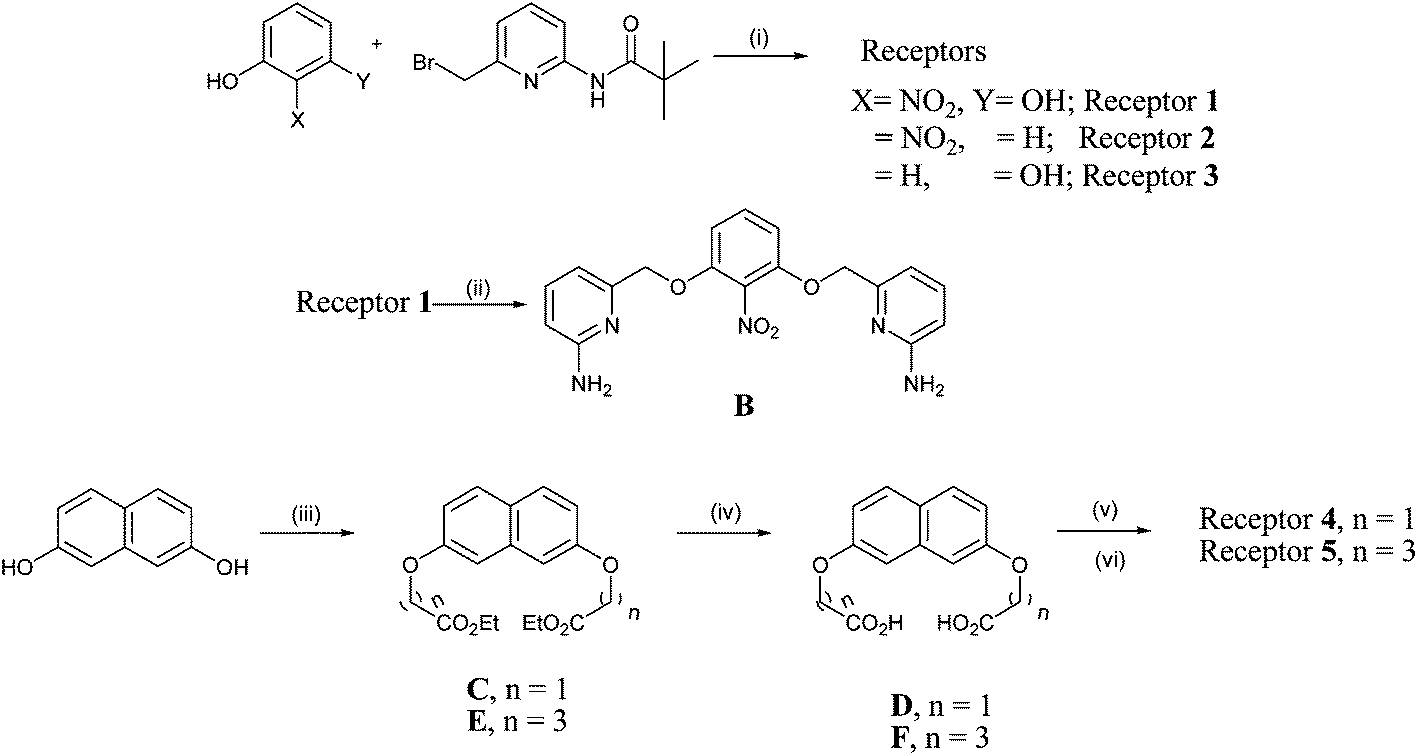
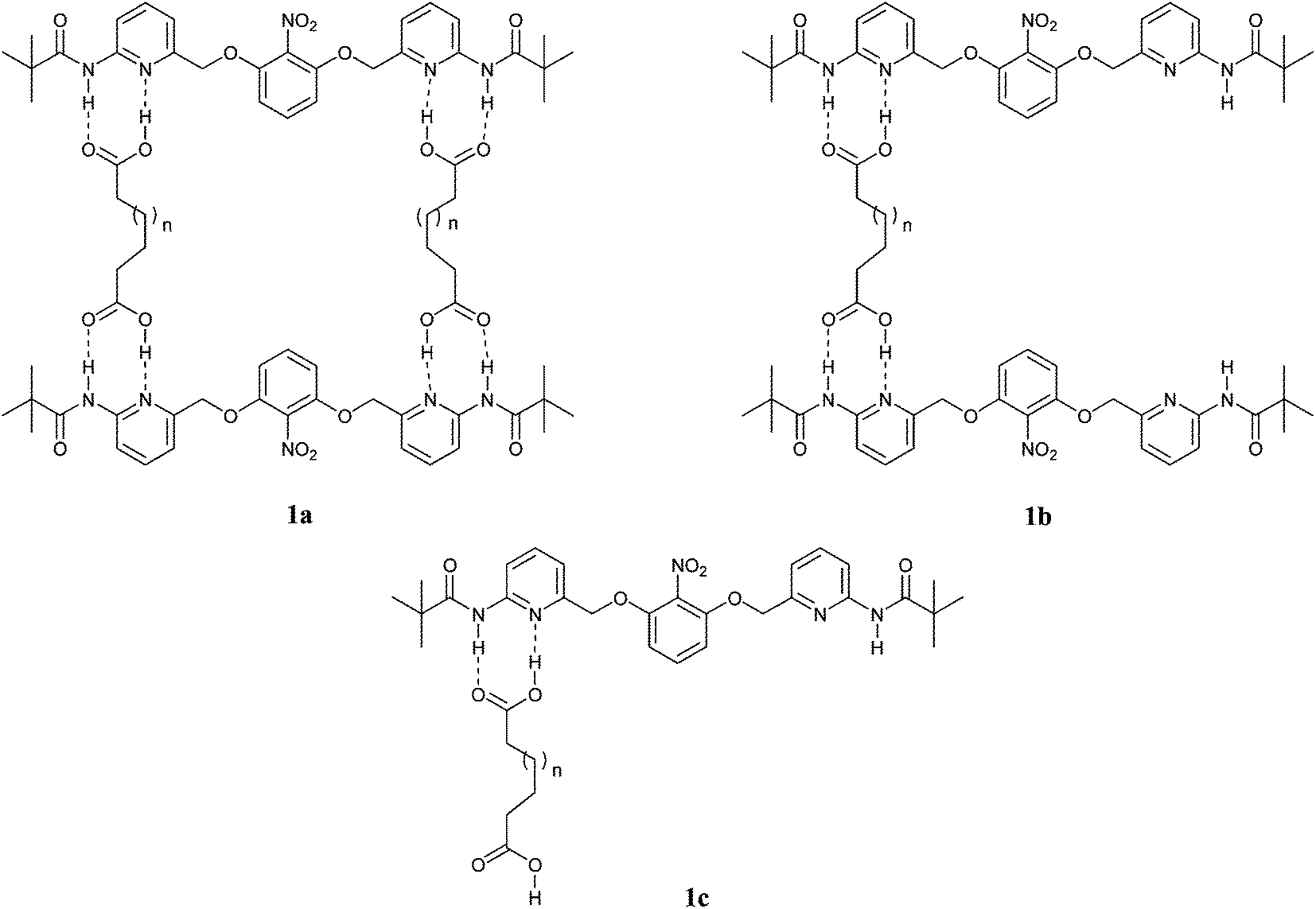
For the recognition of mono and dicarboxylic acids we have synthesized receptors having three point hydrogen bonds6 with different spacers.5a,5b,5g,5h We have also reported the case of hydrogen bond inhibition by N-oxide formation7 in the recognition of dicarboxylic acids, intramolecular hydrogen bond inhibition with dipicolyl urea.5e Macrocyclic receptors5f have also been synthesized for the recognition of dicarboxylic acids. A major portion of the earlier synthesized receptors contain pyridine amide moiety in a suitable position to bind the carboxylic acid group. In this paper, we wish to report the synthesis of a series of ditopic receptor 1 and macrocyclic receptors 4 and 5 containing nitro group (Fig. 1) which behave like monotopic receptors though they have two binding pyridine amide groups for dicarboxylic acids. However, it is interesting to see that though nitro group inhibits binding of one of the carboxyl groups of saturated dicarboxylic acids i.e. it forces the dicarboxylic acids to behave like a monocarboxylic one (Ka is in the order of 102 similar to that of monocarboxylic acid and not 104 as normally expected for dicarboxylic acid), it also probably binds the hydroxy group in malic acid increasing the binding constant from 102 to 103 for the extra hydrogen bond formation. We have also synthesized receptors 2 (without nitro group) and 3 (mono-pyridine amide with adjacent nitro group) for comparison studies (Scheme 1).
 |
| | Scheme 1 Synthesis of receptors 1, 2, 3, 4 and 5. Reagents and conditions (i) K2CO3, acetone, TBAB, r.t., 15 h; (ii) 4(N) KOH in 1:1 ethanol–water, reflux, 10 h; (iii) ethyl chloroacetate (n = 1), ethyl-4-bromobutyrate (n = 3), K2CO3, acetone, r.t., 10 h; (iv) 4(N) KOH in 1:1 ethanol–water, reflux, 12 h; (v) oxalyl chloride, CH2Cl2, DMF (cat.), 3 h; (vi) B, high dilution, THF, NEt3, 14 h. | |
Results and discussions
Binding studies
UV-vis studies. We have studied the binding behavior of the receptors 1, 2, 3, 4 and 5 towards the dicarboxylic and monocarboxylic acids by UV-vis method. Receptor 1 shows λmax at 332 nm, which gradually decreased on addition of the guest solution. Similar trend was observed during the titrations with benzoic acid and propionic acid. Receptor 2 shows better binding and behave like dicarboxylic acid receptor as revealed by its UV-vis spectra after addition of dicarboxylic acids. Receptor 3 behaves like as a usual receptor for monocarboxylic acids. Receptor 4 possesses λmax at 278 nm in its UV-vis spectra. The UV-vis spectra of 4 almost remain unaffected upon addition of mono as well as dicarboxylic acids except DL-malic acid (Fig. 3). Similar observation is found in case of receptor 5 having λmax at 277 nm.
 |
| | Fig. 3 UV-vis spectra of receptor 4 with adipic acid (a); receptor 4 with DL-malic acid (b); receptor 5 with adipic acid (c); receptor 5 with DL-malic acid (d). | |
Model studies. To correlate the binding behavior of receptor 1 in both solutions as well as in solid phases, we have performed a modeling study9 (Fig. 5). The energy minimization predicts the stable geometry of the molecule. It depends on the receptors and the guest. The presence of nitro group is always diverting the positioning of pyridine amide group to remain opposite to the nitro group as found in energy minimized structures in the receptors as well as in the complexes with carboxylic acids in cases of both the receptors 1 and 3 having bulky nitro group adjacent to the pyridine amide binding site staying though a little away by –O–CH2– spacer in between. This opposite geometry of the pyridine amide groups flanked by the nitro group is thus responsible of such opposite orientation due to steric repulsion (Fig. 6).
 |
| | Fig. 5 Energy minimized forms of receptors: (1) receptor 1; (1a) receptor 1 with adipic acid; (1m) receptor 1 with malic acid; (2) receptor 2; (2a) receptor 2 with adipic acid; (2m) receptor 2 with malic acid; (3) receptor 3; (3a) receptor 3 with adipic acid; (3m) receptor 3 with malic acid; (4) receptor 4; (4a) receptor 4 with adipic acid; (4m) receptor 4 with malic acid; (5) receptor 5; (5a) receptor 5 with adipic acid; (5m) receptor 5 with malic acid. | |
 |
| | Fig. 6 ORTEP diagram of receptor 1. | |
From the Emin values (Table 2) it can be concluded that receptor 1 behaves as a receptor of monocarboxylic acid, receptor 2 as a receptor of dicarboxylic acid, receptor 3 as a receptor of monocarboxylic acid, but in 4 and 5 the inhibition property of NO2 group is very much prominent giving rise to more complex stability of receptor–malic acid complex compared to receptor–adipic acid or receptor–benzoic acid or receptor–propionic acid complex. Again in case of receptor 4 energy of the complex of receptor 4 and malic acid is much more lowered compared to its complex with adipic acid. Similar phenomena is observed in the case of receptor 5 which is sound enough from the spectroscopic results and the energy values in theoretical calculation method.
Table 2 Minimum energy (kJ mol−1) of the receptors
| 1 |
28.96 |
1a |
26.99 |
1m |
16.11 |
| 2 |
26.66 |
2a |
1.33 |
2m |
10.23 |
| 3 |
45.22 |
3a |
34.12 |
3m |
40.12 |
| 4 |
12.12 |
4a |
5.12 |
4m |
−30.33 |
| 5 |
10.33 |
5a |
4.43 |
5m |
−45.12 |
X-ray studies. For solid phase recognition study of dicarboxylic acid with receptor 1 and also to confirm the structure of receptor 1, we have been able to grow single crystals of receptor 1 and X-ray studies10 confirmed the structure of receptor 1. Single crystals were obtained by dissolving the material in chloroform followed by slow evaporation at ambient condition.From the crystal structure of receptor 1 it is clear that the NO2 group stays in the opposite direction to that of the pyridine amide moieties which make it behave more like a receptor for monocarboxylic acids even when dicarboxylic acids stay in action. This fact clearly describes macrocycle formation with eventually low yield. So after macrocyclisation due to strain within the cavity receptor 4 and 5 denies to be behaved like a dicarboxylic acid receptor, i.e. in other words it is forced to behave like a monocarboxylic one. Even when we are considering malic acid, which is a hydroxy dicarboxylic acid, the macrocycles are behaving more like a hydroxy monocarboxylic acid receptors.
Receptor 1 crystallizes in monoclinic C2/c space group. Two weak intermolecular C–H⋯O hydrogen bonds11 (H-bond) have been found in the crystal lattice where one layer attached with other through by unusual C–H⋯O hydrogen bonds one is C2–H2⋯O3 and another one is C5–H5A⋯O1 (Table 3). In the hydrogen bonding network two oxygens of the nitro group makes H-bonding with two benzylic hydrogens of the two parallel layers. These two layers are arranged in opposite directions. Here we report, another C–H⋯O hydrogen bonding between the C–H of one pyridine ring with the nitro group of another molecule, which makes a beautiful stairlike structure12 (Fig. 7).
Table 3 Selected hydrogen bond parameters of crystal of receptor 1a
| D–H⋯A |
d(D–H)/Å |
d(H⋯A)/Å |
d(D⋯A)/Å |
θ(D–H⋯A)/deg |
| Symmetry codes: (i) 1/2 − x, 1/2 + y, 1/2 − z; (ii) 1 − x, −y, 1 − z. |
| C2–H2⋯O3i |
0.982(15) |
2.553(16) |
3.3522(14) |
138.6(12) |
| C5–H5A⋯O1ii |
0.964(15) |
2.408(15) |
3.2015(14) |
139.4(11) |
| C7–H7⋯O2 (Intra) |
0.959(15) |
2.385(14) |
2.7407(12) |
101.4(10) |
| C9–H9⋯O3 (Intra) |
0.977(15) |
2.313(15) |
2.8910(13) |
117.1(10) |
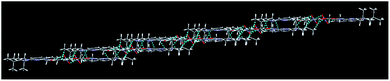 |
| | Fig. 7 Stairlike structure of receptor 1 showing infinite hydrogen bonding arrays. | |
Conclusion
In summary, we have synthesized five receptors for the study of inhibition property of nitro group in hydrogen bonding in molecular recognition. The observations from behavior of macrocyclic receptors 4 and 5 are sound enough to fulfill this aim to demonstrate the effect of nitro group in inhibition of hydrogen bonding in molecular recognition.
Acknowledgements
We wish to express our appreciation to CSIR [Project no. 01(1913)/04/EMR-II] and DST [Project no. SR/S1/OC-13/2005], Govt of India for financial support. R.C. thanks CSIR, Govt of India for a research fellowship. We also thank the Malaysian Government and Universiti Sains Malaysia for the Scientific Advancement Grant Allocation (SAGA) grant no. 304/PFIZIK/653003/A118. The authors extend their appreciation to the Deanship of Scientific Research at the King Saud University for funding the work through the research group project no. RGP-VPP-207.
References and notes
-
(a) O. Almarsson and M. J. Zaworotko, Chem. Commun., 2004, 1889 RSC;
(b) J. A. Mcmahon, B. Moulton, R. D. B. Walsh, N. Rodriguez-Hornedo and M. J. Zaworotko, Cryst. Growth Des., 2003, 3, 909 CrossRef;
(c) R. D. B. Walsh, M. W. Bradner, S. Fleischman, L. A. Morales, B. Moulton, N. Rodriguez-Hornedo and M. J. Zaworotko, Chem. Commun., 2003, 186 RSC;
(d) J. F. Remenar, S. L. Morissette, M. L. Peterson, B. Moulton, J. M. Macphee, H. R. Guzman and O. Almarsson, J. Am. Chem. Soc., 2003, 125, 8456 CrossRef CAS PubMed;
(e) S. L. Childs, L. J. Chyall, J. T. Dunlap, V. N. Smolenskaya, B. C. Stahly and G. P. Stahly, J. Am. Chem. Soc., 2004, 126, 13335 CrossRef CAS PubMed.
-
(a) L. J. Prins, D. N. Reinhoudt and P. Timmerman, Angew. Chem., Int. Ed., 2001, 40, 2382 CrossRef CAS;
(b) J.-M. Lehn, Supramolecular Chemistry: Concepts and Perspectives, VCH, Weinheim, Germany, 1995 Search PubMed;
(c) Crystal design: Structure and function, Perspective in Supramolecular chemistry, ed. G. R.Desiraju, Johnwiley and sons, 2003, vol. 7 Search PubMed;
(d) Comprehensive supramolecular chemistry, ed. J. L.Atwood, J. E. D.Davis, D. D.MacNicol and F.Vogtle, Pergamon, Oxford, UK, 1996, vol. 6, 7, 9 and 10 Search PubMed;
(e) G. R. Desiraju, Science, 1997, 278, 404 CrossRef CAS;
(f) S. Goswami, S. Jana, A. Hazra, H.-K. Fun, S. Anjum and A.-U. Rahman, CrystEngComm, 2006, 8, 712 RSC.
- S. P. Goswami, K. Ghosh and S. Dasgupta, Tetrahedron, 1996, 52, 12223 CrossRef CAS.
- R. J. Fitzmaurice, G. M. Kyne, D. Douheret and J. D. Kilburn, J. Chem. Soc., Perkin Trans. 1, 2002, 7, 841 RSC.
-
(a) S. Goswami, K. Ghosh and S. Dasgupta, J. Org. Chem., 2000, 65, 1907 CrossRef CAS;
(b) S. Goswami, K. Ghosh and M. Halder, Tetrahedron Lett., 1999, 40, 1735 CrossRef CAS;
(c) S. Goswami, S. Dey, H.-K. Fun, S. Anjum and A.-U. Rahman, Tetrahedron Lett., 2005, 46, 7187 CrossRef CAS PubMed;
(d) S. Goswami, S. Jana and H.-K. Fun, CrystEngComm, 2008, 10, 507 RSC;
(e) S. Goswami, S. Jana, S. Dey, H.-K. Fun and S. Chantrapromma, Tetrahedron, 2008, 64, 6426 CrossRef CAS PubMed;
(f) S. Goswami, S. Dey and S. Jana, Tetrahedron, 2008, 64, 6358 CrossRef CAS PubMed;
(g) S. Dey and D. Sain, Supramol. Chem., 2014, 26, 769 CrossRef CAS;
(h) T. Kusukawa, S. Tanaka and K. Inoue, Tetrahedron, 2014, 70, 4049 CrossRef CAS PubMed.
- S. Goswami, K. Ghosh and R. Mukherjee, J. Indian Chem. Soc., 1999, 661 CAS.
- S. Goswami, S. Dey, A. C. Maity and S. Jana, Tetrahedron Lett., 2005, 46, 1315 CrossRef CAS PubMed.
-
(a) Sigmaplot for windows, Version 12.3, Dundas Software, Germany Search PubMed;
(b) H. Benesi and J. H. Hildebrand, J. Am. Chem. Soc., 1949, 71, 2703 CrossRef CAS.
-
(a) Energy minimization was carried out using MMX (PCMODEL Serena Software 1993). Molecular modeling was performed using standard constants and the dielectric constants was maintained at 1.5;
(b) S. Goswami, S. Jana, N. K. Das, H.-K. Fun and S. Chantrapromma, J. Mol. Struct., 2008, 876, 313 CrossRef CAS PubMed.
-
(a) A. L. Spek, PLATON, A Multipurpose Crystallographic tool, Utrecht University, Utrecht, The Netherlands, 2002 Search PubMed;
(b) Different stereoviews of the crystals were generated using Mercury 1.4.1 software Search PubMed.
-
(a) G. R. Desiraju, Acc. Chem. Res., 1991, 24, 290 CrossRef CAS;
(b) A. K. Mahapatra, P. Sahoo, H.-K. Fun and S. Goswami, Asian J. Chem., 2008, 20, 1761 CAS.
- S. Goswami, S. Dey, H.-K. Fun, S. Anjum and A.-U. Rahman, Tetrahedron Lett., 2005, 46, 7187 CrossRef CAS PubMed.
Footnote |
| † Electronic supplementary information (ESI) available. CCDC 764388. For ESI and crystallographic data in CIF or other electronic format see DOI: 10.1039/c4ra06994c |
|
| This journal is © The Royal Society of Chemistry 2014 |
Click here to see how this site uses Cookies. View our privacy policy here.