Extremely selective fluorescence detection of cysteine or superoxide with aliphatic ester hydrolysis

Dhiraj P. Muralea, Hwajin Kimb, Wan Sung Choib, Youngsam Kima and David G. Churchill*a
aDepartment of Chemistry, KAIST, Daejeon, Republic of Korea. E-mail: dchurchill@kaist.ac.kr
bDepartment of Anatomy and Neurobiology, Gyeongsang National University, Jinju, Republic of Korea

Received 9th July 2014 , Accepted 9th September 2014

First published on 12th September 2014


Abstract

A novel fluorescence probe modality demonstrated with fluorescein affords a highly selective aqueous-based detection of cysteine over other biothiols, e.g. homocysteine, with a limit of detection of 11.3 μM.

Low molecular weight bio-thiols are essential molecules in nature and include Cysteine (Cys), homocysteine (hcy) and glutathione (GSH). These amino acids are involved in many physiological processes such as protein synthesis, metabolism, detoxification, signal transduction and gene regulation. Also, they are involved in regulating redox states and in composing the structure of proteins.1 Thus, these biothiols, by their nature, have adopted roles for normal health, but are also involved in the status of numerous different diseases. The structures of Cys and Hcy are very similar (differing by one methylene group); it is a difficult task to develop probes that possess selectivity enough to discriminate biothiols (especially between Cys and Hcy). An imbalance of Cys concentration is thought to relate to the cause of certain diseases; it may lead to neurotoxicity2 and deficiency, causes hair depigmentation, creates edema, slows growth, lethargy, promotes liver damage, muscle weakness, skin lesions as well as muscle and fat loss.3 An elevated level of Hcy in human plasma is related to incidence of Alzheimer's disease and cardiovascular disease; cellular GSH deficiency leads to oxidative stress which is suspected to be a main cause of certain neurodegenerative disorders. Cancers and AIDS are also related to biothiol concentration.4,5 Fluorescent molecular sensors for the detection of biothiols have great inherent advantages in their simplicity, potential selectivity and sensitivity in which the possibility of discerning intracellular and inter-compartmental detection allows for a great advantage over other methods of detection. Among the various types of fluorescent molecular sensors are chemodosimeters (based on the reaction of analyte with probe) which are more selective and sensitive in the detection of biothiols than chemosensors.

There are a number of probes that detect both Cys and Hcy;6 but excellent selective detection especially between Cys and Hcy is rare because of the structural similarities between analytes.7 Recently, in our research group, we have developed a novel rechargeable meso-aryl BODIPY-based chemodosimeter for the selective detection of Cys over other biothiols (Hcy and GSH).8a In a continuation of our interest in detecting biothiols, we have developed a novel route to detect biothiols via ester hydrolysis8b as well as selective detection of Cys, confirmed by chloropropionate ester hydrolysis.8c

Reactive oxygen species (ROS) play a key role in neurodegenerative disease disorders (Alzheimer's and Parkinson's disease), and in environmental chemistry.9 Recently, there have been many published trials about how to detect and understand ROS in vivo/vitro.10 Reactive oxygen species (ROS) include superoxide, hypochlorite, hydrogen peroxide, hydroxyl radical, nitric oxide and peroxynitrite. Among these ROS, detection of superoxide is an important issue because of its short life time. In an earlier report we demonstrated that superoxide can be detected by ester hydrolysis in which an aromatic ester bearing electron withdrawing groups was used. Here, we have shown the same strategy of detection can be used with aliphatic esters too.

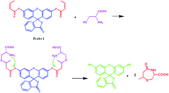
Herein, we report a fluorescein-based probe for the selective detection of Cys over other sulphur-containing amino acids. We designed our probe by a simple esterification reaction of fluorescein and vinyl acetic acid. We began our synthesis from fluorescein and the commercially available vinyl acetic acid by a simple single esterification step. In one facile step involving very convenient reaction conditions, probe 1, bearing two ester moieties, was obtained. Firstly, when we used 0.2 equiv. of DMAP we obtained a mixture of probe 1 and 1a in 1[thin space (1/6-em)]:[thin space (1/6-em)]1 ratio (Fig. S5). So to obtain probe 1 exclusively, we used 2 equiv. of DMAP (Scheme 1). Here, probe 1 was obtained by an isomerization of a vinylic group in making the more stable unsaturated α, β group. This probe has been previously synthesized from butanoic acid.11 We characterized the probe first by 1H and then by 13C NMR spectroscopy; confirmation was also shown by mass spectrometry (ESI). Importantly, the thermodynamic product (1) goes against the initial notion of controlling intermediate ring size in our probe. With a vinylic group such as that found in 1a, the carbonyl of the ester group by the amino group of the covalently-bound Cys would give rise to an important difference based on intermediate ring size. In the case of Cys, the formation of an 8-membered ring would be expected (Fig. S8); an unfavorable 9-membered ring would be expected for homocysteine. However, we now have simply a steric methyl group which appears to play a part in allowing for respective intermediate ring size formation that are 7-(Cys) and 8-membered (Hcy). Despite the smaller ring size, the steering of Cys/Hys selectivity based on this sterical methyl group appears to be effective. We obtained revealing that selective sensing for Cys over Hcy and GSH through a nucleophilic addition–elimination reaction at the vinylic position was possible giving fluorescein as the highly fluorescent product molecule as shown mechanistically below (Fig. 1). The mechanism behind this fluorescence sensing is supposed to be internal conversion (IC) as reported previously.12


image file: c4ra06891b-s1.tif
Scheme 1 Synthesis of probe 1.

image file: c4ra06891b-f1.tif
Fig. 1 Proposed Cys probing mechanism.

Spectroscopic properties of probe 1 were determined under physiological conditions (buffered H2O[thin space (1/6-em)]:[thin space (1/6-em)]DMSO 80[thin space (1/6-em)]:[thin space (1/6-em)]20; pH 7.4 PBS). Firstly, the probe was dissolved in DMSO and then subsequently diluted in 80% PBS buffer, pH 7.4. The detection properties of our new probes can be accessed via UV-vis absorption and emission spectroscopy.

The presence of the vinyl group allowed us to explore the probe with different amino acids. The probe was first tested with sulfur-containing amino acids (L-Cys, Hcy, N-acetyl-L-Cys, Met, GSH), as well as with non-sulfur-containing amino acids (Ala, Arg, Asn, Asp, Gln, Glu, Gly, His, Ile, Leu, Lys, Phe, Pro, Ser, Thr, Trp, Tyr, Val) as aqueous solutions on the order of ∼10 equiv. Here, we used 3 mL of 4.0 × 10⁻⁶ M, 1× PBS buffer, pH 7.4 probe solution and incubated with 10 equiv. amino acid; after 30 min, significant changes in the color of the probe solution was found with Cys.

When assessed quantitatively with analyte via emission studies, a dramatic fluorescence intensity increase was found (Fig. 2). A dramatic enhancement in fluorescence intensity was quantified for Cys (1390-fold), compared to the starting probe. A steady increase in fluorescence intensity was observed when the concentration of Cys was increased (Fig. 3a). The detection limit was estimated to be 11.3 μM, indicating the utility of the probe for detection of Cys in biological systems (Fig. S7). Also, when a time-dependent study was performed in which the probe was exposed to Cys for over 1 h, a steady increase in fluorescence intensity was formed (Fig. 3b). To verify the selectivity and practical utility of the probe, we performed a competition study of Cys solution in the presence of other thiol-containing species: Hcy, N-acetyl-L-Cys, Met, and GSH; interestingly, there was no interference in fluorescence intensity (Fig. S6).


image file: c4ra06891b-f2.tif
Fig. 2 Emission spectra of probe 1 (4.0 × 10⁻⁶ M, buffered H2O[thin space (1/6-em)]:[thin space (1/6-em)]DMSO 80[thin space (1/6-em)]:[thin space (1/6-em)]20; pH 7.4 PBS) with amino acids L-Cys, Hcy, N-acetyl-L-Cys, Met, GSH, Ala, Arg, Asn, Asp, Gln, Glu, Gly, His, Ile, Leu, Lys, Phe, Pro, Ser, Thr, Trp, Tyr, Val (66 μM in water) incubated for 20 min at RT. Slit width for Ex. & Em. = 1.5 nm.

image file: c4ra06891b-f3.tif
Fig. 3 Emission spectra of probe 1 (4.0 × 10⁻⁶ M, buffered H2O[thin space (1/6-em)]:[thin space (1/6-em)]DMSO 80[thin space (1/6-em)]:[thin space (1/6-em)]20; pH 7.4 PBS) with increasing concentrations of L-Cys (33.3–266 μM in water) and incubated for 20 min at RT. (right) Time-dependent emission spectra of probe 1 (2.0 × 10⁻⁶ M, buffered H2O[thin space (1/6-em)]:[thin space (1/6-em)]DMSO 80[thin space (1/6-em)]:[thin space (1/6-em)]20; pH 7.4 PBS) with Cys (200 μM in water, λexci = 490 nm) slit width Ex. & Em. = 1.5 nm.

In order to achieve sensing in more biologically-relevant aqueous conditions, we performed studies in different solvent combinations like 90[thin space (1/6-em)]:[thin space (1/6-em)]10H2O[thin space (1/6-em)]:[thin space (1/6-em)]DMSO, 90[thin space (1/6-em)]:[thin space (1/6-em)]10H2O[thin space (1/6-em)]:[thin space (1/6-em)]EtOH, 80[thin space (1/6-em)]:[thin space (1/6-em)]20H2O[thin space (1/6-em)]:[thin space (1/6-em)]EtOH, 80[thin space (1/6-em)]:[thin space (1/6-em)]20H2O[thin space (1/6-em)]:[thin space (1/6-em)]MeCN and 90[thin space (1/6-em)]:[thin space (1/6-em)]10H2O[thin space (1/6-em)]:[thin space (1/6-em)]MeCN (Fig. S9). As a result, we found 80[thin space (1/6-em)]:[thin space (1/6-em)]20H2O[thin space (1/6-em)]:[thin space (1/6-em)]DMSO is currently the best combination to balance time and fluorescence intensity requirements, as DMSO is promoting this reaction. To support the proposed mechanism, a separate reaction was carried out with probe 1 (1.0 equiv.) and Cys (2.5 equiv.) in 30 mL of a MeOH[thin space (1/6-em)]:[thin space (1/6-em)]H2O (90[thin space (1/6-em)]:[thin space (1/6-em)]10, v/v) solution. The reaction mixture was stirred at RT for 1 h. Then, Et3N (100[thin space (1/6-em)]μL) was added and the solution was stirred for 1 h. The solvent was then removed, and the crude product was checked directly with ESI-MS which revealed a value corresponding to fluorescein as a major product. Finally, the product was confirmed by 1H-NMR spectroscopy. The main product was fluorescein which results in a high emission intensity (520 nm).

To further evaluate the utility of the probe as a sensor for intracellular biothiol detection, SH-SY5Y cell assays were performed. The probe was found to be cell-permeable and selective for the detection of intracellular Cys; this was further supported through the NEM experimental technique. Also, the NEM experiment demonstrated there is no competition of superoxide over Cys in living neuronal cells as NEM is not a scavenger for superoxide (Fig. 4).


image file: c4ra06891b-f4.tif
Fig. 4 Fluorescence microscope images of living SH-SY5Y cells with vehicle (DMSO) and probe (20 μM) with corresponding bright field and fluorescence images (left) and cells pretreated with NEM (right) [scale bar 10 μM].

Finally, the probe with different ROS: there was only a response to superoxide (Fig. 5); 770-fold over other ROS. Superoxide is a strong nucleophile13 which takes part in hydrolysing esters and changes the fluorescence intensity of organic molecules. When manually increasing the concentration of superoxide, there was an increase in the fluorescence intensity; rises in fluorescence intensity also increased with experimental time (Fig. 5). Also, there was no interference or competition involving other ROS with superoxide activity (Fig. S11). From all the sensing data we can confidently construct an “OR” logic gating pattern with Cys and superoxide (Fig. 6).


image file: c4ra06891b-f5.tif
Fig. 5 Emission spectra of probe 1 (4.0 × 10⁻⁶ M, H2O[thin space (1/6-em)]:[thin space (1/6-em)]DMSO 80[thin space (1/6-em)]:[thin space (1/6-em)]20) with (ROS) KO2, H2O2, NaOCl, tBuOOH, ˙OH, ˙OtBu (66 μM in water) incubated for 30 min at RT incubated for 20 min at RT. λexci = 490 nm. Slit width for Ex. & Em. = 1.5 nm.

image file: c4ra06891b-f6.tif
Fig. 6 Emission spectra of probe 1 (4.0 × 10⁻⁶ M, H2O[thin space (1/6-em)]:[thin space (1/6-em)]DMSO 80[thin space (1/6-em)]:[thin space (1/6-em)]20) with increasing concentration of KO2 (33.3–266 μM in water) incubated for 30 min at RT. (right) Time-dependent emission spectra of probe 1 (2.0 × 10⁻⁶ M, H2O[thin space (1/6-em)]:[thin space (1/6-em)]DMSO 80[thin space (1/6-em)]:[thin space (1/6-em)]20) were formed with KO2 (200 μM in water) with λexci = 490 nm (slit width Ex. & Em. = 1.5 nm).

In conclusion, a novel fluorescence probe modality demonstrated with fluorescein affords a highly selective sensor platform for Cys over other relevant and closely-related biothiols. Simple esterification of fluorescein with vinyl acetic acid affords a ratiometric, fluorescent and selective probe for the detection of Cys over other thiol-containing amino acids with a detection limit of 11.3 μM and a ∼1390-fold increase in fluorescence intensity within 20 minutes. This sensing pattern was also confirmed in living neuronal cells. Also, the probe can be used as sensor for selective detection of superoxide over other ROS. As per our knowledge, this is first such probe for the selective detection of Cys and superoxide with “OR” logic gate.

Acknowledgements

D.G.C. is the operator of the Molecular Logic Gate Laboratory and acknowledges research support from the National Research Foundation (NRF) (Grant # 2010–0013660 & 2011–0017280), End Run (Grant # N01140684) and Mr Hack Soo Shin is acknowledged for his help in acquiring NMR spectroscopic data. The research support staff at KAIST facilitating the acquisition of MS data.

Notes and references

  1. (a) J. B. Schulz, J. Lindenau, J. Seyfried and J. Dichgans, Eur. J. Biochem., 2000, 267, 4904–4911 CrossRef CAS; (b) S. Seshadri, A. Beiser, J. Selhub, P. F. Jacques, I. H. Rosenberg, R. B. D'Agostino, P. W. F. Wilson and P. A. Wolf, N. Engl. J. Med., 2002, 346, 476–483 CrossRef CAS PubMed; (c) R. O. Ball, G. Courtney-Martin and P. B. Pencharz, J. Nutr., 2006, 136, 1682S–1693S CAS; (d) R. Hong, G. Han, J. M. Fernandez, B. J. Kim, N. S. Forbes and V. M. Rotello, J. Am. Chem. Soc., 2006, 128, 1078–1079 CrossRef CAS PubMed; (e) Z. A. Wood, E. Schroder, J. R. Harris and L. B. Poole, Trends Biochem. Sci., 2003, 28, 32–40 CrossRef CAS.
  2. X. F. Wang and M. S. Cynader, J. Neurosci., 2001, 21, 3322–3331 CAS.
  3. S. Shahrokhian, Anal. Chem., 2001, 73, 5972–5978 CrossRef CAS.
  4. H. Refsum, P. M. Ueland, O. Nygard and S. E. Vollset, Annu. Rev. Med., 1998, 49, 31–62 CrossRef CAS PubMed.
  5. (a) G. Y. Wu, Y. Z. Fang, S. Yang, J. R. Lupton and N. D. Turner, J. Nutr., 2004, 134, 489–492 CAS; (b) D. M. Townsend, K. D. Tew and H. Tapiero, Biomed. Pharmacother., 2003, 57, 145–155 CrossRef CAS; (c) L. A. Herzenberg, S. C. DeRosa, J. G. Dubs, M. Roederer, M. T. Anderson, S. W. Ela, S. C. Deresinski and L. A. Herzenberg, Proc. Natl. Acad. Sci. U. S. A., 1997, 94, 1967–1972 CrossRef CAS; (d) W. W. Huber and W. Parzefall, Curr. Opin. Pharmacol., 2007, 7, 404–409 CrossRef CAS PubMed.
  6. (a) Y. M. Yang, Q. Zhao, W. Feng and F. Y. Li, Chem. Rev., 2013, 113, 192–270 CrossRef CAS PubMed; (b) X. Chen, Y. Zhou, X. J. Peng and J. Yoon, Chem. Soc. Rev., 2010, 39, 2120–2135 RSC; (c) H. S. Jung, X. Chen, J. S. Kim and J. Yoon, Chem. Soc. Rev., 2013, 42, 6019–6031 RSC; (d) C. Yin, F. Huo, J. Zhang, R. Martinez-Manez, Y. Yang, H. Lv and S. Li, Chem. Soc. Rev., 2013, 42, 6032–6059 RSC.
  7. (a) W. H. Wang, J. O. Escobedo, C. M. Lawrence and R. M. Strongin, J. Am. Chem. Soc., 2004, 126, 3400–3401 CrossRef CAS PubMed; (b) X. F. Yang, Y. X. Guo and R. M. Strongin, Angew. Chem., Int. Ed., 2011, 50, 10690–10693 CrossRef CAS PubMed; (c) H. L. Li, J. L. Fan, J. Y. Wang, M. Z. Tian, J. J. Du, S. G. Sun, P. P. Sun and X. J. Peng, Chem. Commun., 2009, 48, 5904–5906 RSC; (d) H. L. Chen, Q. Zhao, Y. B. Wu, F. Y. Li, H. Yang, T. Yi and C. H. Huang, Inorg. Chem., 2007, 46, 11075–11081 CrossRef CAS PubMed; (e) H. P. Wu, C. C. Huang, T. L. Cheng and W. L. Tseng, Talanta, 2008, 76, 347–352 CrossRef CAS PubMed; (f) J. Mei, Y. J. Wang, J. Q. Tong, J. Wang, A. J. Qin, J. Z. Sun and B. Z. Tang, Chem.–Eur. J., 2013, 19, 613–620 CrossRef CAS PubMed; (g) H. L. Wang, G. D. Zhou, H. W. Gai and X. Q. Chen, Chem. Commun., 2012, 48, 8341–8343 RSC; (h) L. G. Wang, Q. Zhou, B. C. Zhu, L. G. Yan, Z. M. Ma, B. Du and X. L. Zhang, Dyes Pigm., 2012, 95, 275–279 CrossRef CAS PubMed; (i) Z. Guo, S. W. Nam, S. Park and J. Yoon, Chem. Sci., 2012, 3, 2760–2765 RSC; (j) K. H. Hong, S. Y. Lim, M. Y. Yun, J. W. Lim, J. H. Woo, H. Kwon and H. J. Kim, Tetrahedron Lett., 2013, 54, 3003–3006 CrossRef CAS PubMed; (k) X. Yang, Y. Guo and R. M. Strongin, Org. Biomol. Chem., 2012, 10, 2739–2741 RSC; (l) B. Zhu, Y. Zhao, Q. Zhou, B. Zhang, L. Liu, B. Du and X. Zhang, Eur. J. Org. Chem., 2013, 5, 888–893 CrossRef; (m) M. H. Lee, J. H. Han, P. S. Kwon, S. Bhuniya, J. Y. Kim, J. L. Sessler, C. Kang and J. S. Kim, J. Am. Chem. Soc., 2012, 134(2), 1316–1322 CrossRef CAS PubMed; (n) M. H. Lee, J. Y. Kim, J. H. Han, S. Bhuniya, J. L. Sessler, C. Kang and J. S. Kim, J. Am. Chem. Soc., 2012, 134(30), 12668–12674 CrossRef CAS PubMed; (o) T. Anand, G. Suvaraman and D. Chellappa, J. Photochem. Photobiol., A, 2014, 281, 47–52 CrossRef CAS PubMed.
  8. (a) O. G. Tsay, K. M. Lee and D. G. Churchill, New J. Chem., 2012, 36, 1949–1952 RSC; (b) D. P. Murale, H. Kim, W. S. Choi and D. G. Churchill, Org. Lett., 2013, 15, 3630–3633 CrossRef CAS PubMed; (c) D. P. Murale, H. Kim, W. S. Choi and D. G. Churchill, RSC Adv., 2014, 4, 5289–5292 RSC.
  9. (a) A. P. de Silva, H. Q. N. Gunaratne, T. Gunnlaugsson, A. J. M. Huxley, C. P. McCoy, J. T. Rademacher and T. E. Rice, Chem. Rev., 1997, 97, 1515–1566 CrossRef CAS PubMed; (b) T. Gunnlaugsson, M. Glynn, G. M. Tocci, P. E. Kruger and F. M. Pfeffer, Coord. Chem. Rev., 2006, 250, 3094–3117 CrossRef CAS PubMed; (c) E. L. Que, D. W. Domaille and C. J. Chang, Chem. Rev., 2008, 108, 1517–1549 CrossRef CAS PubMed.
  10. (a) F. Yu, P. Li, B. Wang and K. Han, J. Am. Chem. Soc., 2013, 135, 7674–7680 CrossRef CAS PubMed; (b) F. Yu, P. Li, G. Li, G. Zhao, T. Chu and K. Han, J. Am. Chem. Soc., 2011, 133, 11030–11033 CrossRef CAS PubMed; (c) G.-Y. Li, Y.-H. Li, H. Zhang and G.-H. Cui, Commun. Comput. Chem., 2013, 1(1), 88–98 Search PubMed.
  11. X. L. Yan, W. F. Wang, S. F. Zhang, Y. Li, G. Y. Bai and L. G. Chen, Jingxi Huagong, 2005, 22, 440–443 CAS.
  12. P. Zhou, J. Liu, S. Yang, J. Chen, K. Han and G. He, Phys. Chem. Chem. Phys., 2012, 14, 15191–15198 RSC.
  13. I. B. Afanas'ev, Med. Hypotheses, 2005, 64, 127–129 CrossRef PubMed.

Footnote

Electronic supplementary information (ESI) available: Experimental details, in-line spectral data, MS spectra. See DOI: 10.1039/c4ra06891b

This journal is © The Royal Society of Chemistry 2014
Click here to see how this site uses Cookies. View our privacy policy here.