Homogeneous decoration of zeolitic imidazolate framework-8 (ZIF-8) with core–shell structures on carbon nanotubes

JongTae Yooab, SuHyun Leeac, Chang Kee Leea, ChaeRin Kimb, Tsuyohiko Fujigayabd, Hyun Jin Parkc, Naotoshi Nakashima*bd and Jin Kie Shim*a
aKorea Packaging Center, Korea Institute of Industrial Technology, Ojeong-gu, Bucheon 421-742, South Korea
bDepartment of Applied Chemistry, Graduate School of Engineering, Kyushu University, 744 Motooka, Nishi-ku, Fukuoka 819-0395, Japan. E-mail: nakashima-tcm@mail.cstm.kyushu-u.ac.jp; Fax: +81-92-802-2840; Tel: +81-92-802-2840
cCollege of Life Sciences and Biotechnology, Korea University, Seongbuk-gu, Seoul 136-701, South Korea
dInternational Institute for Carbon-Neutral Energy Research (WPI-I2CNER), Kyushu University, 744 Motooka, Nishi-ku, Fukuoka 819-0395, Japan

Received 8th July 2014 , Accepted 26th September 2014

First published on 26th September 2014


Abstract

Considerable attention has focused on the combination of carbon nanotubes (CNTs) and metal–organic frameworks (MOFs) since both nanomaterials have outstanding properties. We describe a method for the homogeneous decoration of a MOF (ZIF-8 was chosen) onto the surfaces of CNTs dispersed by polyvinylpyrrolidones (PVPs) in methanol, which was revealed by a scanning electron microscopic study. The homogeneous coating of the MOF on the CNTs, and nanostructures of the CNT-MOF were controlled by simply changing the concentrations of the MOFs. Furthermore, this method was also applicable to graphene and graphene oxide (GO). CO2 uptakes of the CNT-MOF and graphene-MOF were significantly improved as compared to the nonhomogeneous composites synthesized without the PVP functionalization, and a good reproducibility of the CO2 adsorption was confirmed by the cycling test.

1 Introduction

Carbon nanotubes (CNTs) are nanomaterials with remarkable electrical, thermal, optical and mechanical properties.1–4 They are also an attractive material for gas storage due to their high surface areas (50–1315 m2 g−1).5 Especially, specific nano-spaces in the CNT bundles, such as interstitial channels and external grooves, have significant interactions with aromatic6 and gas molecules, such as Ar,7 Ne,8 He,8,9 CF4,10 H2,11 N2,12,13 O2,13 and CnH2n+2,14,15 and also exhibit selectivity for molecules.6,16,17 However, the rather low gas selectivity and storage capacity on the π-conjugated surfaces of the CNTs has limited their applications to gas sensors and separations. In order to improve such a drawback, covalent18 and non-covalent functionalization19,20 and coating by inorganic materials21 have been carried out.

Metal–organic frameworks (MOFs) with extremely-high surface areas caused by the three-dimensional porous structures including metal centers and organic linkers have emerged as one of the most promising gas absorbent material because the pore sizes and chemical affinities are easily controlled by modifying the metals and linkers.22–28 MOFs have been investigated in many fields including gas absorbents and separations, catalysts29 and drug delivery system (DDS) and fluorescence imaging of cancer cells,30 therefore, the homogeneous decorations of the MOFs are very important for many applications.

Zeolitic imidazolate frameworks (ZIFs), which is one of the MOFs, are known to have the highest thermal and chemical stabilities among the MOF families,31 and their facile synthetic method32 enables complexations with various polymers,33,34 graphene quantum dots (GQDs),35 graphene oxide (GO),36 Cu3(BTC)2-modified CNTs,37 etc. Especially, the ZIF-8 synthesized by 2-methylimidazole (denoted as 2-MeIm) and zinc ions shows good separation properties for CO2/CH4 (ref. 38) and CO2/N2 (ref. 39) compared to other ZIFs, such as ZIF-7, ZIF-22 and ZIF-90.40 Flexible structures opened by rotations of the 2-MeIm would be advantageous to control its morphology, such as core–shell structures. ZIF-8 has been used as a material for the encapsulation of GQDs35 and preparation of composites with polymers,41–46 CNTs47 and graphene oxide.36 However, the formation of core–shell structures using the ZIF-8 as the shell has not yet been reported. We here focus on the homogeneous decorations of the MOFs since such a coating is very important for many applications.

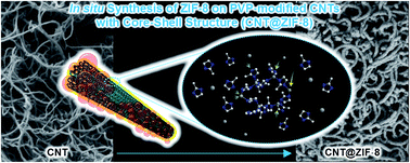
In this study, as shown in Fig. 1, polyvinylpyrrolidone (PVP) was chosen as the structure-directing and stabilizing agent for the preparation of the CNT@ZIF-8 core–shell structure due to its shape-selective properties for inorganic nanowires48–52 and core–shell structures,53–55 and it was expected that its strong interaction and high dispersing property for the CNTs would help to form a homogeneous structure without aggregations of the CNTs.56 Furthermore, graphene and graphene oxide were also used as the core materials for the ZIF-8 shell. This is the first report about the synthesis of extremely well-controlled shell structures of the ZIF-8 coating the surface of CNTs with the high aspect ratio.


image file: c4ra06792d-f1.tif
Fig. 1 A schematic drawing of a CNT@ZIF-8 core–shell structure.

2 Materials and methods

2.1 Materials

Multi-walled carbon nanotubes (MWNTs) (Ctube120, metal oxide <3 wt%, average diameter: ∼20 nm, length: 1–25 μm, CNT Co., Ltd), graphene (3 nm graphene nanopowder, grade AO-1, Graphene Supermarket), graphene oxide (GO) (dry platelets, Graphene Supermarket) and methanol (>99.8%, J. T. Baker®) were used as received. Polyvinylpyrrolidone (PVP) (Mw: ∼360[thin space (1/6-em)]000), 2-methylimidazole (99%) and zinc nitrate hexahydrate (98%) were purchased from Sigma-Aldrich.

2.2 Measurements

The field emission scanning electron microscopy (FE-SEM) and scanning transmission electron microscopy (STEM) were conducted using an SU-8020 (Hitachi, Tokyo, Japan) at 1 kV and 30 kV. Fourier transform infrared (FT-IR) and Raman spectroscopy measurements using a 532 nm laser were carried out by a Varian 660-IR (Varian Medical Systems, Inc., California, USA) and a SENTERRA Raman microscope spectrometer (Bruker Corporation, Billerica, MA, USA), respectively. X-ray diffraction (XRD) measurements in the range of 1° < 2θ < 30° were performed by a SmartLab (Rigaku) at 40 kV and 30 mA (CuKα radiation, λ = 0.154 nm).

2.3 Preparations of CNT, graphene, and GO composites with the ZIF-8

Composites of carbon materials with the ZIF-8 were fabricated by the following in situ ZIF-8 synthesis. The CNTs (30 mg) were added to a PVP methanol solution (2 mg mL−1, 60 mL), then sonicated in a bath-type sonicator (JAC-3010, KODO) for 1 h. After centrifugation (20[thin space (1/6-em)]000g, 1 h), the supernatant was removed, the precipitates were redispersed in methanol (15 mL) to which 2-methylimidazole (2-MeIm) in methanol (22 mg mL−1, 60 mL) was added, then zinc nitrate hexahydrate in methanol (11 mg mL−1, 12 mL) was carefully added to the dispersion while stirring. The generated precipitates were collected after centrifugation (20[thin space (1/6-em)]000g, 0.5 h), then rinsed with methanol, and dried in a vacuum oven at 40 °C.

Graphene and GO composites with the ZIF-8 were also fabricated by the same procedure.

2.4 TGA analysis

CO2 adsorption experiments were carried out by a TGA Q500 (TA Instruments, New Castle, USA) using a previously reported method.57–59 Nitrogen (N2) and carbon dioxide (CO2) gases were used as the purge and furnace gases at the flow rates of 40 and 60 mL min−1, respectively, and all experiments were performed after verification of no weight reduction after N2 flowing for 4 h at 100 °C in order to dehydrate and degas the samples. The temperature of the furnace was raised to 70 °C at the rate of 20 °C min−1, then a furnace gas was changed to the CO2. After the isothermal process for 1 h at each temperature (70, 55, 40, and 25 °C), the furnace gas was changed from the CO2 to N2 during heating from 25 to 70 °C. Recycle test was performed by a similar method at 25 °C, and thermogravimetric analysis (TGA) was carried out in the N2 atmosphere by heating to 900 °C at the rate of 5 °C min−1.

3 Results and discussion

3.1 Preparations of CNT, graphene, and GO composites with the ZIF-8

PVP has been reported not only as a good structure-directing agent for nanowires48–52 and core–shell structures,53–55 but also as a good dispersant for CNTs.56 Recently, the encapsulation of metal nanoparticles into the ZIF-8 has been investigated using the PVP as the capping agent due to its structure-selective property for the nucleation and growth of the ZIF-8 crystals.60 The core–shell structure formation of the CNT@ZIF-8 is shown in Scheme 1, in which the PVP-functionalized CNTs (PVP-CNTs) are redispersed in methanol after the removal of the excess PVPs, and the ZIF-8 was synthesized using the 2-MeIm and zinc ions in the presence of the PVP-CNTs. Graphene and GO were also used as supporting materials in place of the CNTs.
image file: c4ra06792d-s1.tif
Scheme 1 Schematic drawing for the preparation of CNT@ZIF-8 core–shell structures.

As shown in Fig. 2, the ZIF-8 composites with the PVP-CNTs (CNT@ZIF-8) and CNTs (CNT/ZIF-8) clearly exhibited a thicker diameter than that of the pristine CNTs, suggesting the ZIF-8 decoration. In the figure, we recognized that ZIF-8 particles were hardly seen in the CNT@ZIF-8 composite; however in sharp contrast, many ZIF-8 aggregates were observed in the CNT/ZIF-8 composite. It is expected that the PVP worked as a structure-directing agent on the CNT surfaces (see ESI, Fig. S1), and a similar behavior also appeared when using the PVP-functionalized graphene (PVP-G) and GO (PVP-GO). In the ZIF-8 composites with the PVP-G (PVP-G/ZIF-8) and PVP-GO (PVP-GO/ZIF-8), the sizes of the decorated ZIF-8 nanoparticles were smaller and significantly more homogeneous than those of the pristine graphene (G/ZIF-8) and GO (GO/ZIF-8) as can be seen in Fig. 3. As shown in Fig. 4, the scanning transmission electron microscope (STEM) image of the CNT@ZIF-8 clearly shows shell structures on the individually-dispersed CNT surfaces, and the diameter of the decorated ZIF-8 nanoparticles on the PVP-G/ZIF-8 and PVP-GO/ZIF-8 was found to be less than 35 nm.


image file: c4ra06792d-f2.tif
Fig. 2 Scanning electron microscope (SEM) images of the (A) CNT, (B) CNT/ZIF-8, and (C) CNT@ZIF-8. The yellow bars in (B) indicate aggregations of the ZIF-8 nanoparticles. Scale bars: 200 nm.

image file: c4ra06792d-f3.tif
Fig. 3 SEM images of the (A) graphene, (B) G/ZIF-8, (C) PVP-G/ZIF-8, (D) GO, (E) GO/ZIF-8, and (F) PVP-GO/ZIF-8. Scale bars: 200 nm.

image file: c4ra06792d-f4.tif
Fig. 4 Scanning transmission electron microscope (STEM) images of the (A) CNT@ZIF-8, (B) PVP-G/ZIF-8, and (C) PVP-GO/ZIF-8. Scale bars: 100 nm.

All composites showed characteristic peaks in the Raman spectra, which were attributed to the G-band (∼1590 cm−1) and D-band (∼1350 cm−1) of the CNTs, graphene, and GO (see ESI, Fig. S2). We also observed Fourier transform infrared spectroscopy (FT-IR) peaks, which appeared at almost the same wavenumbers as that of the ZIF-8 synthesized without the supported carbon materials (see ESI, Fig. S3 and S4). Furthermore, X-ray diffraction (XRD) patterns of the composites showed patterns similar to the ZIF-8 (see ESI, Fig. S5). Since the formation of the ZIF-8 crystals is evident from the FT-IR and XRD studies together with the results from previous reports,36,61 all the obtained results strongly suggested the formation of ZIF-8-decorated nanostructures.

It was found that during the synthesis of the CNT@ZIF-8 core–shell structures, the concentrations of the 2-MeIm and zinc nitrate hexahydrate played a key role in the shell formation, and the suitable concentrations were ∼22 mg mL−1 of 2-MeIm and ∼11 mg mL−1 of zinc nitrate hexahydrate. However, when using four and eight times higher concentrations, no such CNT@ZIF-8 core–shell structures were formed due to the expected rapid nucleation and growth of the ZIF-8 as shown in Fig. 5. It was found that when using two times higher concentrations, kebab-like structures (see Fig. 5B) were observed in the CNT@ZIF-8. The obtained results indicated that the ZIF-8 formations on the surfaces of the PVP-CNTs can be simply controlled by changing the concentrations of the 2-MeIm and zinc nitrate hexahydrate. In contrast, reaction times did not influence on the nanostructure of the CNT@ZIF-8 even when the experiment was carried out at the suitable concentrations of ∼22 mg mL−1 of 2-MeIm and ∼11 mg mL−1 of zinc nitrate hexahydrate with stirring for over 240 min (see ESI, Fig. S6).


image file: c4ra06792d-f5.tif
Fig. 5 SEM images of the CNT@ZIF-8s prepared from 2-MeIm and zinc nitrate solutions at concentrations of (A) 22 and 11 mg mL−1, respectively, and the (B) 2, (C) 4, and (D) 8 times-concentrated solutions. Scale bars: 300 nm.

3.2 CO2 adsorption properties

The CO2 adsorption capability was examined using a previously reported method.57–59 As shown in Fig. 6, the CO2 uptake in the CNT@ZIF-8 was significantly improved compared to the pristine CNTs and CNT/ZIF-8s. Since it has been reported that homogeneous decorations of nanoparticles with smaller sizes improved the adsorption capacity for hydrogen gases,62 it is expected that the homogeneous decorating of the ZIF-8 on the CNT@ZIF-8, in which the particle size of the ZIF-8 was small, significantly improved the adsorption properties even though the ZIF-8 content in the CNT@ZIF-8 was lower than that in the CNT/ZIF-8 (ESI, Fig. S7). The PVP-G/ZIF-8 also showed a greater CO2 uptake than those of the pristine graphene and G/ZIF-8; however, such an improvement was not observed from the PVP-GO/ZIF-8 although the adsorption rate of the CO2 was improved as compared to the pristine GO (ESI, Fig. S8). The difference of CO2 uptakes between the CNT@ZIF-8 and PVP-G/ZIF-8 was due to the difference of the contents of the ZIF-8 in the composites (ESI, Fig. S7); namely the higher ZIF-8 content in the PVP-G/ZIF-8 provided a higher CO2 uptake than that of the CNT@ZIF-8. Meanwhile, stable reproducibility of the CO2 adsorption on the CNT@ZIF-8 was confirmed from cycling tests without any decrease in the adsorption capacities as shown in Fig. 7. Although the presented CO2 uptake of the ZIF-8 on the CNTs and graphenes is not very sufficient, the uptake amount is expected to be enhanced by optimizing the experimental conditions.
image file: c4ra06792d-f6.tif
Fig. 6 TGA showing mass changes due to the adsorption and desorption of CO2 gases obtained from the (A, blue line) CNT@ZIF-8, (A, red line) CNT/ZIF-8, (A, black line) CNT, (B, blue line) PVP-G/ZIF-8, (B, red line) G/ZIF-8, and (B, black line) graphene.

image file: c4ra06792d-f7.tif
Fig. 7 Cycling of the CO2 adsorption on the CNT@ZIF-8.

4 Conclusions

CNT@ZIF-8 core–shell structures have been successfully fabricated by the in situ ZIF-8 synthesis in the presence of the PVP-functionalized CNTs, and the ZIF-8 nanostructures in the composites were simply controlled by changing the concentrations of the starting chemicals for the synthesis of the ZIF-8. Similar homogeneous decorations were possible when using PVP-functionalized graphene and GO in place of the PVP-functionalized CNTs. Such a homogeneous ZIF-8 decoration was important for the improved CO2 gas adsorption compared to the composites prepared using none-PVP modified CNTs and graphenes. The presented method for fabricating homogeneous ZIF-8 decorations is very simple and the coated ZIF-8 shell structures were easily controlled, suggesting that the method is highly useful for the design and synthesis of many nanomaterials including catalysts, materials for DDS and imaging bionanotechnology, etc.

Acknowledgements

This work was supported by the R&D program of MOTIE/KEIT (no. 100039993, Development of product safety information service solution using nano hydrid smart packaging), Korea.

Notes and references

  1. T. W. Ebbesen, H. J. Lezec, H. Hiura, J. W. Bennett, H. F. Ghaemi and T. Thio, Nature, 1996, 382, 54–56 CrossRef CAS.
  2. M. M. J. Treacy, T. W. Ebbesen and J. M. Gibson, Nature, 1996, 381, 678–680 CrossRef CAS.
  3. Z. Wu, Z. Chen, X. Du, J. M. Logan, J. Sippel, M. Nikolou, K. Kamaras, J. R. Reynolds, D. B. Tanner, A. F. Hebard and A. G. Rinzler, Science, 2004, 305, 1273–1277 CrossRef CAS PubMed.
  4. M. Moniruzzaman and K. I. Winey, Macromolecules, 2006, 39, 5194–5205 CrossRef CAS.
  5. A. Peigney, C. Laurent, E. Flahaut, R. R. Bacsa and A. Rousset, Carbon, 2001, 39, 507–514 CrossRef CAS.
  6. J. Yoo, T. Fujigaya and N. Nakashima, Nanoscale, 2013, 5, 7419–7424 RSC.
  7. Z. J. Jakubek and B. Simard, Langmuir, 2004, 20, 5940–5945 CrossRef CAS.
  8. G. Stan, V. Crespi, M. Cole and M. Boninsegni, J. Low Temp. Phys., 1998, 113, 447–452 CrossRef CAS.
  9. W. Teizer, R. B. Hallock, E. Dujardin and T. W. Ebbesen, Phys. Rev. Lett., 1999, 82, 5305–5308 CrossRef CAS.
  10. O. Byl, P. Kondratyuk, S. T. Forth, S. A. FitzGerald, L. Chen, J. K. Johnson and J. T. Yates, J. Am. Chem. Soc., 2003, 125, 5889–5896 CrossRef CAS PubMed.
  11. S. S. Han, J. K. Kang, H. M. Lee, A. C. T. Van Duin and W. A. Goddard III, Appl. Phys. Lett., 2005, 86, 1–3 Search PubMed.
  12. J. Jiang and S. I. Sandler, Phys. Rev. B: Condens. Matter Mater. Phys., 2003, 68, 245412 CrossRef.
  13. J. Jiang and S. I. Sandler, Langmuir, 2004, 20, 10910–10918 CrossRef CAS PubMed.
  14. S. Vela and F. Huarte-Larrañaga, Carbon, 2011, 49, 4544–4553 CrossRef CAS PubMed.
  15. J. J. Cannon, T. J. H. Vlugt, D. Dubbeldam, S. Maruyama and J. Shiomi, J. Phys. Chem. B, 2012, 116, 9812–9819 CrossRef CAS PubMed.
  16. M. Yamamoto, T. Itoh, H. Sakamoto, T. Fujimori, K. Urita, Y. Hattori, T. Ohba, H. Kagita, H. Kanoh, S. Niimura, K. Hata, K. Takeuchi, M. Endo, F. Rodríguez-Reinoso and K. Kaneko, Adsorption, 2011, 17, 643–651 CrossRef CAS.
  17. M. D. Ellison, M. J. Crotty, D. Koh, R. L. Spray and K. E. Tate, J. Phys. Chem. B, 2004, 108, 7938–7943 CrossRef CAS.
  18. N. Karousis, N. Tagmatarchis and D. Tasis, Chem. Rev., 2010, 110, 5366–5397 CrossRef CAS PubMed.
  19. A. L. Ahmad, Z. A. Jawad, S. C. Low and S. H. S. Zein, J. Membr. Sci., 2014, 451, 55–66 CrossRef CAS PubMed.
  20. R. Mangu, S. Rajaputra and V. P. Singh, Nanotechnology, 2011, 22, 215502 CrossRef PubMed.
  21. H. C. Su, M. Zhang, W. Bosze, J. H. Lim and N. V. Myung, Nanotechnology, 2013, 24, 505502 CrossRef PubMed.
  22. H. K. Chae, D. Y. Siberio-Pérez, J. Kim, Y. Go, M. Eddaoudi, A. J. Matzger, M. O'Keeffe and O. M. Yaghi, Nature, 2004, 427, 523–527 CrossRef CAS PubMed.
  23. M. Eddaoudi, J. Kim, N. Rosi, D. Vodak, J. Wachter, M. O'Keeffe and O. M. Yaghi, Science, 2002, 295, 469–472 CrossRef CAS PubMed.
  24. S. Kitagawa, R. Kitaura and S. I. Noro, Angew. Chem., Int. Ed., 2004, 43, 2334–2375 CrossRef CAS PubMed.
  25. H. Li, M. Eddaoudi, M. O'Keeffe and O. M. Yaghi, Nature, 1999, 402, 276–279 CrossRef CAS PubMed.
  26. N. L. Rosi, J. Eckert, M. Eddaoudi, D. T. Vodak, J. Kim, M. O'Keeffe and O. M. Yaghi, Science, 2003, 300, 1127–1129 CrossRef CAS PubMed.
  27. J. S. Seo, D. Whang, H. Lee, S. I. Jun, J. Oh, Y. J. Jeon and K. Kim, Nature, 2000, 404, 982–986 CrossRef CAS PubMed.
  28. O. M. Yaghi, M. O'Keeffe, N. W. Ockwig, H. K. Chae, M. Eddaoudi and J. Kim, Nature, 2003, 423, 705–714 CrossRef CAS PubMed.
  29. J. Lee, O. K. Farha, J. Roberts, K. A. Scheidt, S. T. Nguyen and J. T. Hupp, Chem. Soc. Rev., 2009, 38, 1450–1459 RSC.
  30. L. He, T. Wang, J. An, X. Li, L. Zhang, L. Li, G. Li, X. Wu, Z. Su and C. Wang, CrystEngComm, 2014, 16, 3259–3263 RSC.
  31. J. Cravillon, S. Münzer, S.-J. Lohmeier, A. Feldhoff, K. Huber and M. Wiebcke, Chem. Mater., 2009, 21, 1410–1412 CrossRef CAS.
  32. K. Zhang, R. P. Lively, M. E. Dose, A. J. Brown, C. Zhang, J. Chung, S. Nair, W. J. Koros and R. R. Chance, Chem. Commun., 2013, 49, 3245–3247 RSC.
  33. C. Zhang, R. P. Lively, K. Zhang, J. R. Johnson, O. Karvan and W. J. Koros, J. Phys. Chem. Lett., 2012, 3, 2130–2134 CrossRef CAS.
  34. X.-L. Liu, Y.-S. Li, G.-Q. Zhu, Y.-J. Ban, L.-Y. Xu and W.-S. Yang, Angew. Chem., Int. Ed., 2011, 50, 10636–10639 CrossRef CAS PubMed.
  35. B. P. Biswal, D. B. Shinde, V. K. Pillai and R. Banerjee, Nanoscale, 2013, 5, 10556–10561 RSC.
  36. R. Kumar, K. Jayaramulu, T. K. Maji and C. N. R. Rao, Chem. Commun., 2013, 49, 4947–4949 RSC.
  37. Z. Xiang, X. Peng, X. Cheng, X. Li and D. Cao, J. Phys. Chem. C, 2011, 115, 19864–19871 CAS.
  38. S. R. Venna and M. A. Carreon, J. Am. Chem. Soc., 2010, 132, 76–78 CrossRef CAS PubMed.
  39. J. Pérez-Pellitero, H. Amrouche, F. R. Siperstein, G. Pirngruber, C. Nieto-Draghi, G. Chaplais, A. Simon-Masseron, D. Bazer-Bachi, D. Peralta and N. Bats, Chem.–Eur. J., 2010, 16, 1560–1571 CrossRef PubMed.
  40. J. R. Li, Y. Ma, M. C. McCarthy, J. Sculley, J. Yu, H. K. Jeong, P. B. Balbuena and H. C. Zhou, Coord. Chem. Rev., 2011, 255, 1791–1823 CrossRef CAS PubMed.
  41. M. Askari and T. S. Chung, J. Membr. Sci., 2013, 444, 173–183 CrossRef CAS PubMed.
  42. A. F. Bushell, M. P. Attfield, C. R. Mason, P. M. Budd, Y. Yampolskii, L. Starannikova, A. Rebrov, F. Bazzarelli, P. Bernardo, J. Carolus Jansen, M. Lanč, K. Friess, V. Shantarovich, V. Gustov and V. Isaeva, J. Membr. Sci., 2013, 427, 48–62 CrossRef CAS PubMed.
  43. R. Jin, Z. Bian, J. Li, M. Ding and L. Gao, Dalton Trans., 2013, 42, 3936–3940 RSC.
  44. V. Nafisi and M. B. Hägg, J. Membr. Sci., 2014, 459, 244–255 CrossRef CAS PubMed.
  45. M. J. C. Ordoñez, K. J. Balkus, J. P. Ferraris and I. H. Musselman, J. Membr. Sci., 2010, 361, 28–37 CrossRef PubMed.
  46. G. M. Shi, T. Yang and T. S. Chung, J. Membr. Sci., 2012, 415–416, 577–586 CrossRef CAS PubMed.
  47. L. Dumée, L. He, M. Hill, B. Zhu, M. Duke, J. Schütz, F. She, H. Wang, S. Gray, P. Hodgson and L. Kong, J. Mater. Chem. A, 2013, 1, 9208–9214 Search PubMed.
  48. G. Xi, Y. Liu, X. Wang, X. Liu, Y. Peng and Y. Qian, Cryst. Growth Des., 2006, 6, 2567–2570 CAS.
  49. F. Yu, W. Zhou, R. M. Bellabarba and R. P. Tooze, Nanoscale, 2014, 6, 1093–1098 RSC.
  50. J. Wang, C. Yang, Z. Huang, M. G. Humphrey, D. Jia, T. You, K. Chen, Q. Yang and C. Zhang, J. Mater. Chem., 2012, 22, 10009–10014 RSC.
  51. J. W. Liu, F. Chen, M. Zhang, H. Qi, C. L. Zhang and S. H. Yu, Langmuir, 2010, 26, 11372–11377 CrossRef CAS PubMed.
  52. J. Wang, X. Wang, Q. Peng and Y. Li, Inorg. Chem., 2004, 43, 7552–7556 CrossRef CAS PubMed.
  53. S. Bhlapibul, K. Pruksathorn and P. Piumsomboon, Renewable Energy, 2012, 41, 262–266 CrossRef CAS PubMed.
  54. M. Tsuji, K. Ikedo, M. Matsunaga and K. Uto, CrystEngComm, 2012, 14, 3411–3423 RSC.
  55. N. Zhang, S. Liu, X. Fu and Y. J. Xu, J. Phys. Chem. C, 2011, 115, 22901–22909 CAS.
  56. M. J. O'Connell, P. Boul, L. M. Ericson, C. Huffman, Y. Wang, E. Haroz, C. Kuper, J. Tour, K. D. Ausman and R. E. Smalley, Chem. Phys. Lett., 2001, 342, 265–271 CrossRef.
  57. E. P. Dillon, C. A. Crouse and A. R. Barron, ACS Nano, 2008, 2, 156–164 CrossRef CAS PubMed.
  58. E. A. Roth, S. Agarwal and R. K. Gupta, Energy Fuels, 2013, 27, 4129–4136 CrossRef CAS.
  59. W. Wang, J. Xiao, X. Wei, J. Ding, X. Wang and C. Song, Appl. Energy, 2014, 113, 334–341 CrossRef CAS PubMed.
  60. G. Lu, S. Li, Z. Guo, O. K. Farha, B. G. Hauser, X. Qi, Y. Wang, X. Wang, S. Han, X. Liu, J. S. DuChene, H. Zhang, Q. Zhang, X. Chen, J. Ma, S. C. J. Loo, W. D. Wei, Y. Yang, J. T. Hupp and F. Huo, Nat. Chem., 2012, 4, 310–316 CrossRef CAS PubMed.
  61. Y. Pan, Y. Liu, G. Zeng, L. Zhao and Z. Lai, Chem. Commun., 2011, 47, 2071–2073 RSC.
  62. L. Wang, N. R. Stuckert, H. Chen and R. T. Yang, J. Phys. Chem. C, 2011, 115, 4793–4799 CAS.

Footnote

Electronic supplementary information (ESI) available. See DOI: 10.1039/c4ra06792d

This journal is © The Royal Society of Chemistry 2014
Click here to see how this site uses Cookies. View our privacy policy here.