Chunyan Zoua,
Qinglong Qiaoab,
Miao Zhaob,
Deqi Maoab,
Danfeng Wanga,
Lei Fenga,
Jingnan Cui*a and
Zhaochao Xu*ab
aState Key Laboratory of Fine Chemicals, Dalian University of Technology, Dalian 116012, China. E-mail: jncui@dlut.edu.cn
bKey Laboratory of Separation Science for Analytical Chemistry, Dalian Institute of Chemical Physics, Chinese Academy of Sciences, Dalian 116023, China. E-mail: zcxu@dicp.ac.cn
First published on 8th September 2014
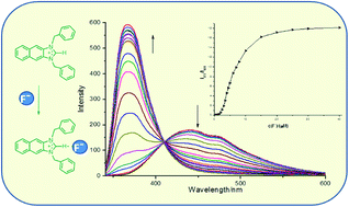
Three imidazolium derivatives 3–5 were designed and synthesized, in which naphthaimidazolium group acted as both fluorophore and anion receptor. Compound 3 exhibited high selectivity for F− in CH3CN solution over all the other anions and acted as a ratiometric fluorescent probe for F− with an enhanced blue-shift in emission. However, the fluorescence of compound 4 and 5 displayed a quenched blue-shift in emission with fluoride ion and could be quenched by some other tested anions, where the degree of quenching depended on the characteristic of the anions. More importantly, only compound 3 could detect F− in DMSO–water (95
:
5, v/v) aqueous solution ratiometrically. Based on the analysis of the results of 1H-NMR and 19F-NMR, it was deduced that compound 3 bound with F− mainly by the force of hydrogen bonding, while compound 4 and 5 coordinated with F− through electrostatic interaction.
In the previous work,11,19,21 naphthaimidazolium group was utilized as the signal source for the detection system, and also as an anion binding receptor. As shown in Scheme 1, the cage compounds 1 and 2 contained three naphthaimidazolium groups inserted between two substituted benzene rings.21 With the addition of 1 equiv. of F−, a strongly increased fluorescent emission of compound 2 centered at 385 nm appeared at the expense of 474 nm. However, the addition of fluoride quenched the fluorescence of compound 1. 19F and 1H NMR spectroscopy, fluorescence emission and theoretical calculations results showed that three (C–H)+⋯F−-type ionic hydrogen bonds formed and from this, the anion–π interaction between the two electron-rich alkylbenzene rings and the fluoride ion existed in compound 2. For compound 1, the electrostatic interaction between the imidazolium and fluoride anion dominated in the recognition process.21
In this study, a two benzyl naphthaimidazolium salts (compound 3), an approximate “half open structure” of compound 1, was designed and synthesized to further study the mechanism. By the change of the substitutions in the 1,3-position of imidazole, another two reference compounds 4 and 5, with one or two electropositive pentafluoro-substituted benzyl groups, were synthesized. (Scheme 2).
:
1, Rf = 0.5) to obtain the compound 11 (0.26 g, yield: 75%). 1H NMR (400 MHz, CDCl3, δ ppm) 8.32 (s, 1H), 8.12 (s, 1H), 8.01 (d, J = 5.9 Hz, 1H), 7.87 (d, J = 5.9 Hz, 1H), 7.68 (s, 1H), 7.40 (d, J = 5.0 Hz, 2H), 7.34 (d, J = 7.8 Hz, 3H), 7.23 (d, J = 6.4 Hz, 2H), 5.43 (s, 2H), 1.55–1.53 (m, 1H). MS (ES API+) m/z [C18H14N2 + H]+ calcd 259.3, found: 259.2.
:
1, Rf = 0.4) (0.28 g, yield: 68%). 1H NMR (400 MHz, CDCl3, δ ppm) 8.32 (s, 1H), 8.12 (s, 1H), 8.01 (d, J = 5.9 Hz, 1H), 7.87 (d, J = 5.9 Hz, 1H), 7.68 (s, 1H), 7.40 (d, J = 5.0 Hz, 2H), 7.34 (d, J = 7.8 Hz, 3H), 7.27–7.17 (m, 2H), 5.43 (s, 2H), 1.31 (t, J = 231.6 Hz, 1H). MS (ES API+) m/z [C18H9N2 + H]+ calcd 349.3, found: 349.1.
:
1, Rf = 0.4) to give light gray white powder (0.11 g, yield: 60%). 3: 1H NMR (400 MHz, CD3CN, δ ppm) 9.19 (s, 1H), 8.31 (s, 2H), 8.10 (dd, J = 6.4, 3.3 Hz, 2H), 7.64 (dd, J = 6.5, 3.2 Hz, 2H), 7.52 (dd, J = 7.8, 1.5 Hz, 4H), 7.46 (m, J = 7.2 Hz, 6H), 5.70 (s, 4H). 13C NMR (100 MHz, CD3CN, δ ppm) 147.24, 134.37, 131.38, 130.64, 129.50, 129.22, 128.83, 128.71, 127.26, 111.90, 50.61. HRMS (TOF LD+) m/z [C25H21N2+] calcd 349.1699, found: 349.1728.
![]() | ||
| Fig. 1 Changes in UV-Vis spectra of compound 3–5 (10 μM) upon the addition of 0–40 equiv. of F− in CH3CN. (a) Compound 3, (b) compound 4 and (c) compound 5. | ||
The UV-Vis titration experiments were performed in CH3CN solution to investigate the behaviour of compound 3–5 upon progressive addition of F−, as shown in Fig. 1. When F− (0–40 equiv.) was progressively added to a solution of 3, a decrease of 325 nm was observed with the appearance of a red-shifted and increased absorption centered at 347 nm.
It suggested the hydrogen bond formed between compound 3 and F− reduced the transfer energy of excited stated charge to redshift the absorption spectra. As for compound 5, the first 4 equiv. F− induced the similar UV-Vis response compared to compound 3, but more F− addition (4–40 equiv.) decreased the intensity at 347 nm while a new absorption at 360 nm appeared (Fig. S2†). Compound 4 displayed the same responses as compound 5 when F− was added gradually. It showed that compound 4 and 5 may experience two steps of complexation with F−.
The fluorescence emission spectroscopy and titration experiments were displayed in Fig. S3† and 2, respectively. The emission spectrum of free 3 displays a broad band with a maximum at 438 nm when it was excited at 331 nm. When F− was added progressively to a solution of 3, a significant increasing fluorescent emission in the 373 nm was observed at the expense of the fluorescent emission centered at 438 nm, which behaved differently from compound 1. A clear isoemission point appeared at 407 nm, and no obvious changes were observed with the addition of other anions. Hence, 3 is a ratiometric fluorescent probe for F−, and the dependence of the ratio of the emission intensities at 373 and 438 nm (I373/I438) on the concentration of F− was shown in the inset of Fig. 2(a). Naphthoimidazolium is a donor–acceptor system and can undergo internal charge transfer (ICT) from naphthalene to imidazolium upon excitation by light, with imidazolium acting as an acceptor. When F− bonds with imidazolium, the electron-withdrawing ability of imidazolium would be weakened and an anion-induced blueshift in emission would occur.19
As for compound 4 and 5, a strong fluorescence quenching were observed with the addition of F−, and the fluorescence decreased to the minimum with about 4 equiv. Adding more F− induced a low fluorescence recovery peaked at 396 nm, which was in accordance with the UV-Vis titration results of compound 4 and 5. Adding of AcO− and H2PO4− caused little emission changes between 4 and 5, and negligible changes occurred before and after adding of other anions. The fluorescence responses of compounds 4 and 5 were quenched by F− while compound 3 behaved as a selective fluorescence increased probe for F−. Based on the former work,11,18 we preliminarily concluded that most amount of F− approached compounds 4 and 5 from the direction close to the naphthoimidazolium part under the force of static electricity for the steric hindrance of the pentafluoro benzyl group, which induced a PET effect from F− to naphthalene to quench the fluorescence, and then small fluorescence recovered until a comparable stable hydrogen bond formed after more than 4 equiv. F− addition. While 3 was attacked by F− from the direction of imidazolium–C2–H more easily for the less hindrance, which would not quench the fluorescence.
To understand the interaction of compound 3–5 with F−, 1H NMR and 19F NMR tests were carried out in CD3CN and the results are shown in Fig. 3, 4 and S4.† As shown in Fig. 3(a), when 1 equiv. of F− was added to the solution of 3, the peak of imidazole–C2–H shifted downfield (δ = 9.19 to 9.50 ppm), which indicates a hydrogen bond was formed between the proton and F−. Upon the addition of F− ≥ 2 equiv., the signal of imidazole–C2–H of compound 3 disappeared, which might be due to the partial deprotonation of imidazole–C–H and/or the formation of imidazole–C2–F complex.19 The aromatic hydrogen almost shifted highfield due to the electrostatic interaction between compound and anion. In the corresponding 19F NMR of compound 3 as shown in Fig. 3(b), the resonance of PF6− appeared as a doublet at δ = 72 and 74 ppm. The sharp singlet at δ = 152 ppm may result from C2–H–F−, and the signal increased with the amount of F−. As for compound 4 and 5, although different responses observed in UV-Vis and fluorescence emission, the 1H NMR and 19F NMR results displayed the similar trends as compound 3: the signal of imidazole–C2–H disappeared with the progressive addition of F−, meanwhile the aromatic hydrogen shifted highfield and the sharp singlet at δ = 152 ppm in 19F NMR increased with the amount of F−. These results indicated that interaction of hydrogen bond and electrostatic force also existed in compound 4 and 5. The sharp singlet appeared at near δ 125 ppm corresponds to the free F− which appeared in the 19F NMR results of compound 4 and 5. Besides, a board singlet at about 150 ppm was almost due to the formation of C2–F bond,19,22 and the MALDI-TOF mass spectra (Fig. 5, S5 and S6†) were examined to prove the mechanism. For example, the peak at m/z 529.07 was assigned to M+ of compound 5, and the peak at m/z 548 was fluorine adduct of 5 as shown in the inset of Fig. 5. Similar results were observed for compound 3 and 4 (see Fig. S5 and S6†). In addition, the aromatic fluorine of compound 4 and 5 shifted highfield which may result from the interaction between F− and the pentafluoro-benzyl groups, such as anion–π interaction for the positive quadrupole moment in the pentafluoro-substituted benzyl groups.
![]() | ||
| Fig. 3 1H NMR (a) and 19F NMR (b) spectra of 3 with fluoride in CD3CN at room temperature. [3] = 2 × 10⁻² mol L−1. | ||
![]() | ||
| Fig. 4 1H NMR (a) and 19F NMR (b) spectra of 5 with fluoride in CD3CN at room temperature. [5] = 1 × 10⁻² mol L−1. | ||
Based on the above discussion, we know that compound 3–5 bound with F− by hydrogen bond and electrostatic force, and excess F− induced deprotonation and addition (Scheme 3).
In addition, the F− binding ability of compound 3–5 in aqueous solution was examined by the fluorescence responses in the presence of F− in DMSO–water (95
:
5, v/v). Fig. 6 shows the fluorescence emission of compound 3–5 with 5 equiv. of various anions. It should be noticed that compound 3 behaved similarly in DMSO–water (95
:
5, v/v) as in CH3CN, while only quenched fluorescence responses of 4 and 5 were observed. These results indicated that compound 3 showed the potential application of detecting F− in aqueous solution.
![]() | ||
Fig. 6 Fluorescence responses of 3 (a), 4 (b) and 5 (c) to various anions (50 μM) in DMSO–water (95 : 5, v/v). [3] = [4] = [5] = 10 μM, λex = 331 nm. | ||
:
5, v/v) as in CH3CN, which indicated the potential application of detecting F− in aqueous solution.
Footnote |
| † Electronic supplementary information (ESI) available: Detailed characterization results of synthesized compounds. See DOI: 10.1039/c4ra06062h |
| This journal is © The Royal Society of Chemistry 2014 |