Selective fluorescence sensing of Mg2+ ions by Schiff base chemosensor: effect of diamine structural rigidity and solvent

P. S. Hariharan and Savarimuthu Philip Anthony*
School of Chemical & Biotechnology, SASTRA University, Thanjavur-613401, Tamil Nadu, India. E-mail: philip@biotech.sastra.edu; Fax: +914362264120; Tel: +914362264101

Received 17th June 2014 , Accepted 26th August 2014

First published on 26th August 2014


Abstract

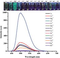
Highly selective strong turn-on fluorescence for Mg2+ (Φ = 0.03 to 0.57) was realized with a simple Salen based Schiff base chemosensor (1a) using dimethylformamide (DMF) or dimethyl sulfoxide (DMSO) as solvent. Importantly, Ca2+ that often interferes in the Mg2+ fluorescence sensing did not show any significant influence on the selectivity. The fluorescence sensing of Mg2+ is highly solvent as well as amine structure dependent. Fluorescence sensing of Mg2+ only by 1,2-phenylenediamine condensed Schiff bases in DMF or DMSO was observed. Different substituents (1b–e) on the salicylaldehyde unit were synthesized and we explored the effect of substitution on Mg2+ sensing and selectivity. Except 1c, other chemosensors showed selective fluorescence sensing of Mg2+ in DMF/DMSO. Interestingly, 1a–e (except 1c) exhibited selective strong turn-on fluorescence for Fe3+ (λmax = 462 nm, Φ = 0.421) in different solvents (DMSO, DMF, THF, CH3CN) after 1 h. The concentration dependent studies showed linear enhancement of fluorescence intensity for Mg2+ with the detection limit of 10−7 M. The practical applications of the chemosensor for selective sensing of Mg2+ in real samples such as pond, tap, river and ground water have also been demonstrated.


Introduction

Magnesium ions (Mg2+) are among the most abundant divalent cations in cells and play pivotal roles in many cellular processes, such as enzyme-driven biochemical reactions, proliferation of cells, and stabilization of DNA conformation.1 The estimated total concentration of Mg2+ in mammalian cells varies between 14 and 20 mM, the majority of it bound to ATP and a smaller amount to proteins, phospholipids, and various phosphometabolites.2 Mg2+ is also involved in many pathological processes, such as congestive heart failure, lung cancer, and muscle dysfunction.3 In contrast, high levels of Mg2+ are contribute to a number of age-related and neuronal diseases ranging from hypertension to Alzheimer's disease.4 The mechanisms by which Mg2+ concentration is regulated at the cellular level and the effects in human health has also been poorly understood due to the scarcity of efficient chemical tools for the study of this ion. As a result, the sensing of Mg2+ has generated increasing interest in the areas of chemical and biological sciences. Many analytical methods have been developed for the detection of Mg2+, including atomic absorption, ion-selective electrodes (ISEs), and NMR in the past few years.5 However, optical methods that track changes of fluorescence or absorption arisen from the Mg2+ induced perturbation of the chromophore, is best suitable for Mg2+ detection in biological systems.

Particularly, molecular chemosensors have drawn significant attention in recent years due to its convenience, real-time response, high sensitivity, selectivity, versatility and relatively simple handling.6 Coumarin based diketone or β-keto acid derivatives are the highly explored fluorescent sensors for selective detection of Mg2+ ions as well as molecular imaging of local changes in intracellular Mg2+ concentration.7 Dong et al. showed Na+ triggered coumarin based Salen fluorescence sensor for Mg2+.8 Crown ether derivatives,9 polymer based ligands10 and nanoparticles,11 have also been successfully employed for selective detection of Mg2+ ions. However, most of these chemosensor molecules require intricate synthetic methodologies and hence are relatively difficult to prepare. The major issues with many of the reported chemosensors for Mg2+ is their low selectivity against Zn2+ and Ca2+. The similar chemical properties of Mg2+ and Ca2+ make it difficult for chemosensor to distinguish them.12 Hence it is still interesting and of importance to design a highly selective and sensitive fluorescent sensor that can recognize Mg2+ without the interference from other metal ions.

The ease of synthesis coupled with tailorability, good biological activities, strong photophysical properties and coordination ability with metal ions has made Schiff bases as one of the most widely explored molecular chemosensors for selective sensing of metal ions.13 Particularly, Salen based π-conjugated tetradentate [O⁁N⁁N⁁O] chelating ligands have been widely employed for selective sensing of various metal ions such as Zn2+, Cu2+, Al3+, La3+, and Pt2+. The interaction of organic ligands with metal ions and resultant solid state structural organization are highly influenced by solvent, temperature, anions and pH.14 A subtle change of organic ligand structure or solvent resulted in completely different molecular organization and properties.14b,c Herein, we report the highly selective fluorescence sensing of Mg2+ ions by simple 1,2-phenylenediamine based Salen Schiff base chemosensor (1a–e) in DMF or DMSO. The chemosensor did not show fluorescence turn-on for Mg2+ other than DMF and DMSO solvent. Chemosensors with different substituent was prepared and studied their role on Mg2+ sensing and selectivity. Importantly, Ca2+ that often interferes in the Mg2+ fluorescence sensing did not show any significant influence on the selectivity. The linear enhancement of fluorescence with concentration of Mg2+ was observed. The chemosensors showed the detection limit of 10−7 M. The practical application of the chemosensor in selective sensing of Mg2+ in real samples such as pond, tap, and ground water have also been demonstrated.

Experimental section

1,2-phenylenediamine, ethylene diamine, (±)-trans-1,2-diaminocyclohexane (99%), salicylaldehyde, 2-hydroxy-4-methoxy benzaldehyde, 2-hydroxy-4-diethylamino benzaldehyde, 2,4-dihydroxy benzaldehyde and 2-hydroxynaphthaldehyde was obtained from Sigma-Aldrich. The spectroscopic grade solvents were obtained from Merck India. All chemicals are used as received. The metal ion solutions used for the fluorescence sensor experiments were prepared using Mill-Q water. The chemosensors were dissolved in organic solvents (DMF, DMSO, acetonitrile, methanol, tetrahydrofuran (THF)). Absorption and fluorescence spectra were recorded using Perking Elmer Lambda 1050 and Jasco fluorescence spectrometer-FP-8200 instruments.

General synthesis of 1a–e

Schiff bases were prepared according to the reported procedure.15 Typically, aldehyde (2.2 mmol, salicylaldehyde or 4-methoxysalicylaldehyde, 4-diethylamino-2-hydroxy benzaldehyde or 2,4-dihydroxy benzaldehyde) was dissolved in ethanol (30 mL) and stirred at room temperature. To this solution, 1,2-phenylenediamine (1 mmol) in ethanol (5 mL) was added in drop-wise under stirring. The immediate appearance of yellow colour indicates the formation of Schiff bases. The solution was allowed stir for another 6 h at room temperature that produced yellow to light yellow coloured precipitates. The formed precipitate was filtered and washed with ethanol and dried under vacuum.
1a (N, N′-bis-(salicylidine)-O-phenylenediamine). Yield = 87%. m.p. 160–162 °C. 1H NMR (400 MHz, d6-DMSO, δ ppm) 12.95 (s, 2H (OH)), 8.94 (s, 2H (CH[double bond, length as m-dash]N)), 7.66–7.68 (d, 2H (aromatic)), 7.40–7.48 (m, 6H (aromatic)), 6.95–7.00 (m, 4H (aromatic)). 13C NMR (200 MHz, d6-DMSO, δ ppm) 166.61, 160.54, 146.41, 133.50, 132.44, 130.24, 126.50, 122.70, 117.64, 116.96. C20H16N2O2 (316.12): calcd C 75.93, H 5.10, N 8.86; found C 75.57, H 5.31, N 8.67. m/z (LC-MS) 317.00 (M + H).
1b (N,N′-bis (4-methoxy salicylidine)-O-phenylenediamine). Yield = 85%. m.p. 174–176 °C. 1H NMR (400 MHz, d6-DMSO, δ ppm) 13.525 (s, 2H (OH)), 8.83 (s, 2H (CH[double bond, length as m-dash]N)), 7.53–7.55 (d, 2H (aromatic)), 7.41–7.44 (m, 2H (aromatic)), 7.34–7.37 (m, 2H (aromatic)), 6.53–6.56 (d, 2H (aromatic)), 6.48 (s, 2H (aromatic)), 3.80 (s, 6H (OCH3)). 13C NMR (200 MHz, d6-DMSO, δ ppm) 168.12, 166.76, 162.53, 144.50, 134.74, 133.94, 128.50, 118.80, 116.31, 55.84. C22H20N2O4 (376.14): calcd C 70.20, H 5.36, N 7.44; found C 70.11, H 5.23, N 7.52. m/z (LC-MS) 377.00 (M + H).
1c (N,N′-bis (4-diethylamino salicylidine)-O-phenylenediamine). Yield = 80%. m.p. 141–143 °C. 1H NMR (400 MHz, d6-DMSO, δ ppm) 13.58 (s, 2H (OH)), 8.61 (s, 2H (CH[double bond, length as m-dash]N)), 7.51–7.52 (d, 2H (aromatic)), 7.42–7.45 (m, 2H (aromatic)), 7.30–7.33 (m, 2H (aromatic)), 6.51–6.54 (d, 2H (aromatic)), 6.34 (s, 2H (aromatic)), 3.31–3.37 (q, 8H), 1.05–1.10 (t, 12H). 13C NMR (200 MHz, d6-DMSO, δ ppm) 163.45, 162.19, 155.12, 144.45, 134.22, 133.00, 128.92, 122.78, 117.78, 116.78, 43.73, 12.53. C28H34N4O2 (458.27): calcd C 73.33, H 7.47, N 12.22; found C 73.42, H 7.30, N 12.41. m/z (LC-MS) 459.20 (M + H).
1d (N,N′-bis (4-hydroxy salicylidine)-O-phenylenediamine). Yield = 80%. m.p. 266–269 °C (decomp.). 1H NMR (400 MHz, d6-DMSO, δ ppm) 13.40 (s, 2H (OH)), 10.30 (s, 2H (OH)), 8.75 (s, 2H (CH[double bond, length as m-dash]N)), 7.42–7.44 (d, 2H (aromatic)), 7.37–7.39 (m, 2H (aromatic)), 7.30–7.33 (m, 2H (aromatic)), 6.37–6.40 (d, 2H (aromatic)), 6.28 (s, 2H (aromatic)). 13C NMR (200 MHz, d6-DMSO, δ ppm) 164.88, 163.42, 161.87, 145.85, 133.33, 128.26, 122.32, 116.23, 115.90. C20H16N2O4 (348.11): calcd C 68.96, H 4.63, N 8.04; found C 68.80, H 4.45, N 8.20. m/z (LC-MS) 349.00 (M + H).
1e (N,N′-bis (2-hydroxy naphthalidine)-O-phenylenediamine). Yield = 80%. m.p. 284–287 °C (decomp.). 1H NMR (400 MHz, d6-DMSO, δ ppm) 15.13 (s, 2H (OH)), 9.70 (s, 2H (CH[double bond, length as m-dash]N)), 8.53–8.55 (d, 2H (aromatic)), 7.96–7.98 (d, 2H (aromatic)), 7.82–7.84 (d, 4H (aromatic)), 7.53–7.58 (t, 2H (aromatic)), 7.44–7.46 (t, 2H (aromatic)), 7.36–7.40 (t, 2H (aromatic)), 7.08 (s, 2H (aromatic)). 13C NMR (200 MHz, d6-DMSO, δ ppm) 166.74, 162.51, 147.90, 135.22, 134.40, 133.74, 133.02, 128.89, 127.78, 124.62, 122.86, 122.12, 118.88, 114.90. C28H20N2O2 (416.15): calcd C 80.75, H 4.84, N 6.73; found C 80.55, H 5.11, N 6.80. m/z (LC-MS) 417.00 (M + H).

Result and discussion

Schiff base chemosensors, 1a–e, were synthesized by quite straightforward condensation reaction between primary amine and an aldehyde precursor in ethanol solution at room temperature (Scheme 1,S1). The fluorescence responses of chemosensor 1a–e for various metal ions such as such as, Mg2+, Ca2+, Cr3+, Al3+, Fe2+, Cu2+, Co2+, Ni2+, Zn2+, Cd2+, Hg2+ and Pb2+ have been investigated in DMF, DMSO, acetonitrile, methanol and THF. The chemosensors and metal ions were dissolved in organic solvents (DMF, DMSO, acetonitrile and ethanol) and water, respectively.
image file: c4ra05827e-s1.tif
Scheme 1 Molecular structures of Schiff base chemosensors.

1a showed weak fluorescence in DMF (λmax = 499 nm, Φ = 0.0092). Addition of Mg2+ ions into 1a lead to the strong enhancement of fluorescence without altering the fluorescence λmax (Φ = 0.1787, Fig. 1). Importantly, other metal ions with 1a did not show any significant fluorescence enhancements. It is noted that Ca2+ addition showed only a small enhancement in the fluorescence intensity. Absorption studies of 1a with Mg2+ in DMF revealed distinct changes that suggest the formation of coordination complex between Mg2+ and 1a (Fig. 2). 1a in DMF showed a strong absorption at 270 nm and 330 nm. The former is assigned to π–π* transition involving the molecular orbitals of imine. As Scheme 2 describes, 1a exist in equilibrium between the two configurations (normal and tautomer) due to excited state intramolecular proton transfer (ESIPT).16 The absorption at 330 nm could be assigned to the transition of O[double bond, length as m-dash]C–C[double bond, length as m-dash]C–(NH).


image file: c4ra05827e-f1.tif
Fig. 1 Digital and fluorescence spectra of 1a in DMF with different metal ions.

image file: c4ra05827e-f2.tif
Fig. 2 Absorption spectra of 1a in DMF.

image file: c4ra05827e-s2.tif
Scheme 2 Mg-complex structure with 1a and tautomer formation by ESIPT.

Addition of Mg2+, Cu2+, Zn2+, Cd2+, Cr3+, Mn2+, Co2+ and Ni2+ reduced the intensity at 330 nm since the metal coordination prevents the ESIPT and tautomer formation (Scheme 2). The metal coordination with imine nitrogen that increase the electron withdrawing property of imine leads to the strong absorption at longer wavelength (380 nm to 440 nm).17 Mg2+ with 1a in DMF showed a strong red shifted absorption at 396 nm. 1a exhibited similar strong selective turn-on fluorescence with higher intensity for Mg2+ in DMSO (Table 1).

Table 1 Quantum yield (Φ) of 1a–e and with Mg2+ and Fe3+ in different solvents. Quinine sulfate in 0.5 M H2SO4 was used as reference
Compound Φ(DMF) ΦDMSO ΦCH3CN ΦTHF
a Measured immediately.b Measured after 1 h.
1a 0.0092 0.021
1b 0.0083 0.019
1d 0.015 0.034
1e 0.003 0.007
1a-Mg2+ 0.1787a 0.3603a
1b-Mg2+ 0.1811a 0.4194a
1d-Mg2+ 0.1861a 0.5767a
1e-Mg2+ 0.025a 0.088a
1a-Fe3+ 0.1318b 0.421b 0.353b 0.431b
1b-Fe3+ 0.0942b 0.406b 0.092b 0.316b
1d-Fe3+ 0.021b 0.193b 0.073b 0.174b
1e-Fe3+ 0.041b 0.073b 0.035b 0.122b


The strong enhancement of fluorescence upon Mg2+ coordination is due to the restriction of C[double bond, length as m-dash]N isomerisation and rigidification of fluorophore structure.18 It is noted that 1a showed strong turn-on fluorescence as well as absorption changes for Mg2+ either in DMF or DMSO. Other solvents such as acetonitrile, methanol and THF did not show any turn-on fluorescence for Mg2+. Absorption studies also showed that Mg2+ did not show any changes in other solvents with 1a (Fig. S1). This suggests that both DMF and DMSO facilitate the formation of stable Mg2+ coordination complex with 1a by involving the coordination. The possible structure of the Mg2+ complex with 1a is shown in Scheme 2. The concentration dependent studies of 1a with Mg2+ are shown in Fig. 3a. A steady fluorescence enhancement was observed with 1a in DMF (10−7 M) up to the addition of 1 equivalent of Mg2+ (10−7 M). These results suggest the formation of 1[thin space (1/6-em)]:[thin space (1/6-em)]1 coordination complex between 1a and Mg2+. It is noted that most of the reported Mg2+ chemosensor showed only micromolar (10−6 M) detection limit.8−11 The selectivity studies of 1a for Mg2+ in presence of other metal ions revealed that Ca2+, Al3+, Cd2+ and Hg2+ had negligible interference. But no turn-on fluorescence was observed for Mg2+ in presence of Zn2+. Transition metal ions (Cr3+, Fe3+, Mn2+, Co2+, Ni2+ and Cu2+) showed strong influence on the Mg2+ selectivity that could be due to the stronger coordination with 1a (Fig. 3b).


image file: c4ra05827e-f3.tif
Fig. 3 (a) 1a (10–6 M in DMF) fluorescence vs. concentration of Mg2+ (10–6 M in water) and (b) interference studies of other metal ions (mM) on the selectivity of Mg2+ (μM).

In order to study the effect of substitutional change on the selectivity and sensitivity of Mg2+, chemosensor 1b–e was synthesized (Scheme 1). Except 1c, other three chemosensors exhibited highly selective strong turn-on fluorescence for Mg2+ in DMF and DMSO (Fig. 4, S2). Surprisingly 1c did not show turn-on fluorescence for any metal ions in explored solvents. 1b showed a weak broad fluorescence with two peaks (480 nm and 504 nm). Addition of Mg2+ showed 10 fold fluorescence enhancements with a small red shift λmax (480 nm to 484 nm, Fig. 4a). 1d chemosensor exhibited weak and two clear fluorescence peaks at 520 nm and 547 nm. Addition of Mg2+ increases the fluorescence by 4 fold without affecting the fluorescence λmax (Fig. 4b). 1e chemosensor showed 30 fold fluorescence enhancements selectively for Mg2+ ions in DMF (Fig. S2). The quantum yield measurements revealed strong fluorescence for chemosensors with Mg2+ in DMSO compared to DMF (Table 1). Among the chemosensors, 1e with Mg2+ exhibited weak fluorescence both in DMF and DMSO and 1d with Mg2+ exhibited strong fluorescence in DMSO. The concentration dependent studies of 1b, 1d, 1e also showed steady enhancement of fluorescence up to the addition of 1 equivalent of Mg2+ (Fig. S3). However, the detection limit of Mg2+ was differed for 1e. 1a, 1b and 1d showed selective detection of Mg2+ up to the level of 10−7 M. But 1e showed selective turn-on fluorescence only for the addition of 10−4 M Mg2+. The selectivity studies of 1b, 1d and 1e for Mg2+ showed that Ca2+, a strong competitor with Mg2+, had little interference on the selectivity with all chemosensor in DMF (Fig. 5, S4). The transition metal ions had strong interference (no fluorescence turn-on) on the selectivity of Mg2+ that is due to the strong coordination character of these metal ions.


image file: c4ra05827e-f4.tif
Fig. 4 (a) 1b and (b) 1d fluorescence spectra with different metal ions in DMF.

image file: c4ra05827e-f5.tif
Fig. 5 Mg2+ interference studies of 1b and (b) 1d in presence of other metal cations in DMF.

The selective fluorescence sensing of Mg2+ by 1a–e is highly solvent dependent. Similarly, Mg2+ fluorescence sensing also depends on the structure of amine moiety in the chemosensor (Scheme S2). For example, use of ethylene diamine or cyclohexane diamine instead of ortho-phenylenediamine did not show any fluorescence sensing for Mg2+ in any solvents including DMF or DMSO. Instead, the ethylene or cyclohexane diamine based Schiff base chemosensor showed highly selective turn-on fluorescence for Zn2+ ions (Fig. S5–8). These results indicate that along with solvent the structural rigidity amine also plays a role in the formation of Mg2+ coordination complex. The practical applicability of 1a for selective sensing of Mg2+ ion in different samples such as river, ground, pond and tap water have also been demonstrated. Mg2+ was dissolved in different water (10−6 M) and addition of 100 μl into 1a in DMF (10−6 m) clearly showed the strong bluish green fluorescence (Fig. 6).


image file: c4ra05827e-f6.tif
Fig. 6 Selective sensing of Mg2+ by 1a in different water. 1a was dissolved in DMF (10–6 M) and Mg2+ (10−6 M) was dissolved in different water. 100 mL of Mg2+ was added into 1a-DMF.

Interestingly, 1a with Fe3+ also exhibited selectively strong blue fluorescence (λmax = 462 nm) in DMF/DMSO but after 1 h (Fig. 7). However, it takes 1 h to produce strong blue fluorescence with Fe3+. 1a-Fe3+ fluorescence is completely different from 1a-Mg2+ fluorescence. The absorption studies also revealed a clear change in the absorption after 1 h. A new peak appeared at 317 nm along with 330 nm peak. It is noted that the absorption peak of 1a has also been changed from broad to sharp (Fig. S9). It is noted that absorption spectra of 1a and 1a with Mg2+ did not show significant variation after one hour. Unlike Mg2+ that showed turn-on fluorescence only in DMSO and DMF, Fe3+ addition into 1a as well as 1b, 1d,e exhibited strong blue fluorescence in DMF, DMSO, acetonitrile and THF after 1 h (Fig. S10, Table 1).


image file: c4ra05827e-f7.tif
Fig. 7 The change of 1a (10−6 M in DMF) fluorescence with Fe3+ (10−6 M in water) with time.

Conclusion

We have demonstrated a highly selective strong turn-on fluorescence for Mg2+ (Φ = 0.03 to 0.57) with simple Salen based Schiff base chemosensors using DMF or DMSO as solvent. The solvent and rigidity of the amine structure was found to be critical for fluorescence sensing of Mg2+. The concentration dependent studies showed linear enhancement of fluorescence for Mg2+ with the detection limit of 10−7 M. Importantly, the chemosensors displayed good Mg2+ selectivity in presence of Ca2+ that often interferes in the Mg2+ fluorescence sensing. Salen chemosensors with different substitution in the salycylaldehyde unit (1b, 1d–e) also exhibited similar Mg2+ fluorescence sensing except small variation in the sensitivity. 1b and 1d showed strong turn-on fluorescence for Mg2+ with similar sensitivity whereas 1e exhibited reduced sensitivity (10−5 M). Thus simple Salen chemosensor have been effectively used to detect biologically important Mg2+ ions by changing the solvent medium. The practical application of the chemosensor in selective sensing of Mg2+ in real samples such as pond, tap, and ground water have also been demonstrated.

Acknowledgements

Financial supports from DST, New Delhi, India DST Fast Track Scheme no. SR/FT/CS-03/2011 (G), SR/FST/ETI-284/2011(c), SASTRA University (TRR Scheme) and instrumentation facility under CRF facility, SASTRA University are acknowledged with gratitude.

Notes and references

  1. A. Romani, Arch. Biochem. Biophys., 2007, 458, 90 CrossRef CAS PubMed; A. Sreedhara and J. A. Cowan, BioMetals, 2002, 15, 211 CrossRef; H. Rubin, Arch. Biochem. Biophys., 2007, 458, 16 CrossRef PubMed; T. Shoda, K. Kikuchi, H. Kojima, Y. Urano, H. Komatsu, K. Suzuki and T. Nagano, Analyst, 2003, 128, 719 RSC.
  2. A. M. P. Romani, Arch. Biochem. Biophys., 2011, 512, 1 CrossRef CAS PubMed; R. D. Grubbs, BioMetals, 2002, 15, 251 CrossRef.
  3. O. B. Stepura and A. I. Martynow, Int. J. Cardiol., 2009, 134, 145 CrossRef CAS PubMed; S. Mahabir, Q. Y. Wei, S. L. Barrera, Y. Q. Dong, C. J. Etzel, M. R. Spitz and M. R. Forman, Carcinogenesis, 2008, 29, 949 CrossRef PubMed; F. H. Nielsen and H. C. Lukaski, Magnesium Res., 2006, 19, 180 Search PubMed.
  4. R. M. Toyuz, Magnesium in clinical medicine, Front Biosci1, 2004, 1278 CrossRef CAS PubMed; Y. Rayssiguier, E. Gueux, W. Nowacki, E. Rock and A. Mazur, Magnesium Res., 2006, 19, 237 Search PubMed; M. Barbagallo and L. J. Dominguez, Arch. Biochem. Biophys., 2007, 458, 40 CrossRef PubMed.
  5. A. V. Zmozinski, A. de Jesus, M. G. R. Vale and M. M. Silva, Talanta, 2010, 83, 637 CrossRef CAS PubMed; A. de Jesus, A. V. Zmozinski, J. A. Barbar, M. G. R. Vale and M. M. Silva, Energy Fuels, 2010, 24, 2109 CrossRef; Z. Kilic, O. Acar, M. Ulasan and M. Ilim, Food Chem., 2002, 76, 107 CrossRef; Y. Luo, B. Zhang, M. Chen, J. Wang, X. Zhang, W. Y. Gao, J. F. Huang and W. L. J. Fu, J. Agric. Food Chem., 2010, 58, 9396 CrossRef PubMed; J. H. Shim, I. S. Jeong, M. H. Lee, H. P. Hong, J. H. On, K. S. Kim, H. S. Kim, B. H. Kim, G. S. Cha and H. Nam, Talanta, 2004, 63, 61 CrossRef PubMed; J. W. Zhu, Y. Qin and Y. H. Zhang, Anal. Chem., 2010, 82, 436 CrossRef PubMed; H. Hifumi, A. Tanimoto, D. Citterio, H. Komatsu and K. Suzuki, Analyst, 2007, 132, 1153 RSC; M. Ishida, Y. Naruta and F. Tani, Angew. Chem., Int. Ed., 2010, 49, 91 CrossRef PubMed; S. Patra and P. Paul, Dalton Trans., 2009, 8683 RSC.
  6. J. Zhang, R. E. Campbell, A. Y. Ting and R. Y. Tisen, Nat. Rev. Mol. Cell Biol., 2002, 3, 906 CrossRef CAS PubMed; A. P. de Silva, J. Eilers and G. Zlokarnik, Proc. Natl. Acad. Sci. U.S.A., 1999, 96, 8336 CrossRef.
  7. T. Fujii, Y. Shindo, K. Hotta, D. Citterio, S. Nishiyama, K. Suzuki and K. Oka, J. Am. Chem. Soc., 2014, 136, 2374 CrossRef CAS PubMed; S. C. Schwartz, B. Pinto-Pacheco, J. P. Pitteloud and D. Buccella, Inorg. Chem., 2014, 53, 3204 CrossRef PubMed; Y. Suzuki, H. Komatsu, T. Ikeda, N. Saito, S. Araki, D. Citterio, H. Hisamoto, Y. Kitamura, T. Kubota, J. Nakagawa, K. Oka and K. Suzuki, Anal. Chem., 2002, 74, 1423 CrossRef; T. Shoda, K. Kikuchi, H. Kojima, Y. Urano, H. Komatsu, K. Suzuki and T. Nagano, Analyst, 2003, 128, 719 RSC; H. Komatsu, N. Iwasawa, D. Citterio, Y. Suzuki, T. Kubota, K. Tokuno, Y. Kitamura, K. Oka and K. Suzuki, J. Am. Chem. Soc., 2004, 126, 16353 CrossRef PubMed; H. M. Kim, P. R. Yang, M. S. Seo, J.-S. Yi, J. H. Hong, S.-J. Jeon, Y.-G. Ko, K. J. Lee and B. R. Cho, J. Org. Chem., 2007, 72, 2088 CrossRef PubMed.
  8. Y. Dong, J. Li, X. Jiang, F. Song, Y. Cheng and C. Zhu, Org. Lett., 2011, 13, 2252 CrossRef CAS PubMed.
  9. G. Farruggia, S. Iotti, L. Prodi, M. Montalti, N. Zaccheroni, P. B. Savage, V. Trapani, P. Sale and F. I. Wolf, J. Am. Chem. Soc., 2006, 128, 344 CrossRef CAS PubMed; J. Kim, T. Morozumi and H. Nakamura, Org. Lett., 2007, 9, 4419 CrossRef PubMed; Y. Liu, M. Han, H.-Y. Zhang, L.-X. Yang and W. Jiang, Org. Lett., 2008, 10, 2873 CrossRef PubMed; X. Xiang, D. Wang, Y. Guo, W. Liu and W. Qin, Photochem. Photobiol. Sci., 2013, 12, 1232 Search PubMed.
  10. A. L. Ding, J. Pei, W. L. Yu, Y. H. Lai and W. Huang, Thin Solid Films, 2002, 417, 198 CrossRef CAS; J. Pei, W. L. Yu, A. L. Ding, W. L. Yu and Y. H. Lai, Macromol. Rapid Commun., 2002, 23, 21 CrossRef.
  11. E. J. Park, M. Brasuel, C. Behrend and M. A. Philbert, Anal. Chem., 2003, 75, 3784 CrossRef CAS.
  12. J. Pesco, J. Salmon, J. Vigo and P. Viallet, Anal. Biochem., 2001, 290, 221 CrossRef CAS PubMed; J. Kim, T. Morozumi, N. Kurumatani and H. Nakamura, Tetrahedron Lett., 2008, 49, 1984 CrossRef PubMed; A. D. Dubonosov, V. I. Minkin, V. A. Bren, E. N. Shepelenko, A. V. Tsukanov, A. G. Starikov and G. S. Borodkin, Tetrahedron, 2008, 64, 3160 CrossRef PubMed; S. P. Gromov, E. N. Ushakov and O. A. Fedorova, J. Org. Chem., 2003, 68, 6115 CrossRef PubMed.
  13. Y. Xu, J. Meng, L. Meng, Y. Dong, Y. Cheng and C. Zhu, Chem. - Eur. J., 2010, 16, 12898 CrossRef CAS PubMed; H. Y. Shrivastava and B. U. Nair, Anal. Bioanal. Chem., 2003, 375, 169 Search PubMed; Y. Zhou, H. N. Kim and J. Yoon, Bioorg. Med. Chem. Lett., 2010, 20, 125 CrossRef PubMed; W. H. Wu, J. F. Sun, S. M. Ji, W. T. Wu, J. Z. Zhao and H. M. Guo, Dalton Trans., 2011, 40, 11550 RSC; C. Gou, S. H. Qin, H. Q. Wu, Y. Wang, J. Luo and X. Y. A. Liu, Inorg. Chem. Commun., 2011, 14, 1622 CrossRef PubMed; I. P. Oliveri and S. Di Bella, J. Phys. Chem. A, 2011, 115, 14325 CrossRef PubMed; Y. Feng, J. H. Cheng, L. Zhou, X. G. Zhou and H. F. Xiang, Analyst, 2012, 137, 4885 RSC; L. Zhou, Y. Feng, J. H. Cheng, N. Sun, X. G. Zhou and H. F. Xiang, RSC Adv., 2012, 2, 10529 RSC.
  14. W. L. Leong and J. J. Vittal, Chem. Rev., 2011, 111, 688 CrossRef CAS PubMed; C.-P. Li and M. Du, Chem. Commun., 2011, 47, 5958 RSC; S. P. Anthony and T. P. Radhakrishnan, Cryst. Growth Des., 2004, 4, 1223 Search PubMed.
  15. D. Chen and A. E. Martell, Inorg. Chem., 1987, 26, 1026 CrossRef CAS.
  16. S. J. Formosinho and L. G. Arnaut, J. Photochem. Photobiol., A, 1993, 75, 21 CrossRef CAS; S. M. Ormson and R. G. Brown, Prog. React. Kinet., 1994, 19, 45 Search PubMed; S. R. Vázquez, M. C. R. Rodríguez, M. Mosquera and F. Rodríguez-Prieto, J. Phys. Chem. A, 2008, 112, 376 CrossRef PubMed; L. Wang, W. Qin, X. Tang, W. Dou and W. Liu, J. Phys. Chem. A, 2011, 115, 1609 CrossRef PubMed.
  17. S. D. Bella, I. Fragala, I. Ledoux, M. A. Diaz-Garcia and T. J. Marks, J. Am. Chem. Soc., 1997, 119, 9550 CrossRef CAS; C. Gou, S. H. Qin, H. Q. Wu, Y. Wang, J. Luo and X. Y. Liu, Inorg. Chem. Commun., 2011, 14, 1622 CrossRef PubMed.
  18. J. S. Wu, W.-M. Liu, X. Q. Zhuang, F. Wang, P. F. Wang, S. L. Tao, X. H. Zhang, S. K. Wu and S. T. Lee, Org. Lett., 2007, 9, 33 CrossRef CAS PubMed.

Footnote

Electronic supplementary information (ESI) available: Colorimetric absorption spectra for metal ions and turn-on fluorescence sensing by 1a–d and 2a–d. See DOI: 10.1039/c4ra5827e

This journal is © The Royal Society of Chemistry 2014
Click here to see how this site uses Cookies. View our privacy policy here.