DOI:
10.1039/C4RA05642F
(Paper)
RSC Adv., 2014,
4, 38293-38299
Highly specific DNA detection from massive background nucleic acids based on rolling circle amplification of target dsDNA†
Received
12th June 2014
, Accepted 30th July 2014
First published on 26th August 2014
Abstract
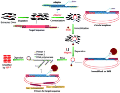
An advanced rolling circle amplification (RCA) strategy based on the target-circularization of the targeted double-stranded DNA (dsDNA) was established. Different from the traditional padlock-RCA, in which single-stranded probe DNA was circularized and amplified as signal amplification, our new approach could amplify a double-stranded DNA target. This special circularization of the target was realized by the ligation of target DNA with a biotin-labelled duplex adaptor containing 9 nt sticky ends by complementary base pairing. High specificity was obtained using two primers targeting the target sequence but not the probe itself in traditional padlock-RCA. With the help of streptavidin magnetic beads that immobilized the ligated dsDNA amplicon, the background nucleic acids contributing the most to non-specific amplification were eliminated. Under optimized conditions, less than 60 copies of the target sequence could be detected in the presence of massive background nucleic acids (>1012 copies of unrelated sequences). The sensitivity and specificity can rival canonical PCR. Without thermal cycles, the reduced handling and simpler equipment requirements render this assay a simple and rapid alternative to conventional methods. Based on these advantages, this method is a promising candidate in practical applications such as detecting contaminated food-borne pathogens in comprehensive food samples.
Introduction
DNA amplification is widely used as an essential approach in molecular biology, biotechnology, and gene detection. As a classic amplification method, PCR has been dominant in DNA diagnosis and detection for years. Its applications in gene engineering, diagnosis of infectious disease and environmental monitoring have been well documented.1 However, false positive results caused by non-target priming with mismatches, requirements for special equipment and sensitivity to temperature fluctuations limit its application as a low-cost and precise DNA detection strategy.2
Accordingly, several isothermal amplification approaches have been developed to overcome the shortcomings of PCR.3–8 Among them, the RCA approach has the advantages of not requiring an exquisite thermal cycler and being facile to conduct high-throughput detection.9,10 The specificity of padlock-RCA, a traditional RCA-based approach for DNA detection, depends on the specific hybridization of two terminal parts of a circularizable DNA (probe) with the target sequence.11 In padlock-RCA, RCA was employed as a signal amplification approach because the target sequence is merely used as a splint to circularize the probe, and the amplification products are the copies of the probe other than the target sequence.12–14 Nonetheless, even in the case that several mismatches are present at a position not close to the ligation site, the probe could still be circularized.15,16 Once the probe was circularized with the non-target sequence, non-specific amplification ensued. Therefore, false positive results may occur because the specificity of those protocols only depends on the circularization step. To detect a given DNA sequence, complicated denaturing and annealing steps need to be conducted.17 Moreover, when the target sequence coexists with a large amount of unrelated nucleic acid, padlock-RCA is inadequate for detecting the target sequence because the specificity merely depends on the circularization of the probe. Once the circular probes are formed on the non-target sequences, the RCA reaction can still conduct and deliver false positive results. Therefore, it is necessary to develop a new RCA strategy to overcome these disadvantages in practical applications.
Herein, we proposed a novel mode of RCA, dubbed as streptavidin magnetic beads (SMB) assisted target-circularization RCA (SMB-assisted TC-RCA). The target sequence, but not the padlock probe, was circularized into a double-stranded DNA circular amplicon utilizing a duplex adaptor. The two primers for hyper-branched RCA can then anneal to the target sequence to distinguish it from the non-target ones. Without denaturing and annealing processes, which were essential for padlock-RCA, the circularization of the target sequence could be realized and provided good reproducibility and specificity than that of the padlock probe. Furthermore, SMB was applied to remove the background nucleic acids to avoid non-specific amplification.
Material and methods
Reagents
All the oligonucleotides used in this work were synthesized by Sangon Company (Shanghai, China). Their sequences are listed in Table 1. All the adaptors used in this work were prepared by mixing two single-stranded oligonucleotides in a buffer solution. Taq DNA ligase, Phi29 DNA polymerase, Pfu DNA polymerase and Streptavidin magnetic beads (SMB) were purchased from New England Biolabs (Ipswich, USA). Fast Digest TspR I (recognition sequence: 5′-NNCASTGNN-3′) and Proteinase K were obtained from Thermo Scientific (Pittsburgh, USA). Deoxyribonucleotides (dNTPs), pUC-18 plasmid and ethidium bromide were purchased from Tiangen (Beijing, China). SYBR Green I was provided by Life Technologies (Carlsbad, USA). All other reagents were of analytical reagent grade. All the aqueous solutions were prepared using Milli-Q purified water (resistance of 18.2 MΩ cm−1).
Table 1 Sequence of the oligonucleotides used in this studya
Name |
Sequence (5′ → 3′) |
length (nt) |
‘p-’ indicates a phosphate modification and ‘b-’ indicates a biotin modification. The solid underline indicates the hybridization sequences between the adaptors and their corresponding target DNAs, and the recognition site of TspR I. All the oligonucleotides serving as primers (P1/P2) were phosphorothioate modified to prevent 3′-exonucleolytic degradation by Phi29 DNA polymerase. The modification sites are marked with ‘*’. By mixing two single strand oligonucleotides (F/R) together in buffer solution containing 2.0 mM Mg2+, the adaptors used in this work were prepared before the ligation reaction. |
105F |
b-TTTTTTTTTTTTTTTTTTTTTAAT ACGACTCACTATAGG![[T with combining low line]](https://www.rsc.org/images/entities/char_0054_0332.gif) ![[A with combining low line]](https://www.rsc.org/images/entities/char_0041_0332.gif) ![[C with combining low line]](https://www.rsc.org/images/entities/char_0043_0332.gif) ![[A with combining low line]](https://www.rsc.org/images/entities/char_0041_0332.gif) ![[G with combining low line]](https://www.rsc.org/images/entities/char_0047_0332.gif) ![[T with combining low line]](https://www.rsc.org/images/entities/char_0054_0332.gif) ![[G with combining low line]](https://www.rsc.org/images/entities/char_0047_0332.gif) ![[A with combining low line]](https://www.rsc.org/images/entities/char_0041_0332.gif) ![[G with combining low line]](https://www.rsc.org/images/entities/char_0047_0332.gif) |
48 |
105R |
p-CCTATAGTGAGTCGTATTACCCC CCCCCCCCCCCTATT![[A with combining low line]](https://www.rsc.org/images/entities/char_0041_0332.gif) ![[G with combining low line]](https://www.rsc.org/images/entities/char_0047_0332.gif) ![[C with combining low line]](https://www.rsc.org/images/entities/char_0043_0332.gif) ![[A with combining low line]](https://www.rsc.org/images/entities/char_0041_0332.gif) ![[C with combining low line]](https://www.rsc.org/images/entities/char_0043_0332.gif) ![[T with combining low line]](https://www.rsc.org/images/entities/char_0054_0332.gif) ![[G with combining low line]](https://www.rsc.org/images/entities/char_0047_0332.gif) ![[G with combining low line]](https://www.rsc.org/images/entities/char_0047_0332.gif) ![[G with combining low line]](https://www.rsc.org/images/entities/char_0047_0332.gif) |
47 |
271F |
b-TTTTTTTTTTTTTTTTTTTTTAAT ACGACTCACTATAGG![[G with combining low line]](https://www.rsc.org/images/entities/char_0047_0332.gif) ![[C with combining low line]](https://www.rsc.org/images/entities/char_0043_0332.gif) ![[C with combining low line]](https://www.rsc.org/images/entities/char_0043_0332.gif) ![[A with combining low line]](https://www.rsc.org/images/entities/char_0041_0332.gif) ![[C with combining low line]](https://www.rsc.org/images/entities/char_0043_0332.gif) ![[T with combining low line]](https://www.rsc.org/images/entities/char_0054_0332.gif) ![[G with combining low line]](https://www.rsc.org/images/entities/char_0047_0332.gif) ![[G with combining low line]](https://www.rsc.org/images/entities/char_0047_0332.gif) ![[T with combining low line]](https://www.rsc.org/images/entities/char_0054_0332.gif) |
48 |
271R |
p-CCTATAGTGAGTCGTATTACCCC CCCCCCCCCCCTATT![[T with combining low line]](https://www.rsc.org/images/entities/char_0054_0332.gif) ![[C with combining low line]](https://www.rsc.org/images/entities/char_0043_0332.gif) ![[C with combining low line]](https://www.rsc.org/images/entities/char_0043_0332.gif) ![[A with combining low line]](https://www.rsc.org/images/entities/char_0041_0332.gif) ![[C with combining low line]](https://www.rsc.org/images/entities/char_0043_0332.gif) ![[T with combining low line]](https://www.rsc.org/images/entities/char_0054_0332.gif) ![[G with combining low line]](https://www.rsc.org/images/entities/char_0047_0332.gif) ![[A with combining low line]](https://www.rsc.org/images/entities/char_0041_0332.gif) ![[G with combining low line]](https://www.rsc.org/images/entities/char_0047_0332.gif) |
47 |
347F |
b-TTTTTTTTTTTTTTTTTTTTTAAT ACGACTCACTATAGG![[C with combining low line]](https://www.rsc.org/images/entities/char_0043_0332.gif) ![[C with combining low line]](https://www.rsc.org/images/entities/char_0043_0332.gif) ![[C with combining low line]](https://www.rsc.org/images/entities/char_0043_0332.gif) ![[A with combining low line]](https://www.rsc.org/images/entities/char_0041_0332.gif) ![[G with combining low line]](https://www.rsc.org/images/entities/char_0047_0332.gif) ![[T with combining low line]](https://www.rsc.org/images/entities/char_0054_0332.gif) ![[G with combining low line]](https://www.rsc.org/images/entities/char_0047_0332.gif) ![[C with combining low line]](https://www.rsc.org/images/entities/char_0043_0332.gif) ![[T with combining low line]](https://www.rsc.org/images/entities/char_0054_0332.gif) |
48 |
347R |
p-CCTATAGTGAGTCGTATTACCCC CCCCCCCCCCCTATT![[A with combining low line]](https://www.rsc.org/images/entities/char_0041_0332.gif) ![[A with combining low line]](https://www.rsc.org/images/entities/char_0041_0332.gif) ![[C with combining low line]](https://www.rsc.org/images/entities/char_0043_0332.gif) ![[A with combining low line]](https://www.rsc.org/images/entities/char_0041_0332.gif) ![[T with combining low line]](https://www.rsc.org/images/entities/char_0054_0332.gif) ![[G with combining low line]](https://www.rsc.org/images/entities/char_0047_0332.gif) ![[C with combining low line]](https://www.rsc.org/images/entities/char_0043_0332.gif) ![[G with combining low line]](https://www.rsc.org/images/entities/char_0047_0332.gif) ![[C with combining low line]](https://www.rsc.org/images/entities/char_0043_0332.gif) |
47 |
506F |
b-TTTTTTTTTTTTTTTTTTTTTAAT ACGACTCACTATAGG![[C with combining low line]](https://www.rsc.org/images/entities/char_0043_0332.gif) ![[T with combining low line]](https://www.rsc.org/images/entities/char_0054_0332.gif) ![[C with combining low line]](https://www.rsc.org/images/entities/char_0043_0332.gif) ![[A with combining low line]](https://www.rsc.org/images/entities/char_0041_0332.gif) ![[C with combining low line]](https://www.rsc.org/images/entities/char_0043_0332.gif) ![[T with combining low line]](https://www.rsc.org/images/entities/char_0054_0332.gif) ![[G with combining low line]](https://www.rsc.org/images/entities/char_0047_0332.gif) ![[A with combining low line]](https://www.rsc.org/images/entities/char_0041_0332.gif) ![[C with combining low line]](https://www.rsc.org/images/entities/char_0043_0332.gif) |
48 |
506R |
p-CCTATAGTGAGTCGTATTACCCC CCCCCCCCCCCTATT![[G with combining low line]](https://www.rsc.org/images/entities/char_0047_0332.gif) ![[C with combining low line]](https://www.rsc.org/images/entities/char_0043_0332.gif) ![[C with combining low line]](https://www.rsc.org/images/entities/char_0043_0332.gif) ![[A with combining low line]](https://www.rsc.org/images/entities/char_0041_0332.gif) ![[G with combining low line]](https://www.rsc.org/images/entities/char_0047_0332.gif) ![[T with combining low line]](https://www.rsc.org/images/entities/char_0054_0332.gif) ![[G with combining low line]](https://www.rsc.org/images/entities/char_0047_0332.gif) ![[G with combining low line]](https://www.rsc.org/images/entities/char_0047_0332.gif) ![[C with combining low line]](https://www.rsc.org/images/entities/char_0043_0332.gif) |
47 |
958F |
b-TTTTTTTTTTTTTTTTTTTTTAAT ACGACTCACTATAGG![[A with combining low line]](https://www.rsc.org/images/entities/char_0041_0332.gif) ![[G with combining low line]](https://www.rsc.org/images/entities/char_0047_0332.gif) ![[C with combining low line]](https://www.rsc.org/images/entities/char_0043_0332.gif) ![[A with combining low line]](https://www.rsc.org/images/entities/char_0041_0332.gif) ![[C with combining low line]](https://www.rsc.org/images/entities/char_0043_0332.gif) ![[T with combining low line]](https://www.rsc.org/images/entities/char_0054_0332.gif) ![[G with combining low line]](https://www.rsc.org/images/entities/char_0047_0332.gif) ![[C with combining low line]](https://www.rsc.org/images/entities/char_0043_0332.gif) ![[A with combining low line]](https://www.rsc.org/images/entities/char_0041_0332.gif) |
48 |
958R |
p-CCTATAGTGAGTCGTATTACCCC CCCCCCCCCCCTATT![[G with combining low line]](https://www.rsc.org/images/entities/char_0047_0332.gif) ![[G with combining low line]](https://www.rsc.org/images/entities/char_0047_0332.gif) ![[C with combining low line]](https://www.rsc.org/images/entities/char_0043_0332.gif) ![[A with combining low line]](https://www.rsc.org/images/entities/char_0041_0332.gif) ![[C with combining low line]](https://www.rsc.org/images/entities/char_0043_0332.gif) ![[T with combining low line]](https://www.rsc.org/images/entities/char_0054_0332.gif) ![[G with combining low line]](https://www.rsc.org/images/entities/char_0047_0332.gif) ![[G with combining low line]](https://www.rsc.org/images/entities/char_0047_0332.gif) ![[C with combining low line]](https://www.rsc.org/images/entities/char_0043_0332.gif) |
47 |
GyrBF |
b-TTTTTTTTTTTTTTTTTTTTTAAT ACGACTCACTATAGG![[T with combining low line]](https://www.rsc.org/images/entities/char_0054_0332.gif) ![[T with combining low line]](https://www.rsc.org/images/entities/char_0054_0332.gif) ![[C with combining low line]](https://www.rsc.org/images/entities/char_0043_0332.gif) ![[A with combining low line]](https://www.rsc.org/images/entities/char_0041_0332.gif) ![[G with combining low line]](https://www.rsc.org/images/entities/char_0047_0332.gif) ![[T with combining low line]](https://www.rsc.org/images/entities/char_0054_0332.gif) ![[G with combining low line]](https://www.rsc.org/images/entities/char_0047_0332.gif) ![[G with combining low line]](https://www.rsc.org/images/entities/char_0047_0332.gif) ![[T with combining low line]](https://www.rsc.org/images/entities/char_0054_0332.gif) |
48 |
GyrBR |
p-CCTATAGTGAGTCGTATTACCCC CCCCCCCCCCCTATT![[T with combining low line]](https://www.rsc.org/images/entities/char_0054_0332.gif) ![[G with combining low line]](https://www.rsc.org/images/entities/char_0047_0332.gif) ![[C with combining low line]](https://www.rsc.org/images/entities/char_0043_0332.gif) ![[A with combining low line]](https://www.rsc.org/images/entities/char_0041_0332.gif) ![[G with combining low line]](https://www.rsc.org/images/entities/char_0047_0332.gif) ![[T with combining low line]](https://www.rsc.org/images/entities/char_0054_0332.gif) ![[G with combining low line]](https://www.rsc.org/images/entities/char_0047_0332.gif) ![[G with combining low line]](https://www.rsc.org/images/entities/char_0047_0332.gif) ![[A with combining low line]](https://www.rsc.org/images/entities/char_0041_0332.gif) |
47 |
105P1 |
GGCAACTATGGAT*G*A |
15 |
105P2 |
ATAACTACGATAC*G*G |
15 |
271P1 |
GTAGCCGTAGTTA*G*G |
15 |
271P2 |
CAAGCAGCAGATT*A*C |
15 |
347P1 |
ATACCAAACGACG*A*G |
15 |
347P2 |
CAAGGCGAGTTAC*A*T |
15 |
506P1 |
ACCTACACCGAAC*T*G |
15 |
506P2 |
ACGCAGGAAAGAA*C*A |
15 |
958P1 |
ACGAGTGGGTTAC*A*T |
15 |
958P2 |
AGATGCGTAAGGA*G*A |
15 |
GyrBP1 |
ATTGTGGAGGGTG*A*C |
15 |
GyrBP2 |
AAGCCTTCTTTAT*C*C |
15 |
Digestion by TspR I restriction enzyme
For sample digestion, 1.0 μL of the target DNA (varies in concentration) mixed with 1.0 μL of oyster genomic DNA (600 ng μL−1) was incubated with 1.0 μL Fast Digest TspR I in 10 μL 1 × Fast Digest buffer (Thermo Scientific, the buffer composition was not disclosed) at 65 °C for 10 min. The solution was then stored at −18 °C for subsequent experiments.
Ligation for circularization
10 μL of digested product was introduced into 20 μL of 1 × ligation buffer containing 10 nM specific biotin labelled adaptors, 10 mM Tris–HCl, 25 mM KAc, 10 mM Mg(Ac)2, 10 mM DTT, 1.0 mM NAD (pH 7.8), and 0.1% Triton. Prior to the addition of Taq DNA ligase, the mixture was incubated at 50 °C for 5 min, and then 2.0 U Taq DNA ligase was added. The mixture was incubated at 45 °C for 12 h. After ligation, 0.6 μL of SMB suspension was added to the solution to immobilize the dsDNA amplicon at room temperature for 5 min. The magnetic separation rack was then employed to isolate the beads from the solution. The beads were washed with 1 × Phi29 DNA polymerase buffer for three times. The products (circular amplicons) were then pipetted to a new Eppendorf tube for the RCA reaction.
RCA reaction
For the RCA reaction, 10 μL of product was mixed with 2.0 μL 10 × reaction buffer [500 mM Tris–HCl, 100 mM MgCl2, 10 mM (NH4)2SO4 and 40 mM DTT (pH 7.9)], 5.0 U Phi29 DNA polymerase, 4.0 μL dNTPs (2.5 mM), 0.5 μL forward primer and 0.5 μL reverse primer (10 μM) in a total volume of 20 μL. The reaction mixture was vortexed and incubated at 30 °C for 16 h. After amplification, Phi29 DNA polymerase was inactivated at 65 °C for 10 min.
Analysis of amplification products
The RCA products with concatenated sequence copies were digested with TspR I. A 2.0 μL aliquot of the product was digested by 1.0 μL Fast Digest TspR I at 65 °C for 2.0 h in 20 μL volume. The digested products (final products) were analyzed on 8.0% PAGE electrophoresis, stained by ethidium bromide and photographed under ultraviolet illumination at 302 nm with a Bio-rad molecular imager.
Preparation of genomic DNA
Vibrio Parahaemolyticus (ATCC 17802) was cultured in 5 mL of brain heart infusion broth at 37 °C overnight. Genomic DNA from each culture was extracted and purified with a Tiangen Fungal/Bacterial DNA extract kit following the manufacturer's protocol and quantified by measuring the optical density at 260 nm with a Nano Drop 2000 (Thermo Fisher Scientific).
Detection of Vibrio Parahaemolyticus in the presence of oyster genomic DNA
The gradient dilution of the abovementioned obtained genomic DNA was applied to obtain Vibrio parahaemolyticus DNA with various concentrations. Vibrio Parahaemolyticus gyrase subunit protein B (GyrB) gene was chosen to be the target DNA sequence. A 1.0 μL aliquot of Vibrio parahaemolyticus DNA (varies in concentration) was mixed with 1.0 μL of oyster genomic DNA (600 ng μL−1) for detection with TC-RCA according to the procedure described above.
Results and discussion
Principle and basic design of SMB-assisted TC-RCA
SMB-assisted target circularization RCA (TC-RCA) was developed to provide an effective method for dsDNA detection in the presence of massive background nucleic acids. As shown in Fig. 1, the 347 bp fragment from pUC-18 was used as an example to demonstrate the principle of TC-RCA. The target dsDNA fragment was circularized using a biotinylated duplex adaptor, which was designed to have two sticky ends complementary to that of the target sequence. After circularization, SMB is used to eliminate the background DNA and DNA ligase. The hyper-branched RCA was carried out with two primers annealing to the target sequence on the circular amplicon. The following three components are essential for obtaining desirable specificity. One is the specific hybridization of the sticky ends between the adaptor and target fragment. The second is specific priming during hyper-branched RCA by two primers complementary to the target sequence. The third is eliminating the interference of background DNA by removing them with SMB. For adaptor and primer design, A T20 linker was introduced to serve as a spacer eliminating the steric effect on the beads.18 A C15 gap was incorporated to promote the rate of DNA synthesis at the beginning stage.19,20 The two primers were phoshphorothioate-modified to prohibit the 3′-exonuclease activity of Phi29 DNA polymerase.21
 |
| Fig. 1 Detailed illustration of the adaptor and circularization fashion. The 347 bp sequence from plasmid pUC-18 with TspR I recognition sites at both the ends was used as the target sequence. The target sequence was cut by TspR I (recognition site: 5′-NNCASTGNN-3′) to release the digested fragment with 9 nt sticky ends. The biotinylated adaptor, whose sticky ends were complementary to that of the target sequence, was introduced to form a duplex circle. The exponential RCA reaction was initiated by two primers specifically targeting the sequence of interest. | |
At the end of the RCA reaction, concatenated copies containing the target sequence and adaptor one after another were generated. The products can be cut by TspR I to obtain the copies of the target DNA with the same size as the original fragment. Hence, the amplified target sequence could be obtained. The scheme in Fig. 2 shows the procedures of the SMB-assisted TC-RCA.
 |
| Fig. 2 Scheme of SMB-assisted TC-RCA. A digestion step conducted by restriction endonuclease TspR I was employed to generate numerous shorter fragments, including the target DNA. Taq DNA ligase then ligates the biotinylated adaptor and the target sequence to form a dsDNA amplicon. SMB was applied to immobilize the amplicon via biotin–streptavidin bond, and the background DNA was eliminated by magnetic separation. Phi29 DNA polymerase with two specific primers was used to drive hyper-branched RCA. Finally, the products consisting of the tandem copies of the dsDNA target were digested by the same restriction endonuclease. Hence, the target DNA meant to be replicated or detected was obtained with the augmentation of at least eight magnitudes. | |
Detection of target sequences in the presence of background DNA
As shown in Fig. 2, our approach involves the following steps: (1) digestion of sample DNA by TspR I; (2) circularization of target DNA by Ligation; (3) removing the background DNA by magnetic beads; (4) hyper-branched RCA amplification; (5) digestion of RCA products by TspR I. First, we used a 347 bp fragment from plasmid pUC-18 as the target sequence. Approximately 600 ng of oyster genomic DNA was added during the first digestion step as background DNA in a 10 μL volume. This was equivalent to 106 copies of oyster genomic DNA.
The double-stranded adaptor 347F/347R was used to circularize the 347 bp sequence (see Table 1 for sequences). Taq DNA ligase was used for its high specificity, and the ligation could be carried out at 45 °C. Hyper-branched RCA was carried out using two primers (P1/P2) and Phi29 DNA polymerase at 30 °C. Phi29 DNA polymerase was applied for its isothermal nature, good processive synthesis ability and robust strand displacement property.22 Table 1 lists the sequence of the oligonucleotides used in this study.
After the RCA reaction, the amplification products were cut by TspR I and analyzed with PAGE electrophoresis. As shown in Fig. 3A, clear bands were observed and the intensity lowered gradually with decreasing copy number. The band position deviated slightly from the typical duplex ladder which had blunt ends because the target sequence featured 9 nt sticky ends. Further analysis showed that the amplification products had the same size as the original 347 bp fragment from pUC-18 (Fig. 3B). This indicated that the target sequence was amplified from the massive background DNA with high specificity and sensitivity. The high specificity can be attributed to the novel target-circularization mode, where a target sequence other than the probe was incorporated into the circular amplicon. By this novel design, the highly specific exponential RCA could be carried out because two primers were complementary to the target sequence but not to the non-target ones. Furthermore, the ligation reaction was sticky-end specific and immune to the interference imposed by background nucleic acid sequences, which may be used as a “candidate template” for primers in PCR.23 The specificity was provided not only by ligation-dependent circularization but also by target-specific amplification. Therefore, two steps of screening enabled our protocol to detect the specific target sequence from massive background DNA under isothermal conditions. Considering that the protocol could amplify the target sequence without triggering unexpected amplification under optimized conditions, SYBR Green I staining was used to directly detect the amplified product (Fig. 3C). Hence, the digestion of the amplification products and electrophoresis analysis could be saved, enhancing the efficiency of the detection.
 |
| Fig. 3 Detection of 347 bp target sequence by SMB-assisted TC-RCA. (A) Final products of the target sequences with various concentrations in the presence of 600 ng oyster genomic DNA. (B) Comparison of the pUC-18 plasmid digested fragments and 347 bp final products. (C) Amplification products analyzed by SYBR Green I staining. A 1.0 μL aliquot of the RCA product solution was mixed with 1.0 μL 10 × SYBR Green I nucleic acid dye and photographed under ultraviolet illumination at 520 nm on the Bio-rad molecular imager. (D) Final products of the reactions with all or partial reaction components. DNA, POL and LIG indicate sample DNA, Phi29 DNA polymerase and Taq DNA ligase, respectively. The concentration of the target sequence was 6 × 10². | |
To confirm whether the amplification was carried out according to our design, control reactions without either of the enzymes were conducted. As expected, RCA products were observed only when both the enzymes existed (Fig. 3D). When DNA ligase was absent, no product was observed after 20 h of primer extension, indicating that no closed circle existed as RCA template. However, when the primer extension proceeded for more than 48 h, disordered products emerged and could not be digested by TspR I (data not shown). The ab initio DNA synthesis probably resulted in products, suggesting that a longer reaction duration should not be applied.24 In addition, the control experiment (Fig. 3D, DNA sample missing) did not produce any product, suggesting that the observed band was the amplification products from the target sequence. A control experiment was performed to confirm the specificity of target circularization. The defined adaptor exclusively circularized its target sequence (Fig. S5, ESI†), while the other digested sequences with similar sticky ends were still linear as their original form. Therefore, the ligation reaction is capable of screening the non-target sequences by specific circularization.
Four other pUC-18 fragments were used as targets to check the generality of the protocol. As shown in Fig. 4, a single clean band was observed for each target sequence. The detection limit for 271 bp, 506 bp and 958 bp was also 60 copies. For the 105 bp fragment, the detection limit was 600 copies, probably because dsDNA features the persistent length of around 50 nm (150 bp), and the 105 bp fragment was not long enough to be circularized efficiently.25 The data suggested that a longer target sequence (>200 bp) should be adopted to avoid false negative results.
 |
| Fig. 4 Detection of the target sequences with various lengths by SMB-assisted TC-RCA. The copy number of the target sequences was 6 × 10² in lanes 1, 4, 7, 10; 60 in lanes 2, 5, 8, 11 and 0 (negative control) in lane 3, 6, 9, 12. | |
To investigate the need to remove the background DNA by SMB, an assay without SMB was performed. Unexpected bands emerged and then intensified with decreasing target sequence copies, especially for negative control (Fig. S1, ESI†). Competition for added adaptors between the target sequence and the non-target ones was expected to account for non-specific amplification. On the other hand, because Taq DNA ligase cannot be easily inactivated, it may still be working during the amplification reaction. However, the conditions in the amplification reaction were unsuitable for Taq DNA ligase, thus nonspecific circular amplicons could be generated and led to non-specific amplifications.26,27 For SMB-assisted TC-RCA, factors causing non-specific amplifications, such as the endonuclease (TspR I: cutting the target circular amplicons) and the thermostable ligase (Taq DNA ligase: ligating non-target sequences), as well as RCA inhibitors, such as NAD+ and unrelated DNA fragments, were eliminated.28 In addition, SMB immobilized adaptors whose sticky ends might be used as potential primers to initiate the non-specific hyper-branched RCA reaction. The target-specific circularization and SMB separation jointly contribute to the high specificity of our protocol. One may doubt that the non-specific adsorption of DNAs to magnetic beads could affect the specific detection. In fact, because the beads were only used in the separation step (Fig. 2), the DNAs absorbed on the magnetic beads could not have an impact on the detection reaction.29 In addition, the amount of DNAs clinching to the beads was tiny compared to the background DNAs (Fig. S2, ESI†). Therefore, the non-specific adsorption of DNAs to magnetic beads was negligible in this protocol.
To compare with the other RCA protocol, an experiment detecting a target sequence using padlock-RCA was carried out. The padlock-probe was designed to target the 36 nt region in the 347 bp sequence. As expected, the padlock protocol was unable to specifically amplify the target sequence in the presence of massive background DNA (Fig S6, ESI†). This was attributed to the fact that the specificity of padlock-RCA merely comes from the ligation reaction, which was unable to exclude non-target circularization once the massive background DNA was present.
Detection of bacterial genomic DNA in the presence of background DNA
The detection of food borne pathogens, such as bacteria and viruses, is indispensable for food security monitoring.30 However, the DNA extracted from pathogens and their hosts are mixed and cannot be separated by routine operations. To detect a specific pathogen's gene sequence, the DNA from their host, considered to be background nucleic acids, may impose intense interference on the detection reaction. As an amplification strategy featuring the anti-interference potential, SMB-assisted TC-RCA is promising to be applied in this area. A duplex adaptor and a set of primers were designed (Fig. 5) to detect a specific gene, GyrB (GeneBank accession: AY527390.1), encoding gyrase subunit protein B (GyrB) of Vibrio Parahaemolyticus in the presence of oyster genomic DNA. A BLAST (http://blast.ncbi.nlm.nih.gov) search of the sticky end sequences and primer targeting sites against oyster (Crassostrea gigas) genome showed no precise match for both the adaptor and primers entirely.
 |
| Fig. 5 Specific adaptor and primers for the detection of pathogenic Vibrio parahaemolyticus. The sticky ends of the adaptor were perfectly complementary with that of the target sequence (565 bp) from the GyrB gene of Vibrio Parahaemolyticus. The primers were designed to amplify the specific target sequence. | |
0.1–100 pg of Vibrio Parahaemolyticus DNA in the presence of 600 ng oyster genomic DNA was detected by SMB-assisted TC-RCA. As shown in Fig. 6A, the band intensity decreased with decreasing target sequence. The detection limit was 1.0 pg, indicating that ca. 180 copies of GyrB gene sequence were amplified practically. The sensitivity was lower than that of the plasmid sequence, probably because the intact genomic DNA sequence (ca. 5 × 10⁶ bp) of the bacterium was exposed to intensive shearing force action during the extraction process, severing the genomic DNA into shorter fragments.31 Some copies of the GyrB gene sequence may also be cut, decreasing the number of available templates. The results were also obtained by SYBR Green I staining at the end of the RCA step (Fig. 6B). This was equivalent to detecting a single bacterial cell out of 104 oyster cells. The innovative circularization enabled the GyrB sequence to incorporate into a circular amplicon and to be amplified specifically. A padlock probe and two corresponding primers were designed to detect bacterial DNA from the massive background DNA. Without background DNA, 103 copies of GyrB gene sequence can be detected. However, when 600 ng background DNA was incorporated, only non-specific amplifications were observed (Fig S7, ESI†). Non-specific circularization of the padlock probe was responsible for false negative results, indicating that the method was not applicable for detecting bacterial DNA from massive background DNA.
 |
| Fig. 6 Detection of Vibrio parahaemolyticus DNA in the presence of background DNA by SMB-assisted TC-RCA. (A) Final products of amplified GyrB gene sequence with various concentrations in the presence oyster genomic DNA (B) Amplification products analyzed by SYBR Green I staining. Aliquot 1.0 μL RCA product solution was mixed with 1.0 μL 10 × SYBR Green I and photographed under ultraviolet illumination at 520 nm on a Bio-rad molecular imager. | |
Comparison with PCR for plasmid and Vibrio Parahaemolyticus detection
We studied the PCR-mediated detection in the presence of massive background DNA. The primers were designed using NCBI primer tools. The selected primers were subjected to Primer-BLAST (http://www.ncbi.nlm.nih.gov/tools/primer-blast) against oyster genomic DNA to ensure that they were specific to the target sequence (plasmid or GyrB). Routine PCR could amplify plasmid sequence without background DNA but failed to detect them in the presence of background DNA (Fig. S3A, ESI†). This may be caused by intense competition for primers between target templates and unrelated nucleic acids, which was 106–108 fold more than the target ones and PCR inhibitor in extracted DNA sample.32 Two or three orders of magnitude gradient dilution that reduced the concentration of background DNA and PCR inhibitors was conducive to PCR detection (Fig. S3B, ESI†). However, when the target sequence had a low copy number (less than 103 copies), PCR failed to amplify the target sequence even with more cycles (more than 40 cycles). This indicated that the gradient dilution could not be applied because of the possibility of causing false negative results. For Vibrio Parahaemolyticus detection, three sets of optimized primers were used (Fig. S4, ESI†). The results were similar to those of plasmid detection, indicating that the background DNAs were adverse to routine PCR detection. Therefore, the data conversely highlights the anti-interference ability of the RCA-based strategy, which logically could be used to analyze the exogenous gene from the host DNA in a genetics study.
To amplify a DNA fragment with a designated length using PCR, the primers need to anneal at fixed sites, which possibly were considered to be an unsatisfactory priming region. This may become a hindrance if the sequence of interest is short and cannot provide desirable priming sites.33 In the proposed protocol, the potential priming sites had much more choice because the primers targeting any site of circular amplicon can be extended.34 Ruling out unacceptable priming sites in terms of Tm, secondary structure and cross priming potential, available binding regions were still sufficient to amplify the specific sequence by SMB-assisted TC-RCA. This feature facilitated us to confirm one amplicon with different sets of primers and exclude the non-specific amplification by cross-referencing all the results.35
Conclusion
SMB-assisted TC-RCA was proved to be applicable to amplify the target sequences under isothermal conditions with good sensitivity and specificity. Compared to PCR, our protocol avoids a complicated thermal cycling program, and heating in a period of time using a simple incubator is sufficient to amplify DNA to detectable levels. Compared to padlock-RCA, denaturing and annealing steps are not required, and the amplification of the target sequence itself with more stability and specificity is accomplished. The high specificity endowed by a specific adaptor and primers enables the detection of the target sequence in the presence of massive background DNA under isothermal conditions. This protocol is promising to be used for detecting harmful bacteria in food material and products, as well as analyze exogenous gene expression in a genetics study. By employing random primer and general primer binding the adaptor region, sequences with the same sticky end could be amplified simultaneously.36
Funding
This work was supported by “Fund for Distinguished Young Scholars” of Shandong province [JQ201204], Program for Changjiang Scholars and Innovative Research Team in University [IRT1188], and “National Youth Qianren Plan”.
Acknowledgements
We thank Prof. Haijin Mou at Ocean University of China for supplying Vibrio parahaemolyticus.
Notes and references
- T. Avni, L. Leibovici and M. Paul, J. Clin. Microbiol., 2011, 49, 665–670 CrossRef PubMed.
- V. V. Demidov, Expert Rev. Mol. Diagn., 2002, 2, 542–548 CrossRef CAS PubMed.
- G. Nallur, C. Luo, L. Fang, S. Cooley, V. Dave, J. Lambert, K. Kukanskis, S. Kingsmore, R. Lasken and B. Schweitzer, Nucleic Acids Res., 2001, 29, e118 CrossRef CAS PubMed.
- T. Notomi, H. Okayama, H. Masubuchi, T. Yonekawa, K. Watanabe, N. Amino and T. Hase, Nucleic Acids Res., 2000, 28 Search PubMed.
- J. Compton, Nature, 1991, 350, 91–92 CrossRef CAS PubMed.
- N. Kurn, P. C. Chen, J. D. Heath, A. Kopf-Sill, K. M. Stephens and S. L. Wang, Clin. Chem., 2005, 51, 1973–1981 CAS.
- M. Vincent, Y. Xu and H. M. Kong, EMBO Rep., 2004, 5, 795–800 CrossRef CAS PubMed.
- G. T. Walker, M. S. Fraiser, J. L. Schram, M. C. Little, J. G. Nadeau and D. P. Malinowski, Nucleic Acids Res., 1992, 20, 1691–1696 CrossRef CAS PubMed.
- M. Nilsson, Histochem. Cell Biol., 2006, 126, 159–164 CrossRef CAS PubMed.
- T. Konry, R. B. Hayman and D. R. Walt, Anal. Chem., 2009, 81, 5777–5782 CrossRef CAS PubMed.
- S. P. Jonstrup, J. Koch and J. Kjems, RNA, 2006, 12, 1747–1752 CrossRef CAS PubMed.
- J. H. Smith and T. P. Beals, PLoS One, 2013, 8, e65053 CAS.
- X. Zhou, Q. Su and D. Xing, Anal. Chim. Acta, 2012, 713, 45–49 CrossRef CAS PubMed.
- J. Y. Marciniak, A. C. Kummel, S. C. Esener, M. J. Heller and B. T. Messmer, BioTechniques, 2008, 45, 275 CrossRef CAS PubMed.
- H. Zhao, X. Ma, M. Li, D. Zhou, P. Xiao and Z. Lu, J. Biomed. Nanotechnol., 2011, 7, 292–299 CrossRef CAS PubMed.
- S. Zhang, Z. Wu, G. Shen and R. Yu, Biosens. Bioelectron., 2009, 24, 3201–3207 CrossRef CAS PubMed.
- M. Szemes, P. Bonants, M. de Weerdt, J. Baner, U. Landegren and C. D. Schoen, Nucleic Acids Res., 2005, 33, e70 CrossRef PubMed.
- A. Hatch, T. Sano, J. Misasi and C. L. Smith, Genet. Anal.: Biomol. Eng., 1998, 15, 35–40 CrossRef.
- A. Tikhomirova, I. V. Beletskaya and T. V. Chalikian, Biochemistry, 2006, 45, 10563–10571 CrossRef CAS PubMed.
- K. R. Lieberman, G. M. Cherf, M. J. Doody, F. Olasagasti, Y. Kolodji and M. Akeson, J. Am. Chem. Soc., 2010, 132, 17961–17972 CrossRef CAS PubMed.
- P. M. Lizardi, X. Huang, Z. Zhu, P. Bray-Ward, D. C. Thomas and D. C. Ward, Nat. Genet., 1998, 19, 225–232 CrossRef CAS PubMed.
- J. R. Nelson, Y. C. Cai, T. L. Giesler, J. W. Farchaus, S. T. Sundaram, M. Ortiz-Rivera, L. P. Hosta, P. L. Hewitt, J. A. Mamone, C. Palaniappan and C. W. Fuller, BioTechniques, 2002, 44–47 Search PubMed.
- W. A. Nix, M. S. Oberste and M. A. Pallansch, J. Clin. Microbiol., 2006, 44, 2698–2704 CrossRef CAS PubMed.
- X. Liang, T. Kato and H. Asanuma, Nucleic Acids Symp. Ser., 2007, 351–352 CrossRef CAS PubMed.
- J. A. Abels, F. Moreno-Herrero, T. van der Heijden, C. Dekker and N. H. Dekker, Biophys. J., 2005, 88, 2737–2744 CrossRef CAS PubMed.
- F. Barany, Proc. Natl. Acad. Sci., 1991, 88, 189–193 CrossRef CAS.
- P. F. Liu, A. Burdzy and L. C. Sowers, Nucleic Acids Res., 2004, 32, 4503–4511 CrossRef CAS PubMed.
- D. Di Giusto and G. C. King, Nucleic Acids Res., 2003, 31 Search PubMed.
- K. Tonosaki, J. Kudo, H. Kitashiba and T. Nishio, Mol. Breed., 2013, 31, 419–428 CrossRef CAS.
- M. Schuppler, Appl. Microbiol. Biotechnol., 2014, 98, 2907–2916 CrossRef CAS PubMed.
- F. M. C. Muller, K. E. Werner, M. Kasai, A. Francesconi, S. J. Chanock and T. J. Walsh, J. Clin. Microbiol., 1998, 36, 1625–1629 CAS.
- Q. Chou, M. Russell, D. E. Birch, J. Raymond and W. Bloch, Nucleic Acids Res., 1992, 20, 1717–1723 CrossRef CAS PubMed.
- C. Larsson, J. Koch, A. Nygren, G. Janssen, A. K. Raap, U. Landegren and M. Nilsson, Nat. Methods, 2004, 1, 227–232 CrossRef CAS PubMed.
- B. Wang, S. J. Potter, Y. G. Lin, A. L. Cunningham, D. E. Dwyer, Y. L. Su, X. J. Ma, Y. D. Hou and N. K. Saksena, J. Clin. Microbiol., 2005, 43, 2339–2344 CrossRef CAS PubMed.
- X. Chen, B. Wang, W. Yang, F. Kong, C. Li, Z. Sun, P. Jelfs and G. L. Gilbert, J. Clin. Microbiol., 2014, 52, 1540–1548 CrossRef PubMed.
- S. Kaocharoen, B. Wang, K. M. Tsui, L. Trilles, F. Kong and W. Meyer, Electrophoresis, 2008, 29, 3183–3191 CAS.
Footnote |
† Electronic supplementary information (ESI) available. See DOI: 10.1039/c4ra05642f |
|
This journal is © The Royal Society of Chemistry 2014 |
Click here to see how this site uses Cookies. View our privacy policy here.