Yam K. Shrestha and
Fei Yan*
Department of Chemistry, North Carolina Central University, Durham, North Carolina, USA. E-mail: fyan@nccu.edu; Fax: +1 919 5305135; Tel: +1 919 5307518
First published on 13th August 2014
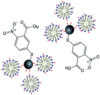
A novel approach, based on surface-enhanced Raman scattering (SERS) and 5,5′-dithiobis-(2-nitrobenzoic acid) (DTNB) functionalized silver nanoparticles, was developed to determine the critical micelle concentration (CMC) of cationic surfactants. A graph was generated by plotting the Raman intensity ratio between the aromatic ring vibration of DTNB at 1558 cm−1 and its symmetric nitro stretching at 1333 cm−1 as a function of surfactant concentration. An abrupt change of slope at a particular concentration was shown to reliably predict the CMC of cetyltrimethylammonium bromide (CTAB).
In recent years, surface-enhanced Raman scattering (SERS) has been rapidly evolving into a practical analytical tool that can be applied to measurements of multiple analytes, even in complex matrices such as food and body fluids.14–27 The objective of this investigation was to explore the application of SERS as an alternative non-invasive approach for the determination of CMCs of a variety of surfactants, especially cationic surfactants. It is hypothesized that the perturbation of the various vibrational bands of special Raman-active tags, as a result of the electrostatic interaction between the negative-charged surface of silver nanoparticles and cationic surfactants, would exhibit a strong correlation with the aggregation extent of the surfactants, and the relative intensity shift could be used to predict the CMC of cationic surfactants.
Herein, we demonstrate the usefulness of SERS for the facile and accurate determination of the CMC of cationic surfactants. Our approach involves surface modification of silver nanoparticles (Ag NPs) by a Raman-active molecule, namely Ellman's reagent (5,5′-dithiobis-(2-nitrobenzoic acid)) or DTNB, which is one of the most commonly used colorimetric agents for the quantification of thiol groups in biological samples.28 Cetyltrimethylammonium bromide (CTAB) and sodium dodecyl sulfate (SDS) were chosen as a model compound for cationic surfactant and anionic surfactant, respectively. It was shown that the CMC of CTAB could be readily determined by analysing the relative Raman intensity ratio between two characteristic vibrational bands associated with DTNB. SERS determination of the CMC of cationic surfactants by DTNB-functionalized Ag NPs involved three steps, as illustrated in Fig. 1. Firstly, highly SERS active colloidal Ag NPs were synthesized via reduction of AgNO3 with hydroxylamine hydrochloride at alkaline pH and at room temperature.29 Specifically, 10 mL of 10 mM AgNO3 solution were added drop wise at a rate of 1 mL min−1 using a burette, to 90 mL of 1.67 mM hydroxylamine hydrochloride solution containing 3.3 mM NaOH under vigorous stirring (1000 rpm), and the resultant mixing solution was kept stirring for 1 hour under ambient conditions. The ability to control the size and morphology of Ag NPs was typically accomplished via the use of surface modifiers such as CTAB and poly (N-vinyl-2-pyrrolidone) (PVP) etc. However, the huge promise for SERS applications offered by the excellent structure variability is so far largely unfulfilled, mostly due to the tedious work in the complete removal of the surfactants and the reduced colloidal stability of the stabilizer-free nanoparticles in aqueous solutions. The advantages of using as-prepared Ag NPs arise primarily from (1) ease of preparation, (2) clean surface for facile thiolation, (3) large extinction coefficient of the surface plasmon absorption (which is approximately 3–4 times higher than that of AuNPs with the same size),30 and (4) its rendering of the surface of the particles a net negative charge (as shown in the reaction below):
| 4Ag+ + 16NH2OH·HCl + 20OH− + 7O2 → 4Ag + 8N2O + 16Cl− + 42H2O |
Secondly, the functionalization of Ag NPs by DTNB was accomplished by mixing 1 mL of a DTNB solution (1 mg mL−1 dissolved in a 1
:
1 v/v ethanol–DI water mixture) with 5 mL of as-prepared Ag NPs. Thiols are well-known ligand molecules as they can form a robust self-assembled monolayer on noble metal nanoparticle surfaces through a strong metal–sulfur covalent bond (the energy gold–sulfur bond is 418 kJ mol−1 and the silver–sulfur bond is 217 kJ mol−1).31 When mixed with Ag NPs, the disulphide group in DTNB can easily break and form an Ag–S bond (Fig. 1A). Thirdly, as the surfactant concentration was raised above a certain level, aggregates of CTAB assemble such that the hydrophobic tails of the CTAB molecules are packed together in the interior of the micelle while the polar or hydrophilic head groups form a boundary zone between the nonpolar core of the micelle and the polar aqueous solution beyond (Fig. 1B). Aggregates of DTAB-functionalized Ag NPs are then adsorbed onto the surface of the CTAB micelles, causing a detectable signal that can be monitored by Raman spectroscopy (Fig. 1C).
As depicted in Fig. 2A, spherical silver nanoparticles displayed a broad absorption peak centred around 450 nm. The morphology and sizes of Ag NPs were shown in Fig. 2B. The diameter of this particular batch of Ag NPs ranged from 50–78 nm. To prepare the samples for scanning electron microscopy (SEM) studies, a drop of the Ag colloids was placed on a piece of silicon wafer substrate and dried over night at room temperature. The samples were then observed using a Nova NanoSEM 630 (FEI) field emission scanning electron microscope to assess the particle size and shape.
Fig. 3 shows the SERS spectra of DTNB in the presence of different amounts of CTAB and SDS. Raman spectra were obtained with a DeltaNu Advantage 200A (DeltaNu, Laramie, WY) equipped with a HeNe laser emitting at 633 nm. The laser power was 3 mW and the spectral resolution was 10 cm−1. Raman spectra were in backscatter directly from an NMR glass tube (with an integration time of 10 s), and baseline-corrected using the software came with the system. The stock solutions of CTAB (1% w/v) and SDS (1% w/v) were prepared by dissolving weighed amounts of each surfactant in appropriate amounts of high purity deionized water (resistivity more than 18 MW cm).
As shown in Fig. 3A, the adsorbed DTNB molecules exhibit strong characteristic Raman signals via the SERS-inducing silver substrate. The Raman spectrum of DTNB is dominated by several strong peaks at 1079, 1333, and 1558 cm−1, which could be assigned as the succinimidyl N–C–O stretch overlapping with aromatic ring modes, the symmetric stretch of the N–O nitro group, and an aromatic ring stretching mode, respectively.32 The latter two peaks were deviated from their reported values (i.e., 1342 and 1566 cm−1), suggesting an orientation change of DTNB in CTAB solutions vs. that in pure water systems. We also investigated the effect of SDS, an anionic surfactant, on the SERS spectra of DTNB after mixing with several serial dilutions of a SDS solution (1% w/v). It was noted that the relative intensities of all the characteristic DTNB Raman peaks remained remarkably consistent even though the SDS concentration varied from 1× to 32× dilution (see Fig. 3B). The Raman intensity of various bands at 1333, 1342 or 1558 cm−1, as measured by the peak height of each corresponding peak, was obtained automatically by the software came with the DeltaNu Raman system, after a baseline correction was performed for each measurement. The fluctuation of the relative Raman intensities in the presence of different amounts of CTAB might be attributed to the distance changes between the relevant functional groups of DTNB molecules and the AgNP surface, which were perturbed by the adsorption of various aggregated structures of CTAB on negatively charged AgNP surfaces. Our observation was consistent with similar studies using attenuated total reflection-Fourier transform infrared (ATR-FTIR) spectroscopy.33,34
Despite the strong similarity between the SERS spectra obtained in the presence of CTAB and those in the presence of SDS, there exists a striking difference between the two sets of spectra, whereby the 1333 cm−1 peak observed in the pure DTNB SERS spectrum was split into a doublet (i.e., 1342 and 1353 cm−1) in the presence of CTAB, but not in the presence of SDS, suggesting that one of the peaks could have the contribution of CTAB. It is reasonable to assume that the peak at 1352 cm−1 in the SERS spectra of DTNB-functionalized Ag NPs originates from the adsorption of CTAB on Ag NP, and could be ascribed to deformation of methyl groups attached to the quaternary nitrogen.35 This suggests that the adsorption of CTAB molecules on negatively-charged Ag NP is established via the quaternary ammonium group.
A careful analysis of the relative peak intensity ratio between vibrational bands at 1333 cm−1 and 1558 cm−1 revealed a sudden change of the slope as a function of the CTAB concentration (see Fig. 4). More strikingly, the CTAB concentration (∼1.1 mM) at the turning point extrapolated from the graph matches the well-accepted CMC value for CTAB in similar solvent conditions.12,36,37 The unique selectivity for the recognition of CTAB over SDS can be explained by the electrostatic interactions between positively-charged CTAB molecules and the as-prepared negatively-charged Ag NPs, and the electrostatic repulsion between negatively-charged SDS molecules and negatively-charged Ag NPs. With increasing concentrations of CTAB in solution, CTAB molecules started to aggregate, and eventually took the shape of a micelle. CTAB micelles in the size of several nm are then adsorbed onto the surface of AgNP aggregates. The steric hindrance among the benzene ring moieties forced DTNB to stand more perpendicularly from the surface of Ag NPs. As a result, the nitro group was extended a little bit further from the surface, which led to a decrease in its Raman intensity. The surface Raman electromagnetic enhancement factor (EF) for individual bands in a self-assembled monolayer of molecules can be approximated using EF = [r/(r + d)],10 whereby d is the distance between the specific bond and the surface of a particle, and r is the radius of the nanosphere.38–40 Whereas in the case of SDS, no extra force existed to induce any orientation change of the same set of functional groups in DTNB.
In summary, we have demonstrated a novel SERS-based method for CMC determination of cationic surfactants. Our approach differs fundamentally from many existing ones in many aspects. First, the intensities of the various vibrational bands were found to show a strong dependence on the aggregation extent of the surfactants, specifically for cationic surfactants. The strong perturbations in the vibrational band intensities of DTNB can be used as a Raman probe in the study of micellar aggregates. Moreover, the variations in the intensity ratio between the aromatic ring vibration of DTNB at 1558 cm−1 and its symmetric nitro stretching at 1333 cm−1 could be used to accurately determine the CMC of CTAB, whilst no such changes were observed for SDS. It is expected that, by tailoring the surface charges of the Raman-active labels and/or the surface of noble metal nanoparticles, our approach can be further developed to become a powerful investigative tool for the study of a variety of amphiphilic species besides surfactants, including polymers, membranes, biological peptides and proteins etc.41
| This journal is © The Royal Society of Chemistry 2014 |