Nanoengineered CdSe quantum dot–montmorillonite composites: an efficient photocatalyst under visible light irradiation

Rajeev C. Chikate*a, Brijesh S. Kadua and Madhura A. Damleb
aNanoscience Group, Department of Chemistry, Post-graduate and Research Centre, MES Abasaheb Garware College, Karve Road, Pune-411004, India. E-mail: rajuchikate29@gmail.com; Fax: +91-20-25438165; Tel: +91-20-41038263
bDepartment of Biotechnology, MES Abasaheb Garware College, Karve Road, Pune-411004, India

Received 3rd June 2014 , Accepted 4th August 2014

First published on 4th August 2014


Abstract

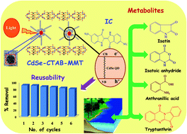
In this paper, we have developed an efficient electrostatic self-assembly strategy for the synthesis of a CdSe–CTAB–MMT composite that contains well-dispersed CdSe quantum dots (CdSe QDs) within the MMT structure. These nanocomposites are characterised by XRD, TEM, Raman, DRS and photoluminescence studies which indicate that 3–5 nm sized CdSe QDs are intercalated within lamellar sheets of MMT. Visible light induced photocatalytic activities of the composites exhibit decolourisation of Indigo Carmine (IC) solution (100 mg L−1) within 30 min. at 1.0 g L−1 catalyst loading. This feature is attributed to separation of photogenerated electron–hole pairs, enhanced interlayer spacing (7.2 A°), higher specific surface area and better adsorption capacity of the MMT. The involvement of reactive oxygen species (ROS) in the photodegradation process is ascertained by addition of selective quenchers such as NaN3 (for singlet oxygen), benzoquinone (for O2˙), ascorbic acid (for OH˙) and KI (for h+). It is observed that singlet oxygen and photogenerated h+ do not contribute towards degradation; rather O2˙ is a prominent species that degrades ∼74% of IC while the remaining part is oxidized by OH˙. The photodegradation pathway involves desulphonation of IC followed by its oxidation to isatin, anthranilic acid, tryptanthrin and isatoic anhydride. The antibacterial studies of degraded IC solution as well as chemoinformatics studies suggest that these metabolites are non-toxic in nature. These catalysts remain active for up to 6 cycles with a marginal decrease in their removal capacity that can be ascribed to inhibition of photocorrosion even after successive exposure to light. Thus, nano-engineered CdSe-composites may be regarded as efficient photocatalysts that have potential applications in sustainable development towards continuous removal of organic dyes from aqueous streams.


1. Introduction

Semiconductor photocatalysis, an environmental benign process, can address issues related to energy shortages and remediation of organic pollutants.1 However, photoefficiency of these catalysts is restricted due to: (i) low quantum yield as a consequence of fast recombination of hole (h+) and electron (e); (ii) poor absorption of visible light and (iii) moderate reaction rates that severely affect the development of a high throughput processes.2,3 To overcome these lacunas, two strategies are adopted: (a) shifting the photoabsorption process in the visible region by doping noble metals4 and (b) separation of photogenerated charge carriers via formation of semiconducting composites.5

Semiconductor assisted degradation of pollutants proceeds through sequence of events that occur on the photocatalytic surface as: (a) generation of e and h+ pairs through absorption of photons (b) their effective separation to avoid recombination process (c) oxidation of the organics by the photo-generated h+ (d) formation of ROS like OH˙, O2˙ and H2O2 via reduction of dissolved oxygen by photogenerated e and (e) oxidation of organics by ROS.6 It is envisaged that these oxidative species are primarily responsible for the degradation of dyes generating secondary metabolites that are identified using chromatographic techniques to elucidate the degradation mechanism.7–12 However, none of these reports have focused on establishing the relationship between ROS and degraded products, rather majority have probed formation of ROS through variety of spectroscopic techniques. For example, generation of O2˙ and OH˙ radicals is established with ESR-DMPO spin-trapping technique where low degradation activity was observed with TiO2 based photocatalysts.13,14 Moreover, kinetics of degradation of dyes in presence of scavenger for h+ revealed that it predominantly influences the degradation while photogenerated e exhibit negligible contributions towards reductive cleavage.15–17 Fluorescence method is also applied for the detection of OH˙ with various TiO2 powders under UV irradiation.18 Therefore, the exact role of ROS in the degradation and subsequent formation of secondary metabolites is still remained speculative.

To address this issue, we have explored two strategies: (i) separation of photogenerated e and h+ pair by forming semiconductor nanocomposites and (ii) probing the role of ROS in the degradation process by monitoring the formation of secondary metabolites. To ascertain these hypotheses, CdSe–MMT nanocomposites are employed as photocatalyst for visible light driven photocatalytic degradation of IC (ESI, Fig. S1). CdSe, an n-type semiconductor with band gap19 in the range 1.65–1.8 eV has received wide applications in labelling, sensors, optometric devices, solar cells and photocatalysis. Although it has extensively been employed for biological applications,20 its immobilization onto a solid support for photocatalytic applications is still remained unexplored. For this purpose, MMT is selected as a support because it possesses (i) high cation exchange capacity (119 meq/100 g),21 (ii) hydroxyl groups on the edges, (iii) beneficial Lewis and Bronsted acidity and (iv) potentiality towards formation of laminar structures through intercalation.22,23 This approach is beneficial for better photoabsorption followed by degradation of organic pollutants as well as for repetitive usage of photocatalysts.24 We have also tried to establish the correlationship between ROS and the degraded products of IC via radical quenching experiments and proposed ROS mediated degradation pathway.

To assess the environmental implications, antibacterial activity of degraded IC solution is carried out against E. coli, B. subtilis and S. aureus. Since the isolation of individual metabolite is not possible at our end, their drug-like activity and related properties are evaluated with chemo-informatics softwares such as Mole Inspiration Properties Calculator (MIPC) and Molsoft to evaluate their environmental hazards.

2. Experimental

2.1. Chemicals

All chemicals were of analytical grade and used as received. IC (Hi-Media Chemicals, UK) Cadmium acetate (Cd(OAc)2. 2H2O, Loba Chemie, UK), Selenium powder (Loba Chemie, UK), Cetyl trimethyl ammonium bromide (Sigma Aldrich); Montmorillonite K-10 (Fluka) and Sodium borohydride (NaBH4, Loba Chemie, UK).

2.2. Pretreatment of MMT

MMT clay was digested with excess 1 M NaCl solution for 24 h at 60 °C and the solid was filtered, washed with acetone then dried at 50 °C. The powder was dispersed in distilled water to prepare the colloidal suspension of clay particles and kept overnight. It was filtered, washed with distilled water and acetone and finally dried under vacuum.

2.3. Synthesis of cadmium selenide (CdSe QD's–CTAB)

0.3 g of NaBH4 in 5 mL water was added to 0.78 g Se in 5 mL water to obtain wine red coloured solution of sodium hydrogen selenide (NaHSe). In other flask, aqueous solution of cadmium acetate (1.3 g in 10 mL water) was mixed with 10 mL CTAB (0.02 M) solution. To this mixture, NaHSe solution was added drop wise with constant stirring at 40 °C and the resulting mixture was kept under stirring for 1 h. Wine red coloured product was centrifuged at 3000 rpm, successively washed with methanol and then dried under vacuum.

2.4. Preparation of CdSe–CTAB–MMT nanocomposite

0.2 g of CdSe was suspended in 15 mL distilled water and the whole mixture was sonicated for 4 h. This solution was added to an aqueous suspension of 1.8 g Na–MMT in 10 mL of water and the whole mixture was stirred at 40 °C for 2 h. The wine red mixture was filtered, washed successively with distilled water and finally with acetone and the product was dried under vacuum. This resulted in 10%-CdSe–CTAB–MMT composite and similar procedure was adopted for the preparation of 5% and 15% compositions with appropriate amounts of CdSe and MMT.

2.5. Characterization of nanocomposites

X-ray powder diffraction (XRD) spectra were recorded on Phillips X'pert MPD X-ray diffractometer using CuKα radiation. Transmission electron microscope (TEM) images were obtained on JEOL electron microscope (model 1200X). Field emission scanning electron microscope (FE-SEM) images were obtained on Hitachi S-4800. Raman spectra were recorded on Horiba JY LabRAM HR 800 micro-Raman spectrometer operating at 17 mW and 632.8 nm excitation. Diffuse reflectance spectra of the solid samples were measured on a Shimadzu UV-VIS-NIR-3101 PC scanning spectrophotometer using an integrated sphere. Photoluminescence (PL) spectra were recorded on a Hitachi F-4500 fluorescence spectrophotometer with the excitation wavelength of 450 nm. UV-VIS spectra were recorded on UV-VIS spectrophotometer (Perkin Elmer, Lambda 25). ICP-OES measurements were carried out on Optima 7000 DV ICP-OES Spectrometer (Perkin-Elmer).

2.6. Photocatalytic measurements

The photocatalytic activities of CdSe–CTAB–MMT composites were evaluated for the degradation of IC (100 mg L−1) prepared in distilled water. 100 mg of the catalyst was added the 100 mL of the IC solution in a 250 mL quartz reactor (ESI, Fig. S2). The mixture was stirred in the dark for 30 min to establish baseline correction after adsorption of dye on catalyst. It was then irradiated with Xenon lamp (500 W Xe lamp with a 420 nm cutoff filter) and 5 mL aliquots were withdrawn at certain time intervals to monitor the absorbance of supernatant at 610 nm.

2.7. Identification of metabolite

The identification of degraded products was carried out on LC-MS (Dionex, Ultimate 3000) chromatographic system interfaced with a PE SCIEX model API3000 Triple Quadruple mass spectrometer equipped with a turbo ion spray. Samples were eluted at a flow rate of 1.5 mL min−1 through an Inertsil ODS 3 V column (5 μm, 150 mm × 4.6 mm) using gradient: from 0/100 (ammonium acetate 10 mM in acetonitrile/water) to 90/10 in 25 min, which was then maintained for 6 min. The interface conditions for the positive ion mode were as follows: nebulizer gas: 8 mL min−1, curtain gas: 12 mL min−1, needle voltage: 4500 V at 450 °C, orifice declustering potential: +10 V and focusing potential: 100 V. The flow from the HPLC-UV was split to allow 200 μL min−1 to enter the turbo ion spray interface.

2.8. Quenching experiments

The quenching experiments with radical scavengers were as follows:

To 100 mL IC solution containing 100 mg nanocomposite, 0.2 mM of appropriate quencher was added and the whole mixture was illuminated for 30 min. using Xenon lamp in a photocatalytic reactor. The mixture was centrifuged at 3000 rpm and the supernatant was subjected for LC-MS analysis.

2.9. Reusability studies

The recycling experiments were carried out on [IC]ini = 100 mg L−1 with 1 g L−1 nanocomposite. After each photodegradation experiment, solution was centrifuged at 3000 rpm successively and the catalyst was carefully separated from liquid phase by a simple decantation. The recovered catalyst was successively washed with water and acetone and then used for subsequent cycles.

2.10. Antibacterial study

Antibacterial activity of IC and its degraded solution was evaluated with E. coli (NCIM-2065), B. subtilis (NCIM-2063) and S. aureus (ATCC-6538). All cultures were grown overnight at 37 °C in an incubator shaker maintained at 120 rpm. The cell suspension was made from the cultures under sterile physiological saline (106 CFU per mL) condition. Nutrient agar plates were prepared by pour plate method using 0.1 mL of cell suspension and 6 mm sized wells were bored inside the gel. 40 μL of both IC and its degraded solution was added in the wells and the plates were incubated at 37 °C for 24 h. Distilled water was used as the control. After 24 h of incubation, the zone of inhibition was measured.

3. Results and discussion

The strategy adopted for the synthesis of nanocomposites is illustrated in Scheme 1. It comprises of three processes: (i) pretreated Na–montmorillonite exfoliates into the separate sheets when dispersed in the distilled water (ii) size quantization of CdSe due to its capping with CTAB (iii) formation of CdSe/CTAB/Clay sandwich-like structure through self-assembly of exfoliated MMT and CTAB coated CdSe QD's via ion-exchange mechanism. Na+ ions present in the interlayers of MMT undergo exchange reaction with homogeneously dispersed CdSe QD's. It results in the formation of laminar structure whose basal spacing is increased due to intercalation of CdSe QD's.
image file: c4ra05280c-s1.tif
Scheme 1 Graphical illustration of the synthesis of intercalated CdSe QD's in MMT sheets.

3.1. Characterisation of nanocomposites

X-ray diffraction patterns of CdSe QD's and its nanocomposites are shown in Fig. 1. The reflections appearing at 25.36°, 42.7° and 50.58° due to (002), (110), (112) planes correspond to Wurtzite structure25 of CdSe (JCPDS 08-0459) while pretreated MMT exhibits characteristic (001) peak at 7.8°. Upon composite formation between Na–MMT and CdSe–CTAB, the peak positions corresponding CdSe Wurtzite structure are retained with broadened nature. It implies that CdSe QD's are homogeneously dispersed26 within the MMT matrix with the particle size in the range of 2–3 nm. Thus, it may be argued that the interlayer spacing of MMT is effectively utilized for the formation of the organically modified CdSe–CTAB–MMT nanocomposites.
image file: c4ra05280c-f1.tif
Fig. 1 X-ray diffraction patterns of CdSe-nanocomposites.

The morphology and the size of CdSe particle are investigated by TEM measurements. CdSe QD's possess spherical morphology of 3 nm size (Fig. 2a) as well as exhibit characteristic (002) plane having identical d-spacing reported for Wurtzite structure.25 On the other hand, these QD's are uniformly distributed within MMT matrix that results in a sheet-like structure (Fig. 2b–d). Considering the enhanced interlayer spacing of MMT, it is observed that disk- or plate like CdSe QD's are properly intercalated (ESI, Fig. S3) within the CTAB–MMT layers with particle size of about 2–3 nm. SEAD patterns of nanocomposites (Fig. 2e–h) reveal that CdSe QD's are homogeneously dispersed within the interlayers of MMT. The specific surface area of Na–MMT, CdSe QD's, 5%- and 10%-CdSe–CTAB–MMT is found to be 34, 24.4, 68.9 and 177.0 m2 g−1 respectively. Higher value 10% CdSe doped MMT implies that the nanocomposite formation leads to generation nanoporous structure.


image file: c4ra05280c-f2.tif
Fig. 2 HR-TEM images of (a) CdSe QD's–CTAB (b) 5%-CdSe–CTAB–MMT (c) 10%-CdSe–CTAB–MMT, (d) 10%-CdSe–CTAB–MMT and SAED patterns of nanocomposites (e–f).

Fig. 3 represents Raman spectra for CdSe QD's and its nanocomposites obtained by Lorenzian fitting methodology. A peak due to a longitudinal-optical mode of CdSe27 is observed at 208 cm−1 (or 6.23 THz) for CdSe QD's while the same mode occurs at 213 cm−1 (or 6.38 THz) for both 5%- and 10%-CdSe–CTAB–MMT. Such a shift with broadening (FWHM = 16 cm−1) for nanocomposite is caused by decrease in size as well as homogenization of CdSe QD’s within MMT matrix. Similarly, 2LO photon peak for composites is also shifted towards higher energy as compared to CdSe QD's (419 cm−1) which can be attributed to the relaxation of momentum conservation28 while the broadening probably originate from negative phonon dispersion.


image file: c4ra05280c-f3.tif
Fig. 3 Raman Spectra of (a) CdSe QD's–CTAB (b) 5%-CdSe–CTAB–MMT and (c) 10%-CdSe–CTAB–MMT.

The absorption onsets from diffuse reflectance spectra (Fig. 4) reveal band gaps in the range 1.83–2.07 eV. It is interesting to note that there exists a blue shift for CdSe-composites as compared CdSe QD's and this observation is consistent with starch,29 oleic acid30 and TOP/TOPO31 capped CdSe nanocrystals. Such a feature can be attributed to ionic interaction of CdSe QD's with CTAB as well as their prohibited agglomeration due to structural rigidity of MMT layers. Thus, synergistic effect of CTAB capping and MMT results in the formation of properly dispersed CdSe QD's that generates distinct reactive sites.


image file: c4ra05280c-f4.tif
Fig. 4 DRS Spectra of (a) CdSe QD's–CTAB (b) 5%-CdSe–CTAB–MMT and (c) 10%-CdSe–CTAB–MMT.

The photoluminescence spectrum of CdSe QD's–CTAB (Fig. 5 a) exhibit a strong emission peak at 572 nm with a full width at half-maximum (FWHM) of 26 nm and is consistent with literature reports on CdSe nanoparticles capped with thiolato-,32 chitosan-,33 PEG-,34 hexadecylamine-35 and oleic acid.36 On the other hand, the PL spectrum of 5% and 10% CdSe composites (Fig. 5b and c) revealed a broad emission at 510 and 528 nm with FWHM of 56 and 64 nm respectively. Broadening in the PL spectra for composites indicates uniform shape, size, and homogeneous dispersion of CdSe QD's within the interlayer space of montmorillonite.37,38 The blue shift of the near band-edge emission for composites is originating from quantum confinement as well as exfoliated nature of MMT sheets.26 It is plausible that such a feature arises due to surface defects that may lead to dissipation of emission energy and subsequent broadening of band-edge emission.39


image file: c4ra05280c-f5.tif
Fig. 5 Photoluminescence spectra of (a) CdSe QD's–CTAB (b) 5%-CdSe–CTAB–MMT and (c) 10%-CdSe–CTAB–MMT.

3.2. Kinetics of IC degradation

The photocatalytic decolourisation of IC with CdSe QD's–CTAB and their nanocomposites is carried out with 100 mg L−1 IC solution and 1 g L−1 photocatalyst. It is observed that almost 92% decolourization occurs within 30 min (Fig. 6) for CdSe–CTAB–MMT composites while CdSe QD's exhibits lesser removal rates. These results are attributed to the pillaring of clay with CdSe QD's that lie between S–G–S layers of the clay.38 Due to pillaring, the change in the basal space enhances the accessibility of IC towards CdSe centres that are responsible for the efficient degradation of dye in presence of light.26 Since photocatalytic degradation of dye is a pH dependent phenomenon, effect of pH on removal capacity in the pH range of 1–8 is carried out on 10%-CdSe–CTAB–MMT composite. Higher removal capacity at pH 7 (ESI, Fig. S4) suggests that there is a deactivation of photocatalyst surface under acidic medium. Thus, further investigations are carried out with this photocatalyst at pH 7 for elucidating the degradation mechanism as well as biological screening of metabolites generated after degradation of IC molecule.
image file: c4ra05280c-f6.tif
Fig. 6 Time profile of IC decolourisation by CdSe QD's–CTAB and CdSe–CTAB–MMT nanocomposites: {[IC]ini = 100 mg L−1, [nanocomposite] = 1 g L−1, at 25 °C}.

3.3. Effect of reactive oxygen species (ROS) on degradation pathway

It is well established that dissolved oxygen (D.O.) in water is the primary source for degradation of organic pollutants.40 In order to ascertain this fact, the degradation of IC carried out in aqueous and N2 purged (1 h) solutions (Fig. 7, inset) which suggests that only 10% of dye is decolorized under anaerobic condition as compared to ∼90% in presence of D.O.41 Such a small amount of decolorization for N2 purged sample may be occurring due to either adsorbed oxygen or hydroxyl groups present on photocatalytic surface.42 However, these findings do not demonstrate the type of oxygen species responsible for oxidative cleavage of dye molecule. To substantiate this hypothesis, various scavengers such as NaN3 (for singlet oxygen),43 1,4-benzoquinone (BQ; for O2˙)44 and ascorbic acid (AA; for OH˙ radical)45 are added to IC solution before it is irradiated with visible light. It is reported that h+ are also responsible for oxidation of organic pollutants.46 This aspect is evaluated by addition KI, a hole quencher, as it would hinder electron–hole recombination process as well as assist in predicting the ease at which the formation of ROS takes place through reduction of dissolved oxygen. Fig. 7 illustrates decolourisation curves for IC without and with addition of 0.02 M quenchers. It is interesting to note that quenching of h+ and singlet oxygen does not affect degradation as the nature of graphs is almost similar to non-quenched solution. Such a feature illustrates that electron–hole recombination process is hindered due to presence of MMT layers that dissipate photogenerated electrons and subsequently facilitates the reduction of adsorbed oxygen on catalytic surface. However, when O2˙ and OH˙ radicals are scavenged, only 20% decolourisation is observed indicating that these two radicals are indeed responsible for oxidative cleavage of IC molecule.
image file: c4ra05280c-f7.tif
Fig. 7 Variation of normalized concentration of IC in solution in the presence of 2 mM of AA, BQ, NaN3, and KI. Inset: % Degradation in presence of D.O. and N2 purged solutions. {[IC]ini = 100 mg L−1, [10%-CdSe–CTAB–MMT] = 1 g L−1, 25 °C}.

To further corroborate the role of O2˙ and OH˙ radicals in the formation of secondary metabolites, HPLC analysis is carried out on decolorized IC solution obtained either with or without addition of quenchers (ESI, Fig. S5). Chromatogram of non-quenched solution (ESI, Fig. S5a) reveals that five metabolites are formed whose molecular masses are identified from MS analyses (ESI, Fig. S6). Isatin (I) is found to be major degradation product (43%) followed by isatoic anhydride (III, 21%), tryptanthrin (II, 11%) and anthranilic acid (II, 10%) with formation of small amount of Indigo blue (IB, 7%). Formation these metabolites (Scheme 2) can be explained as: (i) desulphonation of IC to IB (ii) reductive cleavage of double bond resulting into isatin (I) and anthranilic acid (II) and (iii) their cyclisation to isatoic anhydride (III) and tryptanthrin (IV) through oxidative cleavage.47


image file: c4ra05280c-s2.tif
Scheme 2 Proposed degradation mechanism of IC in presence of D.O. using 10%-CdSe–CTAB–MMT.

On the other hand, LC-MS analysis of BQ and AA treated IC solution results in the formation of ∼90% of IB through desulphonation of IC that primarily occurs in absence of ROS. It is plausible that positively charged CdSe–CTAB–MMT surface along with visible light is responsible for this step. IV is formed when O2˙ is quenched by addition of BQ (ESI, Fig. S5b) while isatin (I) and its anhydride (II) are generated after scavenging of OH˙ by AA (ESI, Fig. S5c). Small amount of IV is also obtained through condensation of anthranilic acid (II) and isatin (I)48 that are formed due to oxidation of IB. It also suggests that ROS are indeed required for complete oxidation of IC; a feature already been observed for variety of photoredox processes. Conversely, quenching of OH˙ leads to formation of partially oxidized metabolites I and III which suggests that O2˙ radical initiates oxidative cleavage of IB across central double bond. The formation of significant amount of O2˙ radical is possibly favoured due to either hindered electron–hole recombination process or photo-excitation of IC molecule. This hypothesis is further corroborated with addition of KI (h+ quencher) where it is observed that quenching of h+ has no effect on degradation process (Fig. 7). It suggests that photoelectrons generated from composites as well as IB are absorbed by dissolved oxygen and it is estimated that ∼64% of O2˙ are formed during the photocatalytic process. It is estimated from the amounts of different metabolites formed due to the reaction between IC and this radical (Scheme 2). However, for complete oxidation of IB to II, strong oxidizing agent such as OH˙ is essential as it is only formed under aerobic condition (ESI, Fig. S5a). Therefore, it may be argued that O2˙ radical is the most prominent ROS species49 responsible for ∼64% degradation with further cleavage occurring in presence of OH˙ radicals. Thus, quenching experiments clearly demonstrate that O2˙ and OH˙ radicals are reactive species involved in the oxidative degradation IC to oxygenated metabolites. To summarize, visible light assisted photocatalytic degradation of IC occurs through (a) desulphonation of IC to IB (b) partial oxidation with O2˙ radical forming isatin and its anhydride and (c) generation of anthranilic acid through OH˙ radical.

3.4. Reusability studies

The effectiveness of a photocatalyst is considered to be based on how it remains active during continuous illumination of light because it is quite likely that it may loose its catalytic activity due to photocorrosion. To assess this aspect, recycling experiments are carried out on recovered photocatalyst with 100 mg L−1 IC solution with 1 g L−1 photocatalyst (Fig. 8). The retention of photocatalytic efficiency during 6 cycles implies that composite surface does not undergo photocorrosion due to successive exposure to visible light. XRD and TEM analyses of recovered photocatalyst after 6th cycle (ESI, Fig. S7) suggest that there exist negligible structural changes as well as the morphology of composite is also retained even after its repetitive usage. Interestingly, when these results are compared with different photocatalysts50–54 employed for IC degradation (Table 1), it is observed that 10%-CdSe–CTAB–MMT composite decolorizes IC at substantially lesser time even at higher concentration of dye reflecting its significantly higher photocatalytic efficiency. Moreover, our composite possess excellent removal efficiency with almost 520 ppm of IC is successfully removed within 3 h. Since cadmium and selenium both being toxic, it is imperative to estimate the leaching of these two species during photocatalytic experiment. It is monitored with ICP-OES measurements of supernatant obtained after 6th cycle of successive usage of 10%-CdSe–CTAB–MMT and the amounts are found to be 2.9 and 4.5 ppb for cadmium and selenium respectively.
image file: c4ra05280c-f8.tif
Fig. 8 Recycling efficiency of 10%-CdSe–CTAB–MMT towards removal of IC.
Table 1 Comparison of photocatalytic decolourisation efficiency of different photocatalysts towards Indigo carmine
Photocatalyst Dosage (g L−1) Dye conc. (mg L−1) Decolourisation time (min) % Decolourisation Ref.
Nb2O5 1.0 4.66 90 100 50
Fe, N co-doped TiO2 2.0 20 180 89 51
Ag/graphene oxide 1.0 10 420 41 52
TiO2 on SiC 1.0 20 30 100 53
CeAlO3 1.0 100 90 100 54
10%-CdSe–CTAB–MMT 1.0 100 12 92 Present work


Thus, it may be argued that designing of such tailored made functional nanomaterials may be regarded as viable strategy towards the development of sustainable photocatalytic process that may possess potential applications towards continuous removal of organic pollutants from aqueous medium.

3.5. Environmental implications

Other implication of this strategy is also focused on whether the secondary metabolites that are generated during the photodegradation of a toxic dye55 possess any environmental hazards. Two strategies are envisaged to address this issue; (i) isolation of individual metabolites through prep-HPLC and evaluate their individual toxicities and (ii) antimicrobial activity of degraded solution as a whole. However, former studies are not possible at our end while the latter approach would result in combined toxicity and not due to individual metabolite.

Antibacterial activity of IC and its degraded solution is carried out against three bacterial strains viz. E. coli, S. aureus and B. subtilis (ESI, Fig. S8). IC exhibits 6–8 mm zone of inhibition at 100 ppm; even at lower concentration (10 ppm), there exists inhibition of growth implying that IC is indeed toxic in nature. However, when 100 ppm degraded solution is exposed to these strains, there is absence of zone of inhibition suggesting that none of the five metabolites possess antibacterial activity. To corroborate this observation, we have employed chemoinformatics software commonly used for predicting drug-like activity for variety of organic molecules. After careful literature search, it is noticed that Molinspiration Property Calculator (MIPC)56 is routinely employed for such a purpose. Employing this tool, the simulated molecular properties such as percentage of absorption, molecular polar surface area (PSA), log[thin space (1/6-em)]P, number of rotatable bonds, number of hydrogen bond donor and accepter atoms of Lipinski's rule of five are evaluated for metabolites I–IV (ESI, Table T1).

Hydrogen bond acceptor (HBA) and donor (HBD) capacities of functional groups indeed play crucial role in identifying its interaction with potential receptors that favour hydrogen bonding. It can be seen from these two parameters that number of hydrogen bond acceptor (≤10) and hydrogen bond donor (≤5) sites for I–IV obey Lipinski's rule of five implying that they possess good absorption or membrane permeability. Moreover for compounds I and II, these values are ≤3 indicating their drug like character. Lipophilicity (log[thin space (1/6-em)]P) is another factor that governs the passive membrane partitioning as well as influencing permeability of molecule for its subsequent absorption. This value is significantly lower for all the metabolites (0.73–2.90) that augurs well for their substantial lipophilicity. Generally, it is observed that higher molecular weight compounds have adverse effects on interactions with receptors and/or DNA. Such an unfavourable situation is not observed for I–IV that also corroborates with Lipinski's rule of five. Molecular polar surface area (PSA) predicts the transport property of compound and is inversely proportional to %ABS. All the metabolites have maximum absorption (>85%) owing to their small polar surface areas. The drug-likeness model score, computed for secondary metabolites using Molsoft server suggests that tryptanthrin (IV) is the only metabolite that posses drug like properties. Thus, it may be argued that photodegradation process evaluated in the present work may be regarded as eco-friendly that does not possess any threat or hazards to environment.

4. Conclusions

Intercalated CdSe–CTAB–MMT nanocomposites with uniform dispersion of CdSe QD's are fabricated by a facile self-assembly approach that significantly enhances visible light induced photocatalytic degradation of IC. The lamellar structure of composites also hinders recombination of photogenerated electrons and holes corroborated with addition of KI; an h+ quencher, while electrons are subsequently utilised for the formation reduced oxygen species. It is observed that O2˙ radical is the prominent species that photodegrades IC to the tune of ∼74% while remaining part is oxidised via OH˙ radicals. The plausible degradation pathway involves desulphonation followed by oxidative cyclization and complete oxidation of IC generating anthranilic acid. Moreover, these nanocomposites are recycled without sacrificing their photocatalytic activity implying that they can be effective towards continuous removal of dyes. The degraded metabolites are found to non-toxic with negligible leaching of cadmium and selenium during photochemical process even after repetitive usage of photocatalysts. Thus, a simple, tailored made and nanoengineered synthetic strategy can be extended for the fabrication of superior visible-light-driven photocatalysts that are robust and may be employed for diverse photochemical applications in an eco-friendly manner.

Acknowledgements

Authors are thankful to Dr A. D. Natu, Head, Dept. of Chemistry and Dr S.G. Gupta, Principal, A. G. College, for constant encouragement and support. DST, New Delhi is acknowledged for providing FIST grant.

References

  1. M. Yin, Z. Li, J. Kou and Z. Zou, Environ. Sci. Technol., 2009, 43, 8361–8366 CrossRef CAS PubMed.
  2. R. Asahi, T. Morikawa, T. Ohwaki, K. Aoki and Y. Taga, Science, 2001, 293, 269–271 CrossRef CAS PubMed.
  3. Z. Zou, J. Ye, K. Sayama and H. Arakawa, Nature, 2001, 414, 625–627 CrossRef CAS PubMed.
  4. M. Alonso, F. Fresno, S. Suarez and J. Coronado, Energy Environ. Sci., 2009, 2, 1231–1257 Search PubMed.
  5. D. Yang, H. Liu, Z. Zheng, Y. Yuan, J. Zhao, E. Waclawik, W. Ke and H. Zhu, J. Am. Chem. Soc., 2009, 131, 17885–17893 CrossRef CAS PubMed.
  6. J. Tang, Z. Zou and J. Ye, Angew. Chem., Int. Ed., 2004, 43, 4463–4466 CrossRef CAS PubMed.
  7. Y. Lin, D. Li, J. Hu, G. Xiao, J. Wang, W. Li and X. Fu, J. Phys. Chem. C, 2012, 116, 5764–5772 CAS.
  8. C. Chen and C. Lu, J. Phys. Chem. C, 2007, 111, 13922–13932 CAS.
  9. G. Liu, X. Li, J. Zhao, H. Hidaka and N. Serpone, Environ. Sci. Technol., 2000, 34, 3982–3990 CrossRef CAS.
  10. C. Chen, H. Fan and J. Jan, J. Phys. Chem. C, 2008, 112, 11962–11972 CAS.
  11. T. Luo, Z. Ai and L. Zhang, J. Phys. Chem. C, 2008, 112, 8675–8681 CAS.
  12. S. Singh, V. Srivastava and I. Mall, J. Phys. Chem. C, 2013, 117, 15229–15240 CAS.
  13. S. Ribbens, I. Caretti, E. Beyers, S. Zamani, E. Vinck, S. Doorslaer and P. Cool, J. Phys. Chem. C, 2011, 115, 2302–2313 CAS.
  14. Y. Hou, X. Li, Q. Zhao, G. Chen and C. Raston, Environ. Sci. Technol., 2012, 46, 4042–4050 CrossRef CAS PubMed.
  15. X. Doorslaer, P. Heynderickx, K. Demeestere, K. Debevere, H. Langenhove and J. Dewulf, Appl. Catal., B, 2012, 111–112, 150–156 CrossRef PubMed.
  16. L. Martinez, J. Faria, J. Rodriguez, C. Rodriguez and A. Silva, Appl. Catal., B, 2012, 113–114, 221–227 CrossRef PubMed.
  17. W. Wang, L. Zhang, T. An, G. Li, H. Yip and P. Wong, Appl. Catal., B, 2011, 108–109, 108–116 CrossRef CAS PubMed.
  18. J. Zhang and Y. Nosaka, J. Phys. Chem. C, 2013, 117, 1383–1391 CAS.
  19. M. Kotkata, A. Masoud, M. Mohamed and E. Mahmoud, Phys. E, 2009, 41, 640–645 CrossRef CAS PubMed.
  20. W. Chan and S. Nie, Science, 1998, 281, 2016–2018 CrossRef CAS.
  21. H. Koyuncu, Appl. Clay Sci., 2008, 38, 279–287 CrossRef CAS PubMed.
  22. Z. Qian, G. Hu, S. Zhang and M. Yang, Phys. B, 2008, 403, 3231–3238 CrossRef CAS PubMed.
  23. B. Hu and H. Luo, Appl. Surf. Sci., 2010, 257, 769–775 CrossRef CAS PubMed.
  24. B. Kadu, Y. Sathe, A. Ingle, R. Chikate, K. Patil and C. Rode, Appl. Catal., B, 2011, 104, 404–414 CrossRef PubMed.
  25. X. Duan, X. Liu, Q. Chen, H. Li, J. Li, X. Hu, Y. Li, J. Ma and W. Zheng, Dalton Trans., 2011, 1924–1928 RSC.
  26. A. Ontam, N. Khaorapapong and M. Ogawa, J. Colloid Interface Sci., 2011, 357, 554–557 CrossRef CAS PubMed.
  27. F. Widulle, S. Kramp, N. Pyka, A. Gobel, T. Ruf, A. Debernardi, R. Lauck and M. Cardona, Phys. B, 1999, 263, 448–451 CrossRef.
  28. I. Campbell and P. Fauchet, Solid State Commun., 1986, 58, 739–741 CrossRef CAS.
  29. J. Li, C. Ren, X. Liu, Z. Hu and D. Xue, Mater. Sci. Eng., A, 2007, 458, 319–322 Search PubMed.
  30. D. Ayele, H. Chen, W. Su, C. Pan, L. Chen, H. Chou, J. Cheng, B. Hwang and J. Lee, Chem.–Eur. J., 2011, 17, 5737–5744 CrossRef CAS PubMed.
  31. I. Robel, M. Kuno and P. Kamat, J. Am. Chem. Soc., 2007, 129, 4136–4137 CrossRef CAS PubMed.
  32. W. Mahmoud and S. Yaghmour, Opt. Mater., 2013, 35, 652–656 CrossRef CAS PubMed.
  33. T. Wang, S. Zhang, C. Mao, J. Song, H. Niu, B. Jin and Y. Tian, Biosens. Bioelectron., 2012, 31, 369–375 CrossRef CAS PubMed.
  34. Y. Sun, B. Zhou, Y. Lin, W. Wang, K. Fernando, P. Pathak, M. Meziani, B. Harruff, X. Wang, H. Wang, P. Luo, H. Yang, M. Kose, B. Chen, L. Veca and S. Xie, J. Am. Chem. Soc., 2006, 128, 7756–7757 CrossRef CAS PubMed.
  35. S. Kim and H. Yang, Curr. Appl. Phys., 2011, 11, 1056–1059 CrossRef PubMed.
  36. S. Bera, S. Singh and S. Ray, J. Solid State Chem., 2012, 189, 75–79 CrossRef CAS PubMed.
  37. Y. Fukushima, Clays Clay Miner., 1984, 32, 320–326 CAS.
  38. S. Hur, T. Kim, S. Hwang, S. Hwang, J. Yang and J. Choy, J. Phys. Chem. B, 2006, 110, 1599–1604 CrossRef CAS PubMed.
  39. M. Vibin, R. Vinayakan, A. John, C. Rejiya, V. Raji and A. Abraham, J. Colloid Interface Sci., 2011, 357, 366–371 CrossRef CAS PubMed.
  40. P. Kamat, Chem. Rev., 1993, 93, 267–300 CrossRef CAS.
  41. P. Novotna, J. Boon, J. Horst and V. Pacakova, Color. Technol., 2003, 119, 121–127 CAS.
  42. P. Ji, J. Zhang, F. Chen and M. Anpo, Appl. Catal., B, 2009, 85, 148–154 CrossRef CAS PubMed.
  43. C. Schweitzer and R. Schmidt, Chem. Rev., 2003, 103, 1685–1758 CrossRef CAS PubMed.
  44. J. Bandara and J. Kiwi, New J. Chem., 1999, 23, 717–724 RSC.
  45. A. Bokare, R. Chikate, C. Rode and K. Paknikar, Environ. Sci. Technol., 2007, 41, 7437–7443 CrossRef CAS.
  46. C. Chen, W. Ma and J. Zhao, Chem. Soc. Rev., 2010, 39, 4206–4219 RSC.
  47. M. Sousa, C. Miguel, I. Rodrigues, A. Parola, F. Pina, J. Melo and M. Melo, Photochem. Photobiol. Sci., 2008, 7, 1353–1359 CAS.
  48. A. Muruganandam and S. Bhattacharya, Indian J. Chem., Sect. B: Org. Chem. Incl. Med. Chem., 2000, 39B, 125–131 CAS.
  49. C. Guo, J. Xu, S. Wang, Y. Zhang, Y. He and X. Li, Catal. Sci. Technol., 2013, 3, 1603–1611 CAS.
  50. A. Prado, L. Bolzon, C. Pedroso, A. Moura and L. Costa, Appl. Catal., B, 2008, 82, 219–224 CrossRef CAS PubMed.
  51. T. Kim, V. González, G. Gyawali, S. Cho, T. Sekino and S. Lee, Catal. Today, 2013, 212, 75–80 CrossRef CAS PubMed.
  52. R. Orozco, H. Rosu, S. Lee and V. Gonzalez, J. Hazard. Mater., 2013, 263P, 52–60 CrossRef PubMed.
  53. C. Solis, I. Ramírez, E. Moctezuma and L. Martínez, J. Hazard. Mater., 2012, 217–218, 194–199 CrossRef PubMed.
  54. P. Deshpande, S. Aruna and G. Madras, Clean: Soil, Air, Water, 2011, 39, 259–264 CrossRef CAS PubMed.
  55. T. Guaraldo, T. Zanoni, S. Torresi, V. Goncales, G. Zocolo, D. Oliveira and M. Zanoni, Chemosphere, 2013, 91, 586–593 CrossRef CAS PubMed.
  56. S. Alegaon, K. Alagawadi, P. Sonkusare, S. Chaudhary, D. Dadwe and A. Shah, Bioorg. Med. Chem. Lett., 2012, 22, 1917–1921 CrossRef CAS PubMed.

Footnote

Electronic supplementary information (ESI) available: Chemical structure of IC, photocatalytic reactor, kinetics, pH effect, mass spectra of metabolites, antibacterial studies and cheminformatics data of metabolites. See DOI: 10.1039/c4ra05280c

This journal is © The Royal Society of Chemistry 2014